Vol. 46, No. 3, pp. 439 - 444, 2005
Chylopericardium is a rare clinical entity in which chylous fluid accumulates in the pericardial cavity. We report a case of primary idiopathic chylopericardium associated with mul-tiple, small cervicomediastinal cystic hygromas occurring in an asymptomatic 43-year-old woman with no history of trauma, thoracic surgery, malignancy, infection or tuberculosis. Echo-cardiography showed a large amount of pericardial effusions and pericardial fluid analysis revealed inappropriately elevated triglyceride. We did not demonstrate communication between the thoracic duct and the pericardial sac by lymphangiography and chest computed tomography. She successfully responded to 30 days of continuous pericardial drainage and 15 days of a medium-chain triglyceride diet after 30 days of total paren-teral nutrition. Follow-up echocardiography 6 months after treatment commencement showed a minimal reaccumulation of pericardial fluid without symptom. We conclude that if a patient is asymptomatic and can well tolerate daily life, surgery including pericardiectomy or ligation of the thoracic duct is not necessarily required.
Key Words: Chylopericardium, pericardial effusions
INTRODUCTION
The accumulation of chyle in the pericardial cavity, therefore called chylopericardium, is an unusual clinical entity.1 It usually arises from
mediastinal neoplasma,2thrombosis of the subcla-vian vein,3 tuberculosis,4 nonsurgical trauma5 or
thoracic or cardiac surgery.6,7 Primary idiopathic chylopericardium, which literally means it has no
known etiology, is even rarer.8-16 It was first
described as clinical entity by Groves and Effler in 1954.8Although several reviews on the subject have been published, its etiology remains unclari-fied. However, many previously reported cases of primary idiopathic chylopericardium were treated by ligating and resecting the thoracic duct, which suggests that leaks from the thoracic lymphatics resulted in the chylous effusion.8,10,11,13-16 We re-port a case of primary idiopathic chylopericar-dium associated with multiple, small cystic hy-gromas in a 43-year-old woman, which was successfully treated using pericardiostomy, con-comitantly with medium chain triglyceride and low fat meal dietary support after total parenteral nutrition.
CASE REPORT
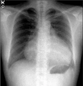
A 43-year-old woman was referred to our hospital for cardiomegaly on chest X-ray (Fig. 1). Physical examinations revealed a healthy ap-pearing woman except for distant heart sounds. All laboratory findings were within the normal range. The patient had no history of trauma, congenital heart disease, thoracic surgery or tuber-culosis. Transthoracic echocardiography revealed a large amount of pericardial effusion without hemodynamic compromise. Diagnostic pericardio-centesis was performed with 500 mL of a milky-colored pericardial fluid aspirated. Routine exami-nation of the pericardial fluid revealed pH 6.8,
Primary Idiopathic Chylopericardium Associated with
Cervicomediastinal Cystic Hygroma
Byoung Chul Cho1, Seok-Min Kang1, Seung Chul Lee1, Jeong Geun Moon1, Dong Hyung Lee1, and Sang Hyun Lim2
1Cardiology Division, Cardiovascular Research Institute, 2Department of Thoracic and Cardiovascular Surgery, Yonsei
University College of Medicine, Seoul, Korea.
Received May 27 2004 Accepted August 23, 2004
Reprint address: requests to Dr. Seok-Min Kang, Cardiology Division, Cardiovascular Research Institute, Yonsei University College of Medicine, 134 Sinchon-dong, Seodaemun-gu, Seoul 120-752, Korea. Tel: 82-2-361-7267, Fax: 82-2-393-2041, E-mail: [email protected]
protein 5.0 g/dL, and LDH 234 IU/L. WBCs and RBCs were uncountable due to cell clumping. Routine bacterial and tuberculous cultures were subsequently reported as negative, as was the cytological examination, and the adenosine deaminase level was 15.5 U/L. Considering these clinical and laboratory findings, the initial clinical impression was of nonspecific chronic pericarditis with effusion. We did not diagnose chyloperi-cardium because we overlooked the clue offered by the color of the pericardial fluid. After peri-cardiocentesis, follow-up echocardiography vealed that minimal pericardial effusion re-mained. She was regularly followed up at our out-patient clinic after discharge. However, echo-cardiography one month later revealed a large amount of pericardial effusion without evidence of hemodynamic compromise (Fig. 2), and she was readmitted to our hospital for further evalua-tion. The patient, who was afebrile on admission, complained only of mild substernal discomfort. Her blood pressure was 90/60 mmHg and pulse rate 72 beats per minute. On physical examination neck veins were distended and peripheral pulses were normal. Her breathing sound was clear without rale, though heart sounds were slightly decreased and pericardial friction rub was inau-dible. The chest X-ray showed marked enlarge-ment of the cardiac shadow with a globar shape, and an electrocardiogram demonstrated a sinus rhythm, with low voltage QRS complexes in the
precordial leads. The laboratory examination
showed; white cell count 7,740/mm3, hemoglobin
13.0 mg/dL, hematocrit 39%, and platelets
238,000/mm3. The ESR was 17 mm/hr and CRP
6.7 mg/dL. Blood chemistry revealed; BUN 12.9 mg/dL, creatinine 0.6 mg/dL, total protein 7.0 g/ dL, albumin 4.7 g/dL, AST/ALT 21/17 IU/L, total cholesterol 196 mg/dL, triglyceride 111 mg/dL, HDL-cholesterol 46 mg/dL, and LDL cholesterol 138 mg/dL. Urinalysis was normal. Antinuclear antibody and anti-viral antibodies including HIV
were all negative. β2-microglobulin and CEA
Fig. 1.Initial chest X-ray showing cardiomegaly.
Fig. 2.Echocardiography showing a large amount of per-icardial effusion.
Fig. 3. Pericardial fluid showing its milky-yellowish ap-pearance.
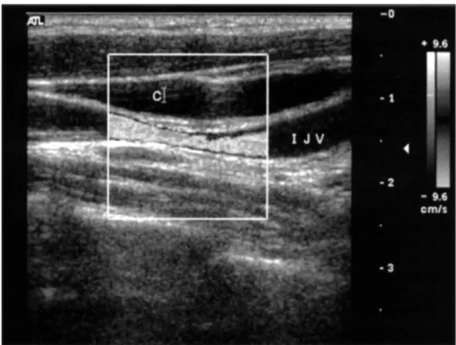
were within the normal range. Repeated pericar-diocentesis was performed, and an immediate gushing out of 700 mL of a milky-yellowish fluid ensued (Fig. 3). An examination of the pericardial fluid showed pH 6.8, protein 7.0 g/dL, glucose 98 mg/dL, LDH 560 IU/L, cholesterol 97 mg/dL, and triglyceride 1078 mg/dL. RBCs and WBCs were uncountable due to cell clumping. The adenosine deaminase level was 17.0 U/L and the CEA level 1.15 ng/mL. Routine cultures for bacteria and tuberculosis were subsequently reported as nega-tive and a cytologic examination showed no tumor cells. These findings with inappropriately elevated triglyceride in the pericardial fluid suf-ficed to diagnose chylopericardium. The patient underwent extensive investigations to determine the secondary causes. Chest computed tomo-graphy revealed a suspicious small cystic lesion on the left neck and mediastinal haziness. The possibility of cystic hygroma with mediastinal extension was highly suggested by radiologists. Neck sonography also showed a 1.9 cm sized multilocular, compressible cystic lesion between the sternocleidomastoid muscle and the internal jugular vein of the left neck. These findings were compatible with cystic hygroma (Fig. 4). The cystic lesion could not be aspirated due to patient's refusal. To demonstrate the presence of any anomalous communication between the lym-phatic channels and the pericardial sac, we per-formed a direct lymphangiography. However, no
direct communication between the pericardial sac and the thoracic duct could be definitely estab-lished. Follow-up echocardiography was per-formed 10 days after the pericardiocentesis and showed a massive reaccumulation of pericardial effusion. 11 days after pericardiocentesis, the patient underwent pericardiostomy. Using the subxyphoid approach, a pericardial window was made with 32 Fr draining tube insertion in an attempt to prevent pericardial fluid reaccumula-tion and cardiac tamponade. Incision of the pericardium resulted in the extrusion of 600 mL or more of milky fluid. The pericardial fluid analysis showed that it had the same nature as the previous sample. When the patient was placed on total parenteral nutrition for 30 days and a subse-quent low fat diet enriched with medium-chain triglycerides for 15 days, the drainage reduced to 30-40 mL per day. On the 45th day after
pericar-diostomy, drainage tube was removed and she was discharged from the hospital on a low fat diet. She remained totally asymptomatic without interval changes of heart size on chest X-ray (Fig. 5) or clinical signs of cardiac tamponade at the 6 month follow-up.
DISCUSSION
Groves and Effler,8 in 1954, reported a case of
chylopericardium in a 31-year-old woman who was found to have an isolated recurrent
accumu-Fig. 4. Neck sonography showing 1.9 cm sized
multilo-cular, compressible cystic lesion between the sternoclei-domastoid muscle and the internal jugular vein of the left neck.
Fig. 5.Six months follow-up chest X-ray showing nearly normal size of heart.
lation of chyle in the pericardium associated with mediastinal cystic hygroma. They used the term “primary idiopathic chylopericardium” in this first case report because no apparent etiologic mechanism was evident. This is such a rare condi-tion that a computerized Medline search from 1960 to 2003 identified fewer than 35 cases of primary idiopathic chylopericardium, including our case. Its etiology is obscure, though in recent years, a few reports have described a lymphatic leak and fistula by combined lymphangiography and computed tomography or intraoperative thoracic ductogram.9,14 These reports clearly
dem-onstrate a leaking flow of contrast media from the thoracic duct to the pericardial sac. This disorder has two possible explanations: (1) the presence of damaged lymphatic valves and the communica-tion of thoracic duct to the pericardial lymphatics resulting in chylous reflux and (2) abnormally elevated pressure in the thoracic duct observed as lymphangiectasia.14,15
Diagnostic modalities essential for evaluating suspected chylopericardium include the follow-ing: A chest X-ray demonstrating cardiomegaly will lead to further investigations and eventually diagnosis. Pericardial effusion is usually diag-nosed by echocardiography or computed tomo-graphy. The nature of the effusion is usually
revealed by pericardiocentesis.9 Computed
tomo-graphy and lymphangiotomo-graphy may be useful in the diagnosis of mediastinal lymphangiomatosis, which is the most common cause of primary chylopericardium.17 Investigation specifically
tar-geting malignant disease, lymphoma and tuber-culosis should also be undertaken.2,4 A history of
trauma caused by thoracic surgery,6,7 the intro-duction of a subclavian venous catheter,3or blunt
trauma5 should be determined.
Lymphangiogra-phy may in certain instances establish fistulous connections and is also useful for delineating the anatomy of the thoracic duct.9,14,21 Primary
chylo-pericardium should be considered in the differ-ential diagnosis of chronic pericardial effusion. As
is pointed out in a review by Akamatsu,14 about
a half of patients are asymptomatic or were noted to have only cardiomegaly by chest X-ray. Cystic hygromas (lymphangiomas) may arise from lymphatic tissue normally present in the area or may grow directly from mesodermal rests that
produce imperfect lymphoid channels.18 These
lesions are usually found in the neck or axilla. It is apparent that lymphangioma may be multi-focal, involving mediastinum, retroperitoneum, and even the skeletal system, and that they produce a number of apparently separate syn-dromes depending on the anatomic location of the lesion.18-20 Our patient had cystic lesion in the
neck, but she refused biopsy for a definitive diagnosis. However, according to the smoothly marginated cystic mass on CT scan and the compressibility of the lesion by sonography, we concluded that this cystic lesion was a cystic hygroma. In our case, we failed to visualize any communication between the thoracic duct and the pericardial sac by lymphangiography and com-puted tomography of the chest. Tucker et al.20 in
1967 reported idiopathic chylothorax in asso-ciation with a cystic lesion of the bone, and Goldstein et al.18 in 1969 reported an association
of a chylopericardium with multiple lymphan-gioma of the bone. Moreover, although they failed to demonstrate a clear communication between the pericardial space and the thoracic duct, they pointed out a generalized abnormality of the lymphatic system in patients with lym-phangiomas. We strongly believe that a cystic hygroma of the neck and chylopericardium reflect a generalized disorder of the lymphatic system in our patient. An abnormal lymphatic system may cause the lymphatic channel pressure elevation and increase the permeability of lymphatic ves-sels, resulting in the reflux of chyle through certain communications between the lymphatic system and the pericardial sac.18-20
The management of chylopericardium is guided by the prevention of cardiac tamponade, and the avoidance of the metabolic, nutritional, and immunologic consequences that occur given a significant chyle leak, and to reduce the
like-lihood of recurrence.8-16 Several different
ap-proaches make be adopted for the treatment of chylopericardium: (1) pericardiocentesis, (2) tho-racostomy drainage, (3) dietary support with medium or short chain triglyceride and low fat meals, (4) pericardiectomy, (5) pericardial win-dow formation, (6) ligation of the thoracic duct above the level of the diaphragm, and (7) a pericardial-peritoneal (Denver) shunt. In contrast
to post-traumatic chylopericardium, the conserva-tive treatment of idiopathic chylopericardium is rarely successful. Surgical treatment is usually required, however, it should be noted that only one case treated conservatively has been reported.12 Nevertheless, initially, a patient should be treated conservatively by pericardial drain placement and the institution of a medium chain triglyceride diet or total parenteral nutrition.10 If
this approach fails, then surgical ligation of the thoracic duct just above the diaphragm combined with window formation or pericardiectomy to ensure adequate drainage has been reported to be definitive treatment.8-11,13-15 Pericardial drainage
with pericardiocentesis or tube thoracostomy with or without dietary management was also found effective, but in many cases reaccumulation occurred.9 Pericardio-peritoneal shunt placement
has been advocated by Chan et al.22 as a simple
and effective alternative to prolonged pericardial drainage or thoracotomy. Although most authors reported surgery as the definitive method of
treatment, Lopez-Castilla et al.12 reported the
successful treatment of a 2-month-old boy by low fat total parenteral nutrition. In our patient, external drainage of the pericardial sac via the pericardiostomy, concomitant with dietary sup-port with medium chain triglyceride and low fat meals, resulted in the complete resolution of pericardial effusion. Six months later, the patient was asymptomatic and the chest X-ray showed a normal cardiac size. This success in our case reasserts the importance of conservative treatment for the management of primary idiopathic chyl-opericardium. If symptoms develop or massive pericardial effusion recurs, thoracic duct ligation will be considered in this patient.
In summary, our case supports that pericar-diostomy combined with total parenteral nutri-tion and a low fat dietary support is a treatment modality for primary idiopathic chylopericar-dium
REFERENCES
1. Crosby IK, Crouch J, Reed WA. Chylopericardium and chylothorax. J Thorac Cardiovasc Surg 1973;65:935-9. 2. Barton JC, Durant JR. Isolated chylopericardium
associated with lymphoma. South Med J 1980;73:1551. 3. Smedts F, Kubat K, Chande H. Chylopericardium and chylothorax, resulting from a catheter to the left sub-clavian vein: an autopsy report. Klin Wochenschr 1989; 67:1214-7.
4. Lorell BH, Braunwald E. Pericardial disease. In: Braunwald E, editor. Heart disease. A textbook of cardiovascular medicine. Philadelphia: W.B. Saunders; 1992. p.1505.
5. DeMeester TR, Lafontaine E. The pleura. In: Sabiston DCJ, Spencer FC, editors. Gibbon's Surgery of the chest. Philadelphia: W.B. Saunders; 1983. p.369-72. 6. Delaney A, Daicoff GR, Hess PJ, Victorica B.
Chylo-pericardium with cardiac tamponade after cardio-vascular surgery in two patients. Chest 1976;69:381-3. 7. Bar-el Y, Smolinsky A, Yellin A. Chylopericardium as a complication of mitral valve replacement. Thorax 1989;44:74-5.
8. Groves LK, Effler DB. Primary chylopericardium. N Engl J Med 1954;250:520-3.
9. Yoon YS, Shim WH, Chung TS, Lee YS. Primary idiopathic chylopericardium: Report of a case and review of the literature. Yonsei Med J 1993;34:98-108. 10. Ossiani MH, McCauley RGK, Patel HT. Primary idiopathic chylopericardium. Pediatr Radiol 2003;33: 357-9.
11. Svedjeholm R, Jansson K, Olin C. Primary idiopathic chylopericardium- a case report and review of the literature. Eur J Cardio-thorac Surg 1997;11:387-90. 12. Lopez-Castilla JD, Soult JA, Falcon JM, Munoz M,
Santos J, Gavilan JL, et al. Primary idiopathic chylo-pericardium in a 2 month old successfully treated without surgery. J Pediatr Surg 2000;35:646-8. 13. Akashi H, Tayama K, Ishihara K, Tanaka A, Fujino T,
Okazaki T, et al. Isolated primary chylopericardium. Jpn Circ J 1999;63:59-60.
14. Akamatsu H, Amano J, Sakamoto T, Suzuki A. Pri-mary chylopericardium. Ann Thorac Surg 1994;58: 262-6.
15. Yuksel M, Yildizeli B, Zonuzi F, Batirel HF. Isolated primary chylopericardium. Eur J Cardio-thorac Surg 1997;12:319-21.
16. Hudspeth AS, Miller HS. Isolated (primary) chyloperi-cardium. J Thorac Cardiovasc Surg 1966;51:528-31. 17. de Menezes IC, Araujo eSG, Damiao A, Telo M,
Martins FM, Macedo MM. Lymphangioma of the mediastinum as a cause of chylopericardium. Acta Med Port 1990;3:119-21.
18. Goldstein MR, Benchimol A, Cornell W, Long DR. Chylopericardium with multiple lymphangioma of bone. N Engl J Med 1969;280:1034-7.
19. Bhatti MAK, Ferrante JW, Gielchinsky I, Norman JC. Pleuropulmonary and skeletal lymphangiomatosis with chylothorax and chylopericardium. J Thorac Surg 1985; 40:398-401.
20. Tucker SM. Bilateral chylothorax with multiple osteo-lytic lesion?: generalized abnormality of lymphatic system. Proc Roy Soc Med 1967;60:17-9.
21. Rankin RN, Raval B, Finley R. Primary chyloperi-cardium: combined lymphangiographic and CT diag-nosis. J Comput Assist Tomogr 1980;4:869-70.
22. Chan BBK, Murphy MC, Rodgers BM. Management of chylopericardium. J Pediatr Surg 1990;25:1185-9.