A Thesis
For the Degree of Master of Veterinary Medicine
Radioprotective Activity of Eckol
in Gamma ray-irradiated Mice
GRADUATE SCHOOL
CHEJU NATIONAL UNIVERSITY
Department of Veterinary Medicine
Eunjin Park
2007. 12.
in Gamma ray-irradiated Mice
Eunjin Park
(Supervised by professor Youngheun Jee)
A thesis submitted in partial fulfillment of the requirement for the degree of Master of Science
2007. 12
This thesis has been examined and approved by
Hyun Jeong Park, Prof. of Veterinary Medicine
Department of Veterinary Medicine
GRADUATED SCHOOL
CHEJU NATIONAL UNIVERSITY
CONTENTS
Table of contents ... 1
List of Figures ... 2
List of Tables ... 4
List of abbreviations ... 5
Abstract ... 6
Introduction ... 7
Materials and Methods ... 10
Results ... 15
Discussion ... 20
References ... 23
Tables ... 27
LIST OF FIGURES
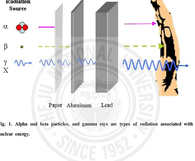
Fig. 1. Alpha and beta particles, and gamma rays are types of radiation associated with
nuclear energy
... 28
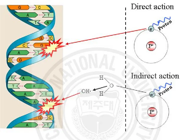
Fig. 2. The direct and indirect actions of radiation
... 29
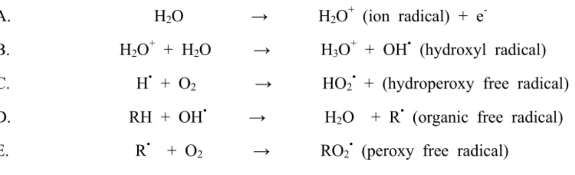
Fig. 3. Production of free radicals
... 30
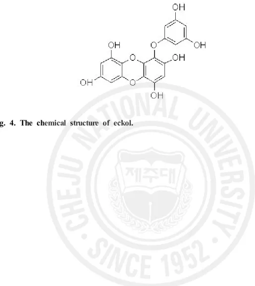
Fig. 4. The chemical structure of eckol
... 31
Fig. 5. The radioprotective effect of eckol on the survival of lethally irradiated mice
... 32
Fig. 6. The effect of eckol on endogenous spleen colonies in 7 Gy-irradiated mice
... 33
Fig. 7. The effect of eckol compound on DNA damage induced by irradiation in lymphocytes was assessed with the alkaline comet assay
... 34
Fig. 8. Eckol compound enhances the proliferative ability suppressed by ionizing radiation of splenocytes .
... 35
... 36
Fig. 10. Eckol rescues small intestinal crypt cells from radiation-induced apoptosis ...
37
Fig. 11. The photo presents p53, Bax, Bcl-2 and PCNA-positive cells with apoptotic
fragments evident by immunohistochemistry ...
38
Fig. 12. Western blot analysis of p53, Bcl-2 and Bax expression modulated by eckol in
the small intestine of a mouse 24 h after 2 Gy irradiation compared to those from non-irradiated and eckol-deprived mice ...
40
LIST OF TABLES
CFUs Colony-forming units
DAB 3,3'-diaminbenzidine
DAPI 4’6-diamidino-2-phenylindole dihydrochloride
DMEM Dulbecco's Modified Eagle's Medium
DPBS Dulbecco’s phosphate-buffered saline
EM radiation Electromagnetic radiation
FITC Fluorescein isothiocyanate
FACS Fluorescence-activated cell sorting
GI syndrome Gastrointestinal syndrome
Gy Gray
H&E Hematoxylin and eosin
HRP Horseradish peroxidase
IR Ionizing radiation
LD Lethal dose
PBS Phosphate-buffered saline
PCNA Proliferating cell nuclear antigen
ROS Reactive oxygen species
Th cells T helper cells
WBI Whole body irradiation
1. ABSTRACT
We have investigated the potential of eckol, a component of the seaweed
Ecklonia cava, for its ability to provide protection from g ray-induced damage. Our results determined that eckol significantly decreased the mortality of lethally irradiated mice. The mechanisms of eckol’s protection were threefold: an improvement in hematopoietic recovery, the repair of damaged DNA in immune cells and an enhancement of their proliferation, which had been suppressed by ionizing radiation. Furthermore, eckol rescued intestinal cells from radiation-induced apoptosis by decreasing the amount of pro-apoptotic p53 and Bax and increasing that of anti-apoptotic Bcl-2. Thus, eckol may be a good candidate for use as a radioprotective agent.
2. INTRODUCTION
Radiation, as used in physics, is energy in the form of waves or moving subatomic particles. Radiation can be classified as ionizing or non-ionizing radiation, depending on its effect on atomic matter. The most common use of the word "radiation" refers to ionizing radiation. Ionizing radiation has enough energy to ionize atoms or molecules while non-ionizing radiation does not. There are several types of ionizing radiation: charged particle, such as alpha and beta particle, non-charged particle, such as neutron, and electromagnetic (EM) radiation such as Gamma rays, x-rays, visible light, and UV. Ionizing radiation differs in frequency and hence in energy. Alpha particles consist of two neutrons and two protons, in the form of atomic nuclei. Because of their relatively large size, alpha particles collide easily with matter and lose their energy quickly. They therefore have little penetrating power and can be stopped by the first layer of skin or a sheet of paper. Beta particles are fast-moving electrons ejected from the nuclei of atoms and have a single negative charge. These particles are much smaller than alpha particles and can penetrate up to 1 to 2 centimeters of water or human flesh. They can be stopped by a sheet of aluminum a few millimeters thick. X- and gamma rays, like light, represent energy transmitted in a wave without the movement of material, just as heat and light from a fire or the sun travels through space. Gamma rays are the most energetic form of such electromagnetic radiation, having the energy level from around 10 keV to several hundred kilo-electron volts, and therefore they are more penetrating than other radiation such as alpha and beta rays [1]. X- and gamma rays can pass through the human body. Mass in the form of concrete, lead or water is used to shield us from them. Neutrons are uncharged particles which are emitted by artificial radionuclides (Fig. 1). Since neutrons have no charge, there is no electromagnetic felid associated with their movement and they do not ionize
positively charged atomic nuclei and are thus able to penetrate close to the nuclei of the material through with they are traveling [2]. Concrete are the most commonly used shields against neutron radiation from the core of the nuclear reactor.
Radiation has a direct or an indirect action on the viable cells [3]. Direct action means that a secondary electron resulting from absorption of photon interacts with DNA directly. On the other hand, electromagnetic radiations (X- and gamma-rays) have an indirect action (Fig. 2.) [3]. They do not produce biological damage themselves, but interact with other atoms or molecules in the cell, particularly water, to produce free radicals that are able to damage the critical targets [3]. Many of these processes involve reactive oxygen species (ROS), such as hydrogen peroxide (H2O2), superoxide anion
(O2·), hydroxyl radicals (OH·) and singlet oxygen (Fig. 3.) [1,3,4]. ROS are relevant components of living organisms that, besides their essential role in the regulation of different important physiological functions, when present in excess are capable to affect cell function and viability through a chain process called oxidative stress [5]. The free radicals react with cellular macromolecules, such as DNA, RNA, proteins, membrane, etc, and lead to lipid peroxidation, protein oxidation, base modifications, DNA strand breaks and genomic instability ultimately resulting in cell death [6]. Exposure to ionizing radiation induces the production of reactive oxygen species (ROS) such as superoxide, hydroxyl radicals, singlet oxygen and hydrogen peroxide [1]. These free radicals react with critical cellular components such as DNA, RNA, protein, membrane,
etc, and cause cell dysfunction and death [2,3].
In the recent past, many synthetic agents such as WR2721 (amifostine), OK-432 and ethiofos have been investigated for their efficacy as protectors from radiation-induced tissue damage [7.8]. However, these agents have the potential to cause serious side effects such as performance decrement, nausea, vomiting, hypotension, allergy and death [9,10]. The marked toxicity of these agents drove researchers to look for safer, less expensive and more effective radioprotectors. Natural agents extracted
from plants are known to be effective in accelerating recovery from disease or injury without obvious side effects. These nontoxic agents that can protect cells and tissues against ionizing radiation have considerable potential as adjuncts to successful radiotherapy.
Ecklonia cava (E. cava), an abundant brown algae (Laminariaceae) from Jeju
Island in Korea, has phlorotannin components with known biological activities. Phlorotannin is reported to reduce allergy by controlling histamine release [11], provide an anti-inflammatory effect [12] and protect cells from oxidative stress by scavenging intracellular ROS [13]. Phlorotannin, which is found in marine algal polyphenols in the form of phloroglucinol polymers, such as eckol, 6,6’-bieckol, dieckol and phlorofucofuroeckol, were identified in Ecklonia species [14]. We previously reported that eckol inhibited oxidative cell damage by scavenging ROS and modulating a cellular signal pathway in lung fibroblast cells [14]. Based on this result, we hypothesized that eckol could have beneficial effects on radiation-induced oxidative damage in vivo. To test this hypothesis, we examined the effect of eckol on the mortality of mice given lethal whole-body irradiation (WBI). We also investigated the mechanisms by which eckol increased survival. These studies described here demonstrated that, in a mouse model, eckol reduced mortality following lethal WBI by accelerating hemopoietic recovery. Additionally, inradiosensitive immune cells, eckol enhanced the repair and the proliferative functions suppressed by ionizing radiation. Eckol also reduced the damage to lymphocytes’ DNA and inhibited apoptosis in radiosensitive cells by decreasing the expression of pro-apoptotic p53 and Bax and correcting the radiation-induced decrease of anti-apoptotic Bcl-2.
3. MATERIALS AND METHODS
Preparation and treatment of eckol compound
Eckol (Fig. 4) compound was obtained from Dr. Nam Ho Lee (Cheju National University, Jeju, Korea). The eckol compound dissolved in phosphate-buffered saline (PBS, pH 7.4) was injected intraperitoneally (i. p.) into mice 1 day before, at the time of and 3 days after irradiation. Additional non-irradiated and irradiated groups were mice injected i.p. with PBS at the same volumes as test mice.
Mice
C57BL/6 mice were purchased from Orientbio, Inc. (Sungnam, Korea) and were 6- to 8-weeks-old and weighed 18-25g when used for the experiments. These mice were housed in conventional animal facilities with a NIH-07-approved diet and water ad
libitum at a constant temperature (23 ± 1ºC) according to the guidelines for the Care
and Use of Laboratory Animals of the institutional ethical committee. Mice were randomly divided into the following four groups: a non-irradiated normal group, an irradiated control group, an eckol only-treated group and an eckol plus irradiated group.
Irradiation with 60Co g -ray
Each mouse was placed individually in a close-fitting Perspex box (3×3×11 cm) then exposed to WBI with a source 60Co irradiator (Theratron-780 teletherapy unit,
Applied Radiological Science Institute, Cheju National University). Briefly, the mice received 9 Gy WBI for the survival experiment, 7 Gy for the splenic colony-forming assay and 2 Gy for the other experiments with a dose rate of 1.5 Gy/min in the box and source-surface distance (150 cm) as previously reported [15,16].
To determine whether the eckol compound conferred a survival advantage after lethal WBI, mice so-treated were observed daily for up to 30 days after irradiation. At 31 days after WBI, surviving mice were euthanized by cervical dislocation.
Endogenous colony forming units (CFUs)
An endogenous CFU assay was performed to confirm the effect of eckol compound on the hematopoietic ability of spleens from mice given ionizing radiation. The mice (3 mice/group) were sacrificed 10 days after irradiation. Their spleens were removed and examined with the naked eye to score for macroscopic colonies on splenic surfaces [17].
Preparation of primary splenocytes
Spleens removed from test mice were the source of single cell suspensions obtained by using a cell strainer. The cells were lysed with ACK buffer (containing 0.84% ammonium chloride) for 10 at room temperature and washed with Dulbecco’s phosphate-buffered saline (DPBS, Gibco BRL). The splenocytes were suspended in RPMI-1640 medium (Gibco BRL) supplemented with 10% fetal bovine serum (Gibco BRL) and 1% antibiotics (100 U/ml penicillin-streptomycin, Gibco BRL). The cell viability was determined by Trypan blue dye exclusion (Sigma Aldrich, St. Louis, MI, USA), and the purified cells (viability > 90%) were directly used for the experiments.
Alkaline comet assay
An alkaline comet assay was used to determine the oxidative DNA damage induced by ionizing radiation. The basic alkaline technique was performed as described by Kang et al [18]. Results observed with a fluorescence microscope were analyzed by using a komet 5.5 program (Kinetic Imaging, Liverpool, UK). The percentage of fluorescence in the tail DNA of 50 cells per slide was recorded.
3H-thymidine incorporation assay
To assess whether eckol could stimulate the regeneration and proliferation of splenocytes damaged by irradiation, a thymidine incorporation assay was performed. The lymphocytes were isolated at 3 and 9 days after irradiation, and these cells (4×10⁵cells /
well) were seeded in 96-well round-bottom microtiter plates (Nunc, Copenhagen, Denmark). After subsequent incubation in triplicate with 200 ml culture medium for 72 h at 37 °C in humidified air containing 5% CO2, 10 l of Con A (Sigma) were added into wells at a final concentration of 5 g/ml Con A. After incubation, each well was pulsed for a final 18 h with 1 Ci of 3H-methylthymidine (specific activity 42Ci/mmol; Amersham, Arlington Heights, IL, USA). An automatic cell harvester gathered the cells onto glass fiber filters. The amount of radioactivity incorporated into DNA was determined in a liquid scintillation spectrometer (MicroBeta TriLux, Perkin Elmer, Wallac, Germany).
Flow cytometry assay
To further define the specific cell population stimulated by eckol, the splenocytes were analyzed by FACS analysis. Mice were sacrificed at 3 days after WBI, and their spleens were removed. The splenocytes (1 × 10⁶) were harvested and, to inhibit
nonspecific staining, combined with an anti-mouse IgG solution (Caltag Lab, Furlingame, CA, USA) in PBS for 15 min at 4°C. The cells were then stained with FITC-labeled CD3 (145-2C11) antibody as a specific marker for pan T cells and FITC-labeled CD45R/B220 (RA3-6B2) antibody as a specific marker for pan B cells for 15 min at 4°C. All antibodies were purchased from BD Biosciences (San Jose, CA, USA). Twenty thousand viable cells per mouse (as determined by light scatter profiles) were analyzed using a BD FACS CaliburTMflow cytometer (BD Biosciences). In a cytogram, three main clusters could be seen that related to the spleen’s major cell types: lymphocytes,
monocytes and polymorphonuclear granulocytes. We compared numerical changes in these cells from untreated mice to those from eckol-treated mice.
Hematoxylin and eosin (H&E) staining and immunohistochemistry of small intestinal crypt cells
To identify the effect of eckol compound on the ionizing radiation-triggered apoptosis of small intestinal crypt cells, small intestines were separated from mice at 1 day after WBI and fixed in 10% formalin buffer. The blocks were then embedded in paraplast wax to prepare 5 mm sections for H&E staining. Apoptotic crypt cells with such morphological characteristics as cell shrinkage, chromatin condensation, margination and cellular fragmentation were counted using a light microscope [19,20]. Counts from small intestine sections of each mouse and 50 crypts per intestinal section were recorded. For immunohistochemical localization of p53 protein, tissue sections were incubated with normal horse serum then reacted with antibody to p53 (1:200 dilutions, Calbiochem, Darmstadt, Germany), Bcl-2 (1:500, Santa Cruz Biotechnology, Santa Cruz, CA, USA), β-actin (1:500, Santa Cruz Biotechnology) or bax (1:500, Cell Signaling Technology Inc, Beverly, MA, USA) for 1 hour. Next came incubation of the sections with a second antibody, biotinylated anti-mouse IgG (Vector, Burlingame, CA, USA), and with horseradish peroxidase (HRP)-labeled VECTASTAIN Elite ABC KIT (Vector). HRP-binding sites were detected with 3,3'- diaminbenzidine (DAB; Vector) and a final counterstain with hematoxylin. The statistical significance of measurements from each sample was assessed by Graph PAD in a Plot computer program (GPIP, Graph PAD Software Inc., San Diego, CA, USA) and EXCEL software. Standard error bars are shown for all data points except where the error is equal to or less than the symbol size.
Western blot analysis
To elucidate the mechanism by which eckol protects radiosensitive cells from radiation-induced apoptosis at the molecular level, expression patterns of various proteins associated with apoptosis were studied in the intestines of mice given WBI. Tissues lysate extracts from their small intestines were prepared, and the equal amount of cellular protein (60 µg/well) was loaded onto 10-12% SDS-PAGE gels and immunoblotted onto a nitrocellulose membrane (Bio-Rad, Hercules, CA, USA). The membrane were exposed to anti-p53 (1:500, Calbiochem), Bcl-2 (1:500, Santa Cruz Biotechnology), β-actin (1:500, Santa Cruz Biotechnology) or Bax (1:500, Cell Signaling Technology Inc.), followed by incubation with HRP-conjugated anti-rabbit IgG or anti-mouse IgG (Santa Cruz Biotechnology). The blots were developed by enhanced chemiluminescence reagents (WestOne, iNtRON, Sungnam, Korea) according to the manufacturer’s instructions.
Statistical analysis
The results are reported as means S.E. or S.E.M. The results were analyzed by using Student’s t-test, and P < 0.05 or P < 0.01 were considered significant.
4. RESULTS
Eckol improves the survival rate in mice after lethal irradiation
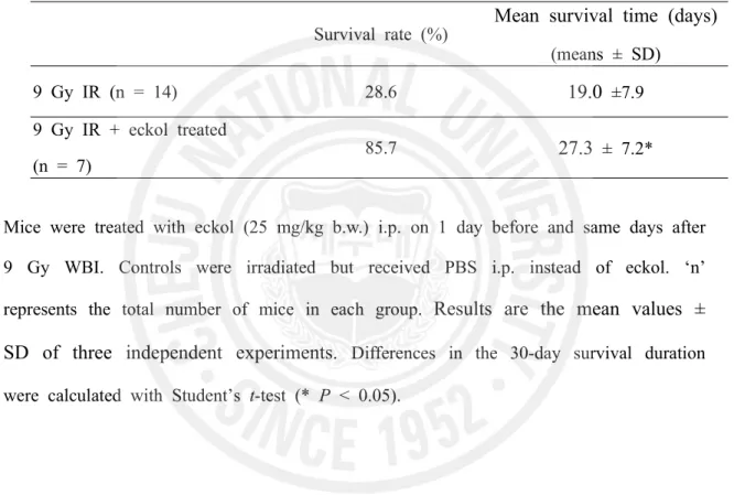
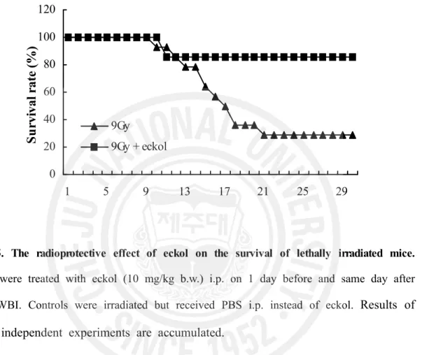
To test whether eckol compound can protect lethally irradiated mice from related injury, we first determined that the survival rates of mice treated with eckol at concentrations of 25, 10 or 5 mg/kg b.w. were 86, 60 and 40%, respectively. Clearly, the low 5 mg/kg concentration of eckol was less effective than the 10 or 25 mg/kg doses. Therefore, in subsequent survival tests, the dose of eckol used was 10 mg/kg b.w. injected i.p. 1 day before and same day after 9 Gy WBI. As a result, only 28.6% (4/14) of the irradiated, untreated group survived for 30 days. In comparison, 85.7% (6/7) of mice given eckol plus irradiation survived for 30 days, denoting the latter’s significantly diminished death rate (Table 1, Fig. 5, P < 0.05). The LD50/30 was 8Gy for this group. No deaths occurred in the non-irradiated group or eckol only-treated group for 30 days. These results demonstrated the eckol compound’s safety and marked radio-protective efficacy by prolonging the survival of lethally irradiated mice.
Eckol enhances hematopoietic ability
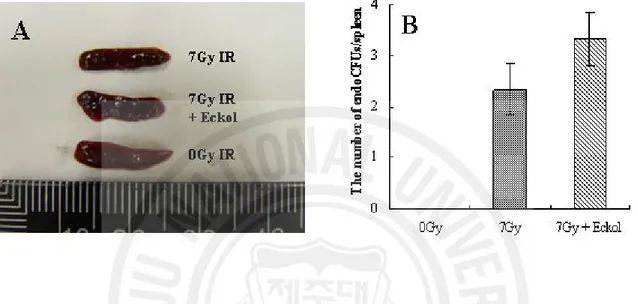
Subsequently, to confirm the radioprotective effect of eckol compound on hematopoietic stem cells damaged by ionizing radiation, an endogenous CFU assay was performed. The spleen’s content of endogenous CFUs is considered to be an indicator of hematopoiesis, which is a critical survival factor [17]. In the eckol plus irradiation groups vs. the irradiation control groups, the number of endogenous CFUs was 2.3 ± 0.5 and 3.5 ± 0.5, respectively (P < 0.05). Eckol-only treatment did not alter these results in non-irradiated groups (data not shown). Therefore, eckol compound increased the number of CFUs about 1.6-fold, indicating the restoration of hematopoietic ability (Fig. 6) and the spleen’s stem cell compartments.
Eckol reduces the DNA damage of lymphocytes
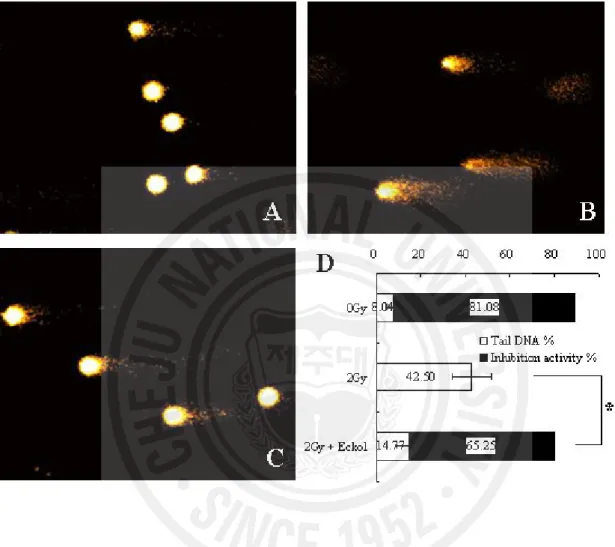
Since eckol is known to have ROS scavenging activity [14], we studied the influence of eckol compound on DNA damage from ionizing radiation in lymphocytes. Because the alkaline comet assay is an effective method for monitoring the extent of DNA strand breakage [21], we used this assay to measure the post-irradiation DNA damage in the tails of eckol-treated and untreated control mice. The percentage of tail DNA in lymphocytes of mice exposed to 2 Gy γ-radiation increased to 42.50 ± 8.73 from 8.04 ± 4.08 %. However, eckol treatment significantly decreased the percentage of tail DNA in lymphocytes damaged by irradiation to 14.77 ± 5.65 % (Fig. 7, P < 0.05). This decrease of DNA damage in our eckol plus irradiated group denotes the compound’s cytoprotective effect in vivo, possibly owing to its ROS scavenging activity.
Eckol induces the proliferation of splenocytes
To verify the survival benefit mediated by eckol compound in radiosensitive immune cells, the proliferation of splenocytes was assessed by measuring their incorporation of 3H-thymidine at 3 and 9 days after mice were irradiated. Eckol significantly increased
thymidine incorporation by splenocytes, i.e., as much as 6.2-fold (3 days) and 2.0-fold (9 days), respectively, in comparison to that by irradiated mice deprived of eckol (Fig. 8, P < 0.05). Therefore, since eckol effectively prevented the radiation-induced suppression of splenocytes’ proliferation, and prolonged the recipients’ survival, more frequent eckol administration may have had an even greater benefit.
Eckol stimulates increases in populations of T and B cells
To define the precise cell population(s) stimulated by eckol, the splenocytes were analyzed with flow cytometry. Three days after irradiation, lymphocytes, monocytes and granulocytes, which were damaged by exposure to WBI, increased numerically (14.9 %, 90.4% and 85.6 %, respectively) in eckol-treated mice (Fig. 9A). Eckol-only treatment
did not alter these results of non-irradiated groups (data not shown). Subsequently, the numbers of CD3+ pan T cells and CD45R/B220+ pan B cells underwent marked
up-regulation in eckol-treated groups compared with untreated control groups after irradiation (Fig. 9B). In particular, the number of CD3+ pan T cells was elevated 44.3%
above that of eckol-untreated controls. Moreover, amounts of CD45R/B220+ pan B cells were enlarged by 27.6% compared with irradiated control. These results demonstrated that eckol could enhance immunoprotection by increasing the recovery rate of specific populations of immune cells.
Eckol reduces the apoptosis of intestinal crypt cells
Since the gastrointestinal system is a major site of cell death induced by ionizing radiation, and intestinal crypt cells are one of the most sensitive cells to irradiation, we next investigated the potential of eckol compound to rescue intestinal crypt cells from radiation-induced apoptosis. The frequency of spontaneous apoptosis in crypt cells of non-irradiated animals was not significantly different from that of eckol only-treated animals (data not shown). One day after WBI, the frequency of apoptotic fragments in crypt cells was reduced in the eckol-treated group (Fig. 10B) compared with untreated group (Fig. 10A). As shown in Fig. 10C the number of apoptotic fragments per intestinal c rypt in the irradiated control group was 4.51 ± 0.30, whereas a 16.63% reduction as was observed in the eckol plus irradiated group (3.76 ± 0.22, P < 0.05).
Eckol modulates the immunohistochemical localization and intensity of apoptosis-related proteins in jejunal crypt cells
According to above data, we investigated the localization of apoptosis-regulated proteins (p53, Bax, and Bcl-2) and PCNA in the intestinal cells under eckol-treated and untreated conditions using by immunohistochemistry. Immunohistochemisty provided similar results for the modulation of pro-apoptotic p53 (Fig. 11A and B) and Bax
expression (Fig. 11C and D) in intestinal crypt cells that paralleled the frequency of their apoptosis in eckol-treated mice. In irradiated conditions without eckol treatment, p53 (Fig. 11A) and Bax (Fig. 11C) immunoreactivities were highly overexpressed on apoptotic cells of radiosensitive crypts. The types of p53 and Bax-expressing cells were essentially the same as those seen in the irradiated intestinal cells. In contrast, p53 (Fig. 11B) and Bax (Fig. 11D) immunoreactivities were generally decreased on crypt cells under eckol-treated conditions. On the other hand, the staining intensity of anti-apoptotic Bcl-2 (Fig. 11E) and PCNA (Fig. 11G) was detected faintly or weakly on laminar propria cells in villi core and crypts in irradiated groups compared with that of normal conditions. However, in parallel with frequency of apoptosis in crypt cells, the Bcl-2 (Fig. 11F) and PCNA (Fig. 11H) proteins were increased in intestinal cells after eckol treatment and their immunoreactivities were increased strongly on round and oval cells of laminar propria in villi core and crypts. In addition, immunostaining of anti-apoptotic proteins after eckol treatment showed a similar pattern in normal conditions (data not shown). Control sections, in which anti-p53, Bax, Bcl-2, and PCNA antibodies were omitted, gave no specific staining (data not shown). Thus, the overexpression of Bcl-2 and PCNA increased in fibrocytes and immune-related cells of laminar propria after eckol treatment were involved deeply in gastrointestinal cells repair. These results suggest that eckol compound improved the viability of radiosensitive intestinal crypt cells by inhibiting radiation-induced apoptosis, and this factor may be an important key for the radioprotection of irradiated mice.
Eckol modulates the expression of apoptosis-related proteins in jejunal crypt cells
To pinpoint the mechanism of eckol compound’s ability to inhibit apoptosis after irradiation in vivo, we investigated expression levels of pro- and anti-apoptotic proteins, i.e., p53, Bax and Bcl-2 in small intestines of irradiated mice from the untreated and eckol-treated groups. Western blot analysis of intestinal tissues obtained 1 day after
exposure to 2 Gy g-rays irradiation showed a significantly increased expression of the pro-apoptotic proteins, such as p53 and Bax with a decrease in the anti-apoptotic proteins classed as a Bcl-2 family in irradiated control mice compared to non-irradiated group (Fig. 12). However, eckol apparently reduced the increased expression level of p53 and Bax after WBI and induced an increase in the expression level of Bcl-2. Since the reduction of apoptosis in intestinal crypt cells after eckol treatment was closely associated with the decrease of p53 protein, eckol compound presumably inhibited the apoptosis of cells in the small intestine in vivo by blocking the up-regulation of pro-apoptotic p53 and Bax and the down-regulation of anti-apoptotic Bcl-2 after irradiation.
5. DISCUSSION
In this study, we demonstrated that eckol effectively protected mice from lethal irradiation and was safe and well tolerated at high concentrations. The survival of lethally irradiated animals can be prolonged by such varied manipulations as inhibiting the generation of free radicals or intensifying their removal, enhancement of DNA repair, replenishment of dead hematopoietic cells and stimulation of immune cell formation or activity [22]. Until now, no report has described this compound’s protective effect against radiation-induced tissue damage or the underlying mechanism of that effect
in vivo. Therefore, the present study was conceived to test eckol for these properties
under several experimental conditions.
Radiation induced oxidative damage to DNA, includes several types of base damage and single- or double-strand breaks. DNA strand breaks are potentially mutagenic and/or carcinogenic when improperly repaired and lethal if left un-repaired [23]. Therefore, understanding the cause and reducing the damage are important for avoiding such complications. By using comet assay, we observed that eckol treatment significantly decreased the percentage of tail DNA in lymphocytes of irradiated mice, indicating that eckol could protect against the radiation-induced damage that might cause cancer. These functions of eckol are likely factors in the prolonged survival of the lethally irradiated animals documented here.
Hematopoietic stem cells are the most sensitive cells to ionizing radiation and/or chemotherapeutic drugs during cancer therapy. However, myelosuppression and hematopoietic dysfunctions are the most common clinical complications following radio- or chemotherapy [24]. The destruction of lymphoid and hematopoietic systems causes pancytopenia as well as a predisposition to infection, hemorrhage, septicemia and death. Therefore, an important adjunct to cancer treatment is to promote the recovery of hematopoiesis [25]. In our experiments, eckol-treated mice enjoyed a recovery from
WBI, as established by their larger number of splenic CFUs in comparison to irradiated controls lacking eckol. The enhancement of endogenous CFUs in eckol plus irradiation-treated mice indicates that eckol protects and/or stimulates the proliferation of hematopoietic stem cells, constituting an important mechanism of eckol’s survival benefit with potential clinical applications.
Furthermore, eckol infusion increased the thymidine incorporation ability of splenocytes. Compared to irradiated controls, the proliferative ability of splenocytes from eckol-treated mice was higher 3 days after irradiation; moreover, this 3-day-peak was higher than that on 9 days after irradiation. Thus, eckol not only significantly and rapidly increased the proliferative ability of splenocytes that had been reduced by irradiation but also a second treatment might have yielded a booster effect on proliferative activity. With these results, we next analyzed the cellular subpopulations of proliferating splenocytes. After eckol treatment, the numbers of lymphocytes, monocytes and granulocytes increased significantly compared to those in irradiated controls. Especially abundant in the treated mice were CD3+ (all mature) and CD45R/B220+ (pan
B) cells. These results indicated that eckol could protect against radiation-induced damage by stimulating the proliferation and maturation (differentiation) of immune cells. This stimulatory effect of eckol with respect to the differentiation and maturation of T and B cells could, of course, promote the synthesis of cytokines and antibodies. Considering that immunosuppression is a major complication of radiation exposure, thereby facilitating opportunistic infections [19], the immunostimulatory capacity of eckol may be a particularly important mechanism of its radioprotective efficacy.
In many cases, radiation-induced DNA damage can kill exposed cells, and radiation-induced cell death has been identified as apoptosis [26]. Cells are regulated by a complex balance in a signal transduction pathway between the pro-apoptotic factors such as p53 and Bax and anti-apoptotic factors such as the Bcl-2 family [27,28]. Especially, the most replicative cells such as spermatocytes, lympho-hematopoietic
elements and intestinal crypt cells are prone to radiation-induced damage. In this study, eckol reduced the death rate of intestinal crypt cells in irradiated mice to a statistically significant extent. The reduction of p53 and Bax and induction of Bcl-2 protein are notable for their relationship to apoptosis and DNA repair [28,29]. In agreement with previous reports, the mechanism of this radioprotection by eckol may have been the decrease of p53 and Bax proteins in small intestine. In addition, Bcl-2 has been associated with the prevention of apoptosis. Our results suggest that eckol modulates p53- and Bcl-2-dependent pathways, thereby blocking radiosensitive cells from entry into apoptosis. These effects would help to prevent the death of irradiated animals associated with gastrointestinal malabsorption, bleeding and tissue destruction.
In summary, the foregoing results demonstrate that eckol provides the benefit of strengthening hematopoiesis, enhancing proliferation of immune cells, preventing DNA damage by scavenging free radicals and reducing radiation-induced apoptosis via inhibition of pro-apoptotic protein expression and superior expression of an anti-apoptotic protein. Combined, these attributes could contribute to survival despite exposure to lethal radiation; therefore, we propose eckol as a candidate for adjuvant therapy to alleviate radiation-induced injuries to cancer patients.
6. REFERENCES
[1] Kovács, E., Keresztes, Á. (2002) Effects of gamma and UV-B/C radiation on plant cell. Micron. 33, 199-210.
[2] Alison, P., Casarett. (1968) Radiation biology practice Hall. Inc. 19-20.
[3] Konings., A.W.T., (1992) Plenary and Symposia Lectures. Edited by E. W. Gerner and T. C. Cetas, Arizona Board of Regents, Hyperthermic oncology, vol. 2. pp. 109-113.
[4] Richter, C., Gogvadze, V., Laffranchi, R., Schlapbach, R., Schweizer, M., Suter, M., Walter, P., Yaffee, M. (1995) Biochim. Biophys. Acta. 1271, 67-74.
[5] Halliwell, B. (1992) Reactive oxygen species and the central nerveous system. J. Neurochem. 59, 1609-1623.
[6] Waselenko, J.K., MacVittie, T.J., Blakely, W.F., Pesik, N., Wiley, A.L., Dickerson, W.E., Tsu, H., Confer, D.L., Coleman, C.N., Seed, T., Lowry, P., Armitage, J.O., Dainiak, N., (2004) Strategic National Stockpile Radiation Working Group. Medical management of acute radiation syndrome: recommendations of the strategic national stockpile radiation working group. Ann. Intern. Med. 140, 1037–1051.
[7] Weiss, J.F. (1997) Pharmacologic approaches to protection against radiation-induced lethality and other damage. Environ. Health Perspect. 105, 1473-1478.
[8] Mashiba, H., Yoshinaga, H., Matsunaga, K., Gojobori, M., Furusawa, M. (1979) Effect of immunochemotherapy on lymphocyte response of patients with gastrointestinal cancer. J. Surg. Oncol. 12, 275-279.
[9] Bogo, V., Jacobs, A.J., Weiss, J.F. (1985) Behavioral toxicity and efficacy of WR-2721 as a radioprotectant. Radiat. Res. 104, 182-190.
[10] Satoh, S., Suzuki, A., Okamura, H., Nishimura, T. (1982) Case of malignant melanoma of the external genotalis responding satisfactorily to a combination of
local injection of OK-432 and chemotherapy. Gan To Kagaku Ryoho. 9, 140-145.
[11] Sugiura, Y., Matsuda, K., Yamada, Y., Nishikawa, M., Shioya, K., Katsuzaki, H., Imai, K., Amano, H. (2006) Isolation of a new anti-allergic phlorotannin, phlorofucofuroeckol-B, from an edible brown alga, Eisenia arborea. Biosci. Biotechnol. Biochem. 70, 2807-2811.
[12] Shin, H.C., Hwang, H.J., Kang, K.J., Lee, B.H. (2006) An antioxidative and antiinflammatory agent for potential treatment of osteoarthritis from Ecklonia cava. Arch. Pharm. Res. 29, 165-171.
[13] Kang, K.A., Lee, K.H., Chae, S., Zhang, R., Jung, M.S., Ham, Y.M., Baik, J.S., Lee, N.H., Hyun, J.W. (2006) Cytoprotective effect of phloroglucinol on oxidative stress induced cell damage via catalase activation. J. Cell. Biochem. 97, 609-620.
[14] Kang, K.A., Lee, K.H., Chae, S., Zhang, R., Jung, M.S., Lee, Y., Kim, S.Y., Kim, H.S., Joo, K.H., Park, J.W., Ham, T.M., Lee, N.H., Hyun, J.W. (2005) Eckol isolated Eklonia cava attenuates oxidative stress induced cell damage in lung fibroblast cells. FEBS Lett. 579, 6295-6304.
[15] Goel, H.C., Prakash, H., Ali, A., Bala, M. (2007) Podophyllum hexandrum modulates gamma radiation-induced immunosuppression in Balb/c mice: implications in radioprotection. Mol. Cell. Biochem. 295, 93-103.
[16] Goel, H.C., Prasad, J., Singh, S., Sagar, R.K., Agrawala, P.K., Bala, M., Sinha, A.K., Dogra, R. (2004) Radioprotective potential of an herbal extract of Tinospora cordifolia. J. Radiat. Res. 45, 61-68.
[17] Zhou, Y., Mi, M.T. (2005) Genistein stimulates hematopoiesis and increases survival in irradiated mice. J. Radiat. Res. 46, 425-433.
[18] Kang, K.A., Zhang, R., Piao, M.J., Lee, K.H., Kim, B.J., Kim, S.Y., Kim, H.S., Kim, D.H., You, H.J., Hyun, J.W. (2007) Inhibitory effects of glycitein on
hydrogen peroxide induced cell damage by scavenging reactive oxygen species and inhibiting c-Jun N-terminal kinase. Free. Radic. Res. 41, 720-729.
[19] Kerr, J.F., Wyllie, A.H., Currie, A.R. (1972) Apoptosis: A basic biological phenomenon with wide-ranging implications in the tissue kinetics. Br. J. Cancer 26, 239-257.
[20]Jee, Y.H., Jeong, W.I., Kim, T.H., Hwang, I.S., Ahn, M.J., Joo, H.G., Hong, S.H., Jeong, K.S. (2005) p53 and cell-cycle-regulated protein expression in small intestinal cells after fast-neutron irradiation in mice. Mol. Cell. Biochem. 270, 21-28.
[21] Gandhi, N.M., Gopalaswamy, U.V., Nair, C.K. (2003)protection by disulfiram: protection of membrane and DNA in vitro and in vivo against γ-radiation. J. Radiat. Res. 44, 255-259.
[22] Arora, R., Gupta, D., Chawla, R., Sagar, R., Sharma, A., Kumar, R., Prasad, J., Singh, S., Samanta, N., Sharma, R.K. (2005)Radioprotection by plant products: present status and future prospects. Phytother. Res. 19, 1-22.
[23] Nair, C.K., Parida, D.K., Nomura T. (2001) Radioprotector in radiotherapy. J. Radiat. Res.21-37.
[24] Chen, T., Burke, K.A., Zhan, Y., Wang, X., Shibata, D., Zhao, Y. (2007) IL-2 facilitates both recovery of endogenous hematopoiesis and the engraftment of stemcells after ionizing radiation. Exp. Hematol. 35, 203-213.
[25] Neta, R., Douches, S., Oppenheim, J.J. (1986) Interleukin 1 is a radioprotector. J. Immunol. 136, 2483-2485.
[26] Haimovitz-Friedman, A. (1998) Radiation-induced signal transduction and stress response. Radiat. Res. 150, S102-108.
[27] Rodel, C., Haas, J., Groth, A., Grabenbauer, G.G., Sauer, R., Rodel, F. (2003) Spontaneous and radiation-induced apoptosis in colorectal carcinoma cells with different intrinsic radiosensitivities: survivin as a radioresistance factor. Int. J.
Radiat. Oncol. Biol. Phys. 55, 1341-1347.
[28] Kastan, M.B., Onyekwere, O., Sidransky, D., Vogelstein, B., Craig, R.W. (1991) Participation of p53 protein in the cellular response to DNA damage. Cancer Res. 51, 6304-6311.
[29] Orlandi, A., Bianchi, L., Costanzo, A., Campione, E., Giusto, S., L., Chimenti, S. (2004) Evidence of increased apoptosis and reduced proliferation in basal cell carcinomas treated with tazarotene. J. Invest. Dermatol. 122, 1037-1041.
Survival rate (%)
Mean survival time (days)
(means ± SD)9 Gy IR (n = 14) 28.6
19.0 ±7.9
9 Gy IR + eckol treated
(n = 7) 85.7
27.3 ± 7.2*
Table 1. The protective effect of eckol compound in lethally irradiated mice
Mice were treated with eckol (25 mg/kg b.w.) i.p. on 1 day before and same days after 9 Gy WBI. Controls were irradiated but received PBS i.p. instead of eckol. ‘n’ represents the total number of mice in each group.
Results are the mean values ±
SD of three independent experiments.
Differences in the 30-day survival durationFig. 1. Alpha and beta particles, and gamma rays are types of radiation associated with nuclear energy.
Fig. 2. The direct and indirect actions of radiation. The structure of DNA shown schematically; the letters A, T, G, and C represent adenine, thymine, guanine, and
cytosine, respectively. In direct action, a secondary electron resulting from absorption of an X-ray photon interacts with DNA to produce an effect. In indirect molecule to produce an OH· radical, which in turn produces the damage to DNA [5].
A. H2O → H2O+ (ion radical) + e
-B. H2O+ + H2O → H3O+ + OH• (hydroxyl radical) C. H• + O2 → HO2• + (hydroperoxy free radical) D. RH + OH• → H2O + R• (organic free radical) E. R• + O2 → RO2• (peroxy free radical)
Fig. 3. Production of free radicals. Most of the free radicals are very short-lived (less than 10-10 sec) and readily combine with each other. Because free radicals contain
unpaired electrons, they are very reactive and can oxidize or reduce the biological molecules within the cell.
0 20 40 60 80 100 120 1 5 9 13 17 21 25 29
S
u
rv
iv
al
r
at
e
(%
)
9Gy 9Gy + eckolFig. 5. The radioprotective effect of eckol on the survival of lethally irradiated mice.
Mice were treated with eckol (10 mg/kg b.w.) i.p. on 1 day before and same day after 9Gy WBI. Controls were irradiated but received PBS i.p. instead of eckol.
Results of
three independent experiments are accumulated.
Fig. 6. The effect of eckol on endogenous spleen colonies in 7 Gy-irradiated mice. (A)
Photograph of spleens from non-irradiated, irradiated and eckol-treated plus irradiated mice. (B) CFU counts observed on splenic surfaces at 10 days after 7 Gy irradiation. Eckol (10 mg/kg b.w., i.p.) was injected i.p. 1 day before and the day of irradiation. Experiments were repeated three times with a minimum of three animals in each group. Data represent the mean ± S.E.M.
Fig. 7. The effect of eckol compound on DNA damage induced by irradiation in lymphocytes was assessed with the alkaline comet assay. (A) 0 Gy irradiation, (B) 2 Gy
irradiation, (C) 2 Gy irradiation plus eckol (10 mg/kg b.w., i.p.) treatment, (D) Columns present the percentage of tail DNA and inhibition of activity in each group, scored according to the Komet 5.5 program. Each data point is expressed as the mean ± SE of about 50 cells per mouse. DNA damage inhibition activity (%)=[(A-B)/A]×100, A: DNA tail percentage of irradiation control group, B: DNA tail percentage of eckol plus irradiation or 0Gy irradiation group (*P < 0.05).
Fig. 8. Eckol compound enhances the proliferative ability suppressed by ionizing radiation of splenocytes. Splenocytes were separated at 3 (A) and 9 (B) days after 2 Gy
irradiation. Eckol was given i.p. at 1 day before and the day of irradiation. Background proliferation depicted in (A) was 550 ± 120 cpm and in (B) 411 ± 92 cpm. As a positive control, Concanavalin A (Con A)-induced proliferation in (A) was 78,010 ± 14,200 cpm and in (B) 31,004± 4,292 cpm. Proliferative responses shown were assessed in triplicate wells for each experiment. Data are means S.E.M. of cpm and represent triplicate experiments. (*P < 0.05).
Fig. 9. Eckol contributes to the restoration of immune cell populations in the spleen.
(A) The columns indicate lymphocytes (a), monocytes (b) and granulocytes (c). Each data point represents the mean ± S.E.M. (*P < 0.05, **P < 0.01). (B) Phenotypic analysis of increased lymphocyteproliferation after eckol treatment. The expression level of each cell type was measured by FACS analysis using anti-CD3 and CD45R/B220 mAbs. The results represent three separate experiments (n = 3).
Fig. 10. Eckol rescues small intestinal crypt cells from radiation-induced apoptosis. The
cells show chromatin condensation into crescentic caps at the nuclear periphery, nuclear disintegration and shrinkage of cell volume by H&E (arrowheads). (A) 2 Gy irradiation, Bars = 30 mm, (B) 2 Gy irradiation + eckol (10 mg/kg b.w., i.p.) treated, Bars = 30 mm. (C) Columns represent the number of apoptotic fragments per small intestinal crypt in each group. The mice were sacrificed, and small intestines were obtained 24 h after 2 Gy g-ray irradiation. Values are means ± S.E. of 50 crypt sections per intestine and 5 small intestine sections from each mouse. (* p < 0.05).
fragments evident by immunohistochemistry. IR; 2 Gy irradiation, IR+eckol; 2 Gy
irradiation + eckol (10 mg/kg b.w., i.p.) treated, (A-D, G and H) Bars = 20.5 mm, (E and F) Bars = 30 mm. The mice were sacrificed, and small intestines were obtained 24 h after 2 Gy g-ray irradiation.
Fig. 12. Western blot analysis of p53, Bcl-2 and Bax expression modulated by eckol in the small intestine of a mouse 24 h after 2 Gy irradiation compared to those from non-irradiated and eckol-deprived mice. β-actin expression was used to demonstrate that