419
Schilder’s disease in a young child with tumefactive
demyelinating brain lesion
1
Hyo Jeong Kim
MD,
2Sang Mi Lee
MD, 2Heung Dong Kim
MD PhD,
2Joon Soo Lee
MD PhD,
2Hoon-Chul Kang
MD PhD1Department of Pediatrics, Konyang University College of Medicine, Daejeon; 2Division of Pediatric
Neurology, Department of Pediatrics, Severance Children’s Hospital, Yonsei University College of Medicine, Seoul, Korea
Abstract
Schilder’s disease is a rare sporadic demyelinating disease of the brain. We report a girl with Schilder’s disease who had undergone Kasai operation for biliary atresia. The patient presented with acute right hemiparesis. Brain magnetic resonance imaging (MRI) showed a single large tumefactive white matter lesion. A diagnosis of Schilder’s disease was based on clinical features and MRI findings. The patient showed dramatic clinical improvement and significant regression of the lesion in response to high-dose intravenous methyl prednisone, and remained free of relapse of other neurologic problems during the 3-year follow-up.
Neurology Asia 2013; 18(4) : 419 – 421
Address correspondence to: Hoon-Chul Kang, M.D., Ph.D., Division of Pediatric Neurology, Department of Pediatrics, Pediatric Epilepsy Clinics, Severance
Children’s Hospital, Yonsei University College of Medicine, 50 Yonsei-ro, Seodaemun-gu, Seoul 120-752, Korea. Tel: 82-2-2228-2075, Fax: 82-2-393-9118, E-mail: [email protected]
INTRODUCTION
Schilder’s disease, also known as myelinoclastic diffuse sclerosis, was first described by Schilder in 1912.1 It is a sporadic subacute demyelinating brain disease that usually affects children. This rare disease results in formation of one or two bilateral large tumefactive plaques.2 The clinical presentation is atypical for pediatric multiple sclerosis, and radiologic findings often mimic brain tumor or abscess. Clinical course or treatments for Schilder’s disease have not been clearly established. However, some radiological characteristics and clinical features suggest differential diagnosis from other demyelinating diseases, brain tumors, or abscess. Here we contribute to the understanding of Schilder’s disease by presenting the case of a child with Schilder’s disease who underwent a Kasai operation for biliary atresia.
CASE REPORT
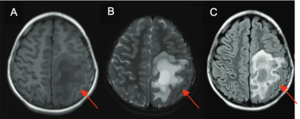
A girl 6 years and 9 months of age who had previously undergone a Kasai operation at one month of age due to biliary atresia presented with acute right side hemiparesis. A year prior to the incident, she presented with tingling sensation both toes and clumsy gait for about 3 days. The symptoms disappeared spontaneously. Brain magnetic resonance imaging (MRI) showed a tumefactive demyelinating lesion with
ill-defined peripheral enhancement in the left fronto-parietal white matter (Figure 1). A demyelinating disease such as multiple sclerosis was suspected, but brain MRI was not typical for multiple sclerosis. Therefore, we diagnosed this patient with Schilder’s disease while performing further evaluation. Oligoclonal bands in cerebrospinal fluid (CSF) were negative, and a spine MRI showed no abnormalities. The liver disease indicators including aspartate aminotransferase (AST), alanin aminotransferase (ALT), and gamma glutamyl transferase (GGT) were increased to 60 IU/L, 269 IU/L, and 234 IU/L, respectively, and liver cirrhosis was detected. Her vitamin E level, which was obtained during convalescence was 9.4 mg/L (3.8-18.4 mg/L) and her cholesterol level was 187 mg/dL (100-240 mg/dL).
The patient was treated with methyl prednisolone (10 mg/kg/day) intravenously for 10 days. Hemiparesis disappeared about fifth day of treatment, allowing the patient to walk independently, and the size of the brain lesion decreased on MRI (Figure 2). However, she showed intention tremor in her right arm and dystonia in her right foot although hemiparesis has resolved, which were noted during the methyl prednisolone treatment. After methyl prednisolone therapy, oral prednisone (1 mg/kg/day) was given. The oral prednisone was gradually tapered over 6 months. The patient fully recovered from intention tremor and dystonia 2 months after
Neurology Asia December 2013
420
oral prednisone treatment, with no relapse of neurological symptom during the 3-year follow-up.
DISCUSSION
The diagnostic criteria of Schilder’s disease proposed by Poser are as follows.2 It is a subacute myelinoclastic disorder resulting in formation of one, or more commonly, two roughly symmetric bilateral plaques measuring at least 2×3 cm in two of three dimensions, involving the centrum semiovale. These must be the only lesions demonstrable by clinical, paraclinical, or imaging studies. There must be no peripheral nervous system lesion. Adrenal function and very long-chain fatty acids must be normal. The histopathology must be identical to multiple sclerosis. Our patient fulfills the Poser’s criteria, except the absence of histopathology, as brain biopsy was not performed. The tests of adrenal function were not available, but the patient did not show symptoms of adrenal crisis or adrenal
insifficiency. As suggested in recent studies3, improved MRI findings of biopsy-proven cases of Schilder’s disease have enabled noninvasive diagnosis of Schilder’s disease. In our patient, a good response to steroid pulse therapy was also supportive of the diagnosis.
MRI in Schilder’s disease show a hypointense lesion on T1 weighted images and a hyperintense lesion on T2 weighted images.3 Open ring contrast enhancement caused by an acute inflammatory process in the white matter is characteristic.4 Other findings are the increased signal on fluid attenuated inversion recovery (FLAIR) and an increase in the apparent diffusion coefficient (ADC). These radiologic features are also supportive of the diagnosis of Schilder’s disease.
In our patient, a possible diagnosis of multiple sclerosis was considered based on the 2010 revisions to the McDonald criteria of MS. She had two episodes of clinical attacks and one brain MRI lesion for dissemination in space.5 However, the single large tumefactive lesion was atypical for
Figure 1. Initial brain MRI shows demyelinating lesion at left fronto-parietal white matter. A) T1-weighted image; B) T2-weighted image; C) T2 FLAIR image.
Figure 2. Brain MRI after 3 months shows resolution of lesions seen in Figure 1, with scarring and a small focal area of cavitation. A) T1-weighted image (hypointense); B) T2-weighted image (hyperintense); C) T2 Flair image (hyperintense).
!"
#
"
421 multiple sclerosis, but consistent with Schilder’s
disease.
In rare cases of acute disseminated encephalomyelitis (ADEM), brain MRI shows a large single lesion, predominantly white matter. However, we believe that the clinical presentation of ADEM must include encephalopathy, which is defined as one or more of the following, behavioral change and alteration in consciousness.6,7 Our patient had two historical evidences of ataxia and hemiparesis without any encephalopathy. As for the relationship between biliary atresia and Schilder’s disease, biliary atresia causes cholestasis, or difficulty in secretion of bile acid. Therefore, impaired absorption of fat-soluble vitamins is common in patients with biliary atresia. Vitamin E deficiency is known to be associated with various neurologic abnormalities such as ataxia, weakness, areflexia, impaired vision, loss of proprioception, and vibratory sensation and variable ophthalmoplegia.8,9 In our patient, we unfortunately could not determine the vitamin E level at the moment of hemiparesis. The serum vitamin E level determined several months after the event was normal and the serum vitamin E to total cholesterol ratio was also normal. In conclusion, Schilder’s disease should be considered in children with acute or subacute neurological symptom, and large tumefactive lesion in MRI.
ACKNOWLEDGEMENT
This work was supported by National Research Foundation grant funded by the Korea government (MEST, 2010-0020353).
DISCLOSURE
Conflict of interest: None
REFERENCES
1. Schilder P. Zur Kenntnis der sogennanten diffusen Sklerose. Z Gesamate Neurol Psychiatr 1912; 10:1-60. 2. Poser CM, Goutiers F, Carpentier MA, Aicardi J.
Schilder’s myelinoclastic diffuse sclerosis. Pediatrics 1986; 77:107-12.
3. Bacigaluppi S, Polonara G, Zavanone ML, Campanella R, Branca V, Gaini SM, Tredici G, Costa A. Schilder’s disease: non-invasive diagnosis?: A case report and review. Neurol Sci 2009; 30:421-30. 4. Masdeu JC, Quinto C, Olivera C, Tenner M, Leslie
D, Visintainer P. Open-ring imaging sign: highly specific for atypical brain demyelination. Neurology 2000; 54:1427-33.
5. Chris HP, Stephen CR, Brenda B, et al. Diagnostic criteria for multiple sclerosis: 2010 revisions to the McDonald criteria. Ann Neurol 2011; 69:292-302.
6. Krupp LB, Banwell B, Tenembaum S. Consensus definitions proposed for pediatric multiple sclerosis and related disorders. Neurology 2007; 68: S7-S12.
7. Tenembaum S, Chamoles N, Fejerman N. Acute disseminated encephalomyelitis: A long-term follow-up study of 84 pediatric patients. Neurology 2002; 59: 1224-31.
8. Rosenblum JL, Keating JP, Prensky AL, Nelson JS. A progressive neurologic syndrome in children with chronic liver disease. N Engl J Med 1981; 304:503-8.
9. Satya-Murti S, Howard L, Krohel G, Wolf B. The spectrum of neurologic disorder from vitamin E deficiency. Neurology 1986; 36:917-21.