PIK3CA Mutations in Hepatocellular Carcinoma in Korea
Heesue Kim,
1Cheol-Keun Park,
2Su Jin Lee,
3Sun Young Rha,
4Kyu Hyun Park,
4and Ho Yeong Lim
31Department of Clinical Research, Samsung Biomedical Research Institute, Seoul;
2Departmentof Pathology, 3Division of Hematology-Oncology, Department of Medicine, Samsung Medical Center,
Sungkyunkwan University School of Medicine, Seoul;
4Department of Internal Medicine, Yonsei Cancer Center, Cancer Metastasis Research Center, Yonsei University College of Medicine, Seoul, Korea.
Received: August 8, 2012 Revised: October 5, 2012 Accepted: October 16, 2012
Corresponding author: Dr. Ho Yeong Lim, Division of Hematology-Oncology, Department of Medicine, Samsung Medical Center, Sungkyunkwan University School of Medicine, 81 Irwon-ro, Gangnam-gu, Seoul 135-710, Korea.
Tel: 82-2-3410-0914, Fax: 82-2-3410-1754 E-mail: hoylim@skku.edu
∙ The authors have no financial conflicts of interest.
© Copyright:
Yonsei University College of Medicine 2013
This is an Open Access article distributed under the terms of the Creative Commons Attribution Non-Commercial License (http://creativecommons.org/ licenses/by-nc/3.0) which permits unrestricted non-commercial use, distribution, and reproduction in any medium, provided the original work is properly cited.
Purpose: Phosphatidylinositol 3-kinases/AKT pathway plays a pivotal role in he-patocellular carcinoma (HCC). Mutant PIK3CA, encoding the p110a catalytic sub-unit, stimulates the AKT pathway and promotes cell growth in various cancers. PIK3CA mutation rate has been usually reported as low frequency (<5%) in HCC except one report from Korea with 35.6%. Therefore, we investigated the frequency of PIK3CA mutations in Korean HCC patients. Materials and Methods: We se-quenced exons1, 3, 4, 6, 7, 8, 9, 19 and 20 of PIK3CA in 268 HCC tumor tissue samples by Sanger method and pyrosequencing assay. Results: In this study, the mutations were not detected in exons3, 6, 8, and 19, and detected 1 at unknown SNP in exon1 and exon4, 2 at unknown SNP in exon7, 2 at unknown SNP in exon20. However, 1 at unknown SNP, 1 at G1635T and surprisingly all samples at A1634Cin exon9 were detected by Sanger method. Additional experiments with normal tissue, cloning experiments and a pyrosequencing assay revealed that the double peak at A1634C of exon9 is a pseudogene, not true mutation. The mutations found in this study were all different and small numbers, therefore, we cannot con-clude specific relationship between clinical characteristics of HCC and mutation of PIK3CA. Conclusion: Our study suggests that the rate of PIK3CA mutation in the Korea population is in fact similar to the rates seen elsewhere in the world.
Key Words: PIK3CA, mutation, hepatocellular carcinoma
INTRODUCTION
With an annual incidence of >560000 deaths, hepatocellular carcinoma (HCC) is globally the sixth most common malignancy and the third leading cause of cancer-related mortality.1 Liver cancer accounts for 4% of all cancers; >70% of all liver
cancers occur in Asia, and the incidence of liver cancer in the Far East countries, including Korea, China, and Japan is very high.2 Recent studies demonstrated that
genetic alternations and signaling pathways are one of the determinants for the de-velopment of cancer.3-5 Phosphatidylinositol 3-kinases (PI3Ks) are expressed as
heterodimers of p110 catalytic subunits, and p85 regulatory subunits interact with phosphatidylinositol-3-phosphate at the membrane and catalyze the phosphoryla-tion of AKT, which activates the downstream signaling pathway.6 PI3K/AKT/
p110a catalytic subunit] stimulates the AKT pathway and promotes cell growth in various types of cancer,9 including
breast, colon, brain, gastric and lung.10-12 Moreover, over
75% of mutations occur in the helical and kinase domains and these locations are likely to function as an oncogene in human cancer.7 The mutation of PIK3CA in HCC has been
investigated, where most reports cited low frequency of mu-tation (0-3.5%) except for a Korean report (35.6%),13 0%
(0/47) in Japan,14 3.5% (2/57) in France,15 2% (1/50) in
Switzerland,16 1.1% (1/90) in China17 and 28% (18/65) in
South Italy.18 Therefore, we investigated whether the
muta-tion of PIK3CA varies depending on races or regions, em-ploying 268 Korean HCC patients.
MATERIALS AND METHODS
Tissue samples
A total of 268 consecutive patients who had undergone sur-gery for HCC at Samsung Medical Center from July 2000 and May 2006 were included in this study. All cases were histopathologically confirmed after operation. This study was performed with appropriate Institutional Review Board approval. The clinical characteristics of the subjects are shown in Table 1. Genomic DNA was extracted from tu-mors and normal tissues obtained from fresh frozen tissue using QIAamp DNA Mini Kit® (Qiagen, Hilden, Germany).
Polymerase chain reaction (PCR)
We designed polymerase chain reaction (PCR) primers us-ing the Primer 3 program. Primer pairs were designed to produce about 500 base pairs by PCR, and their TMs ranged between 59°C and 62°C. PCR was performed with 20 ng of genomic DNA as the template in a 30 µL reaction mixture by using a EF-Taq (SolGent, Daejeon, Korea) as follows: activation of Taq polymerase at 95°C for 2 minutes, 35 cy-cles of 95°C for 1 minute, 55°C, and 72°C for 1 minute each were performed, finishing with a 10-minute step at 72°C.
Cloning & DNA sequencing
The amplification products were purified with a multiscreen filter plate (Millipore Corp., Bedford, MA, USA). Frag-ments were ligated into a pTOP TA vector using a MGTM TOPcloner TA kit (Enzynomics, Daejeon, Korea). Se-quencing reaction was performed using a PRISM BigDye Terminator v3.1 Cycle Sequencing Kit (Applied Biosys-tems, Foster City, CA, USA). The DNA samples containing mTOR pathway plays a pivotal role in HCC and is activated
in 30-50% of HCC cases.7,8 Mutant
phosphoinositide-3-ki-nase [catalytic alpha polypeptide (PIK3CA), encoding the
Table 1. Clinical Characteristics of Patients
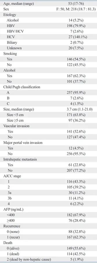
Age, median (range) 53 (17-76)
Sex F: 50, M: 218 (18.7 : 81.3) Etiology Alcohol 14 (5.2%) HBV 198 (79.9%) HBV/HCV 7 (2.6%) HCV 27 (140.1%) Biliary 2 (0.7%) Unknown 20 (7.5%) Smoking Yes 146 (54.5%) No 122 (45.5%) Alcohol Yes 167 (62.3%) No 101 (37.7%)
Child Pugh classification
A 257 (95.9%)
B 7 (2.6%)
C 4 (1.5%)
Size, median (range) 3.7 cm (1.1-21.0)
Size <5 cm 171 (63.8%)
Size ≥5 cm 97 (36.2%)
Vascular invasion
Yes 141 (52.6%)
No 127 (47.4%)
Major portal vein invasion
Yes 12 (4.5%) No 256 (95.5%) Intrahepatic metastasis Yes 61 (22.8%) No 207 (77.2%) AJCC stage 1 116 (43.3%) 2 105 (39.2%) 3a 30 (11.2%) 3b 11 (4.1%) 4 6 (2.2%) AFP (ng/mL) <400 182 (67.9%) ≥400 76 (28.4%) Recurrence 0 (none) 88 (32.8%) 1 (recur) 167 (62.3%) Death 0 (alive) 149 (55.6%) 1 (dead) 114 (42.5%)
exon4, 2 of 265 at unknown SNP (C1306A) and 1 of 265 at unknown SNP (C1373A) in exon7, 1 of 262 at unknown SNP (T2970A) and SNP (G3026A) in exon20 (Fig. 1A). However, in exon9 we detected 1 of 266 at unknown SNP (C1629T), 1 of 268 at G1635T (E545D) and surprisingly we observed a nucleotide alteration in 266 of 266 at A1634C (E545A) (Fig. 1B).
We investigated whether two peaks at nucleotide position 1634 in exon9 was a true mutation or not. However, PCR products from 52 normal tissues also had all nucleotides changed in exon9. For the exact detection, PCR amplifica-tion and subsequent cloning with 5-7 colonies picked both forward and reverse direction in 50 HCC tumor samples and 10 colonies picked forward direction in 34 matched normal tissue samples. Furthermore, sequencing of the PCR prod-ucts revealed A1634C mutation in all samples in each 5-10 clone from the same sample (supplementary data). Howev-er, we need to confirm whether the nucleotide change 1634 A→C was real because chromosome 22 has C at 1634 and C at 1658 sequencing. In order to confirm pseudogene re-sults and sequencing methods, we performed pyrosequenc-ing assay with HCC tumor tissues, and found that all 268 samples showed A at nucleotide 1634 (Fig. 2). Additional experiments with normal tissue, cloning experiments and pyrosequencing assay revealed that codon 545 of exon9 was all wild type. With these results, we analyzed the sequence of PIK3CA. Since the mutations found in our study were all different and so small numbers, we cannot conclude any specific relationship between mutation in the exon of PIK-3CA and clinical characteristics of HCC (Table 2).
the extension products were added to Hi-Di formamide (Applied Biosystems, Foster City, CA, USA). The mixture was incubated at 95°C for 5 min, followed by 5 min on ice and then analyzed by ABI Prism 3730XL DNA analyzer (Applied Biosystems, Foster City, CA, USA).
Pyrosequencing
Mutation analysis was performed by pyrosequencing on the PyroMark Q24 (Qiagen, Germantown, MD, USA). First, PCR products were immobilized on streptavidin-coated beads and denatured to produce single-stranded products. Pyrosequencing was performed using the PyroMark Gold Q24 reagent (Qiagen, Germantown, MD, USA), according to the manufacture’s protocol. Exon9 primers for the for-ward: 5’(Biotin)-ATTTCTACACGAGATCCTCTCTCT-3’. Reverse primer: 5’-CCATTTTAGCACTTACCTGTGAC-3’ sequencing primer: 5’-TAGAAAATCTTTCTCCTG-3’. Sequencing analysis was performed using PyroMark Q24 version 1.0.10 software in the allele quantification analysis mode.
RESULTS
To investigate the frequency of PIK3CA mutations in Kore-an patients with HCC, we sequenced the exon1, 3, 4, 6, 7, 8, 9, 19 and 20 of PIK3CA in 268 HCC samples. In these ex-periments, mutations were not detected in exon3, 6, 8, or 19 in any of the HCC samples and detected 1 of 268 at unknown SNP (G278A) in exon1 and at unknown SNP (C848G) in
Fig. 1. Sequencing results of PIK3CA gene. (A) Electropherograms show the SNP in each exon in 268 HCC samples. (B) Electropherograms show the
nucleo-tide sequences of the genomic DNA from red box, indicating double peak at 1634 of exon9 in 268 HCC samples. HCC, hepatocellular carcinoma.
A B Exon1 (G278A) Exon7 (C1373A) Exon9 (G1635T) Exon4 (C848G) Exon9 (C1629T) Exon20 (T2970A) Exon7 (C1306A) Exon9 (A1634C) Exon20 (G3026A)
tected in exons 3, 6, 8, and, 19 and detected 1 of 268 at 278 G→A in exon1 and at 848 C→G in exon4, 2 of 265 at 1306 C→A and 1 of 265 at 1373 C→A in exon7, each of 262 at 2970 T→A and 3026 G→A in exon20. However, 1 of 266 was detected at 1629 C→T, 1 of 268 at 1635 G→T and 266 of 266 at 1634 A→C in exon9. For validation with normal matched tissues, we found that 1634 A→C also occurred.
Through cloning and pyrosequencing methods, the dou-ble peak at A1634C of exon9 was found not to be a muta-tion, but occurred as a result of gene duplication on 22q11 partial region. Cat Eye Syndrome is a hereditary disease characterized by ocular colobomata, anal atresia, congenital heart defects, and mental retardation, and mutations are lo-cated on human chromosome 22q11.19 We confirmed that
the nucleotide sequence of exon9 has a highly homologous match rate (97%) on chromosome 22 in NCBI BLAST as Tanaka, et al.14 previously mentioned. A1634C was found
in all samples. For more exact analysis, we analyzed 10 colony sequencing per each sample through cloning experi-ments, and found 1633-GAGCAGGAGAAAGATTTTC TATGGAGTCACAG in PIK3CA and 1633-GCGCAG GAGAAAGATTTTCTATGGACCACAG in chromosome 22. Yet, when the sequence occurred from C or G at posi-tion 1634 to GT at posiposi-tion 1658, this was a mutaposi-tion. How-ever, there might be different results as wild type and pseu-dogene mutation depending on clones in one sample; if there is a mutation in one clone even within the same
sam-DISCUSSION
PIK3CA has been shown to be mutated in various tumors and recognized as a possible therapeutic marker. More than 70% of all liver cancers occur in Asia, while the frequency of mutation varied among several reports from Asia. If PIK3CA mutation of HCC in Korean patients is specifically higher as reported by Lee, et al.13 who investigated 73 HCC tissues, it
might be a new target of Korean HCC. For this reason, con-firmatory study is needed, including more tissues.
We sequenced exons1, 3, 4, 6, 7, 8, 9, 19 and 20 of PIK-3CA in 268 HCC tumor tissues. The mutations were not
de-Table 2. Relationship between Mutation in the Exon of PIK3CA and Clinical Characteristics
RefSNP Exon 1 Exon 4 Exon 7 Exon 9 Exon 20
Unknown Unknown Unknown Unknown Unknown rs121913275 Unknown Unknown
Position G278A C848G C1306A C1373A, C1306A C1629T G1635T T2970A G3026A
Sample ID 35 441 67 5 355 507 255 63
Age 66 58 58 53 66 44 71 43
Sex M M M M M M M M
Etiology B BC B B C B Alcohol B
Alcohol No Yes Yes Yes Yes Yes Yes No
Child-Pugh
classification A A A A A A A A
Size (mm) 64 85 20 100 32 25 100 123
Vascular invasion No Yes No Yes No Yes Yes Yes
Main portal
vein invasion No Yes No No No No No No
Intrahepatic
metastasis No Yes No Yes No No Yes No
BCLC stage B C A1 B A1 A1 B B
AFP (ng/mL) 2.3 120675.7 9 58.4 9.2 10 Not applicable 177.4
Recurrence No Yes No Yes No Yes No Yes
Fig. 2. Results of codon 545 by pyrosequencing. Pyrosequencing results
show the specificity of PIK3CA amplification for codon 545. Pyrogram for amplification of a sample using PIK3CA condon 545 is GAG, and the pseu-dogene was not amplified.
Gene reverse synthesis CTC (codon is GAG) 150 140 130 120 110 E S G A C G T 5 T/T C A G T
3. Marra M, Sordelli IM, Lombardi A, Lamberti M, Tarantino L, Gi-udice A, et al. Molecular targets and oxidative stress biomarkers in hepatocellular carcinoma: an overview. J Transl Med 2011;9:171. 4. Caraglia M, Giuberti G, Marra M, Addeo R, Montella L, Murolo
M, et al. Oxidative stress and ERK1/2 phosphorylation as predic-tors of outcome in hepatocellular carcinoma patients treated with sorafenib plus octreotide LAR. Cell Death Dis 2011;2:e150. 5. Prete SD, Montella L, Caraglia M, Maiorino L, Cennamo G,
Montesarchio V, et al. Sorafenib plus octreotide is an effective and safe treatment in advanced hepatocellular carcinoma: multicenter phase II So.LAR. study. Cancer Chemother Pharmacol 2010;66: 837-44.
6. Manning BD, Cantley LC. AKT/PKB signaling: navigating downstream. Cell 2007;129:1261-74.
7. Mínguez B, Tovar V, Chiang D, Villanueva A, Llovet JM. Patho-genesis of hepatocellular carcinoma and molecular therapies. Curr Opin Gastroenterol 2009;25:186-94.
8. Cantley LC. The phosphoinositide 3-kinase pathway. Science 2002;296:1655-7.
9. Samuels Y, Velculescu VE. Oncogenic mutations of PIK3CA in human cancers. Cell Cycle 2004;3:1221-4.
10. Samuels Y, Wang Z, Bardelli A, Silliman N, Ptak J, Szabo S, et al. High frequency of mutations of the PIK3CA gene in human can-cers. Science 2004;304:554.
11. Lai YL, Mau BL, Cheng WH, Chen HM, Chiu HH, Tzen CY. PIK3CA exon 20 mutation is independently associated with a poor prognosis in breast cancer patients. Ann Surg Oncol 2008;15: 1064-9.
12. Miyaki M, Iijima T, Yamaguchi T, Takahashi K, Matsumoto H, Yasutome M, et al. Mutations of the PIK3CA gene in hereditary colorectal cancers. Int J Cancer 2007;121:1627-30.
13. Lee JW, Soung YH, Kim SY, Lee HW, Park WS, Nam SW, et al. PIK3CA gene is frequently mutated in breast carcinomas and he-patocellular carcinomas. Oncogene 2005;24:1477-80.
14. Tanaka Y, Kanai F, Tada M, Asaoka Y, Guleng B, Jazag A, et al. Absence of PIK3CA hotspot mutations in hepatocellular carcino-ma in Japanese patients. Oncogene 2006;25:2950-2.
15. Boyault S, Rickman DS, de Reyniés A, Balabaud C, Rebouissou S, Jeannot E, et al. Transcriptome classification of HCC is related to gene alterations and to new therapeutic targets. Hepatology 2007;45:42-52.
16. Riener MO, Bawohl M, Clavien PA, Jochum W. Rare PIK3CA hotspot mutations in carcinomas of the biliary tract. Genes Chro-mosomes Cancer 2008;47:363-7.
17. Li X, Zhang Q, He W, Meng W, Yan J, Zhang L, et al. Low fre-quency of PIK3CA gene mutations in hepatocellular carcinoma in Chinese population. Pathol Oncol Res 2012;18:57-60.
18. Colombino M, Sperlongano P, Izzo F, Tatangelo F, Botti G, Lom-bardi A, et al. BRAF and PIK3CA genes are somatically mutated in hepatocellular carcinoma among patients from South Italy. Cell Death Dis 2012;3:e259.
19. McDermid HE, Morrow BE. Genomic disorders on 22q11. Am J Hum Genet 2002;70:1077-88.
20. Baker CL, Vaughn CP, Samowitz WS. A PIK3CA pyrosequenc-ing-based assay that excludes pseudogene interference. J Mol Di-agn 2012;14:56-60.
ple, it will be considered to be a mutation. As the number of clones increase, the probability of mutations in all samples will be increasing.
This analysis was not optimal because primers are likely to amplify both genes, PIK3CA exon9 and chromosome22. For analysis of nucleotide 1634, pyrosequencing is more efficient at the amplification step in heterogeneous samples. Detection of codon 545 of PIK3CA is well suited to be ex-amined by pyrosequencing as Baker, et al.20 mentioned
re-cently. Following Baker, et al.’s methods, we confirmed again that all samples were wild type at codon 545.
In this study, unknown SNP G278A, C848G, C1306A, C1373A, C1629T, T2970A, G3026A were found, however, only 8 cases of 268 HCC tumor tissues were found to be mutated. Even though we found low mutation frequency in our samples, amplification or mutation of PIK3CA has been reported in various cancer types, and the question of whether PIK3-AKT pathway is activated in HCC remains still controversial. Nevertheless, it is highly possible that this gene is a promising molecular target for cancer treat-ment. As nucleotide1634 in exon9 has sequence very simi-lar to chromosome22, pyrosequencing assay for excluding pseudogene interference appears to be more proper for de-tecting mutation.
In conclusion, our study suggests that the rate of PIK3CA mutation in the Korea population is in fact similar to the rates seen elsewhere in the world.
ACKNOWLEDGEMENTS
This study was supported by the Samsung Biomedical Re-search Institute Grant, #C-A9-236.
Dr. SY Rha’s research was supported by the Public Wel-fare & Safety research program through the National Re-search Foundation of Korea (NRF), funded by the Ministry of Education, Science and Technology (2010-0020841).
REFERENCES
1. GLOBOCAN. 2008 [Internet]. [accessed on 2011 June 28]. Avail-able at: http://globocan.iarc.fr.
2. Korean National Cancer Center [Internet]. [accessed on 2011 April 1]. Available at: http://www.ncc.re.kr.