30
Hormonal Changes of the Brain-Dead Organ Donors: A 3-Year Experience
Yong Seon Choi, M.D., Sungwon Na, M.D., Seung Youn Kang, M.D. and Shin Ok Koh, M.D.
Department of Anesthesiology and Pain Medicine, Anesthesia and Pain Research Institute, Yonsei University College of Medicine, Seoul, Korea
Background: Success of transplantation is critically dependent upon the quality of the donor organ and optimal management. Recently, hormonal replacement therapy has been reported to result in rapid recovery of cardiac func-tion and enable significantly more organs to be transplanted, while some other studies show conflicting results. The aim of this study is to comprehensively evaluate changes in basal circulating hormonal levels of the brain-dead or-gan donors.
Methods: We reviewed the records of all brain-dead patients between January, 2004, and June, 2007. Hemodyna-mic variables, plasma hormone levels were recorded at following time points: admission to the ICU (T1, baseline), 30 minutes (min) after first apnea test (T2), 30 min after second apnea test (T3), before operation for harvesting (T4). Hormonal measurements included cortisol, adrenocorticotrophic hormone, triiodothyronine (T3), thyroxine, free thyroxine, thyroid-stimulating hormone, growth hormone, and testosterone.
Results: Nineteen patients were included in this study. Comparisons of hemodynamic parameters and hormonal levels to baseline values revealed no significant changes throughout the study period. When the patients were div-ided into 2 groups according to the requirement of norepinephrine (either>0.05 or ≤0.05μg/kg/min), patients re-quiring >0.05μg/kg/min of norepinephrine had T3 level below the normal range at significantly more time points of measurement (7 vs. 0).
Conclusions: In this comprehensive assessment of hormonal levels in brain-dead organ donors, we could not ob-serve any significant changes during the ICU stay. Replacement therapy of T3 may be considered in patients requir-ing >0.05μg/kg/min of norepinephrine.
Key Words: Brain-dead organ donors, Hormonal replacement therapy, Thyroid hormone levels, Vasopressor
Correspondence to: Shin Ok Koh, Department of Anesthesiology and Pain Medicine, Yonsei University College of Medicine, 134, Sinchon-dong, Seodaemun-gu, Seoul 120-752, Korea. Tel: 82-2-2228-2420, 2403, Fax: 82-2-312-7185 E-mail: [email protected]
INTRODUCTION
Despite advances in medical management strategies, trans-plantation is still being considered as the only definitive treat-ment for patients with end-stage cardiac, pulmonary and hep-atic disease.1) Organ transplantations from brain-dead donors have increased since the first successful kidney transplantation in 1979 and 597 transplants were performed from 141 brain-dead donors in 2006 in Korea. However, the demand for transplantable organs continues to exceed the supply. The suc-cess of transplantation is critically dependent upon the quality of the donor organ and optimal management. Optimal manage-ment of brain-dead donors is important for the maximal uti-lization and also right timed operation, because hemodynamic
stability is a key part of successful organ recovery.
A characteristic feature of brain death is the sympathetic storming, causing extreme hypertension and tachycardia, fol-lowed by loss of sympathetic tone and massive vasodilatation leading to hemodynamic instability.2) Also, various functional components of the pituitary and hypothalamic regulatory sys-tems may also become affected as ischemia spreads, resulting in decreased circulating levels of triiodothyronine (T3), corti-sol/adrenocorticotrophic hormone (ACTH), insulin and arginine vasopressin.2-5) Recently, hormonal replacement therapy has been reported to result in rapid recovery of cardiac function in both experimental animals and humans, and to enable sig-nificantly more organs to be transplanted,6-8) while some other studies show conflicting results.9,10) In this study, we inves-tigated the hormonal changes during organ donor management in intensive care unit (ICU) and evaluated correlation of the hormonal levels with hemodynamic parameters and the amount of vasopressor needed to maintain blood pressure in the
brain-dead donors. The aim of this study is to comprehensively evaluate changes in basal circulating hormonal levels of the brain-dead organ donors.
MATERIALS AND METHODS
We reviewed the records of all brain-dead patients that suc-cessfully donated organs between January, 2004, and June, 2007. We examined donors with regard to age, sex, diagnosis, number of organs donated, type of organ donated, the presence or absence of brain-death associated complications. We adopted a new policy of aggressive donor management for potential or-gan donors since January, 2004. This policy involves early identification of potential organ donors, admission to the surgi-cal ICU, and management by a dedicated team using a pre-defined protocol. The protocol for aggressive donor manage-ment includes the following elemanage-ments: (i) pulmonary artery catheterization to monitor hemodynamic status and tissue perfu-sion, (ii) aggressive intravenous fluid resuscitation, (iii) vaso-pressor infusion in case of mean arterial pressures under 70 mmHg despite fluid resuscitation, (iv) identification of brain death related complications and prompt interventions. However, this protocol did not include hormone replacement therapy at that time. Neurogenic pulmonary edema was treated suppor-tively utilizing high positive end expiratory pressure ventilation; diabetes insipidus was treated with fluid replacement and vaso-pressin infusion; disseminated intravascular coagulopathy and thrombocytopenia were treated with blood and blood products; electrolyte abnormalities and acidosis were treated accordingly; and hypothermia was either avoided or if present treated with external warming methods. Dopamine was used as the primary vasopressor to potential donors with hemodynamic instability; norepinephrine was used when requirements for dopamine ex-ceeded dosage of 10μg/kg/min. In addition, to evaluate the need for additional hormone-replacement therapy, high-dose vasopressor was defined when the dose of norepinephrine ex-ceeded 0.05μg/kg/min.
Brain-death diagnosis was confirmed when an irreversible catastrophic structural brain lesion resulted in unresponsiveness to noxious pain stimuli and abolition of brainstem reflexes (pa-pillary light responses, corneal reflexes, vestibuloocular tests, tracheobronchial stimulation) in the absence of hypothermia, metabolic or electrolyte disturbances, and depressant drugs. Testing for apnea was performed using guidelines after all oth-er brain-death critoth-eria had been fulfilled.11,12) The interval be-tween two evaluations for diagnosis of brain death was 6
hours when age of brain-dead donor was more than 6 years. Hemodynamic variables, arterial blood gas analyses and ven-tilator mode were recorded at the following points: admission to the ICU (T1), 30 minutes (min) after first apnea test (T2), 30 min after second apnea test (T3), before operation for har-vesting (T4). Hemodynamic measurements included mean arte-rial pressure (MAP), central venous pressure (CVP), pulmonary capillary wedge pressure (PCWP), and cardiac index (CI). Blood sampling for the hormone levels was performed at the same points. Hormonal measurements included cortisol, ACTH, growth hormone (GH), testosterone and the thyroid hormone levels as measured by T3, thyroxine (T4), free thyroxine (fT4) and thyroid-stimulating hormone (TSH). However, blood sam-pling for cortisol and ACTH was performed twice at the morning of ICU day 1 and at the evening of ICU day 2; tes-tosterone was sampled only in male patients. Determination of T3, T4, fT4, TSH, cortisol (sensitivity: 0.02μg/dl) and testoster-one was performed using a chemiluminescent immunoassay (ADVIA Centaur, Simens, New York, USA), and Determina-tion of ACTH (sensitivity: 1.2 pg/ml) and GH was performed using an immunoradiometric assay (Gamma counter COBRA II, Packard, Meriden, USA). Operative harvesting of the allog-rafts generally occurred within 12 hours of the declaration of clinical brain death.
Statistical analyses were performed with SPSS 13.0 (SPSS Inc., Chicago, IL, USA). All data are expressed as mean±stan-dard deviation (SD) or number of patients. Changes between time points within the group were compared using univariate analysis of variance with post hoc comparisons using the Dunnette's test. A p value of less than 0.05 was considered statistically significant.
RESULTS
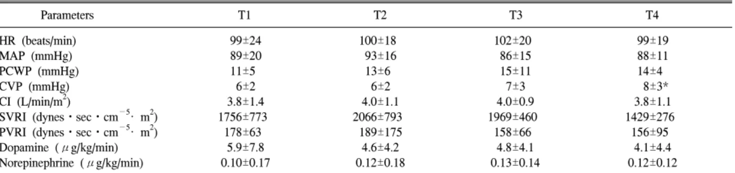
A total of 23 patients were admitted to the ICU after clin-ical brain death during the three-year period. Four patients were excluded due to incomplete data collection of hormonal levels. The other 19 patients (14 men, 5 women), having a mean age of 32±13 yrs (range, 12 to 63 yrs), were included in this study. The cause of brain death was cerebrovascular disease in 11 cases (58%), cerebral anoxia related to cardiac arrest in 5 cases (26%), and head trauma in 3 cases (16%). Comparisons of hemodynamic parameters to baseline values revealed no significant changes throughout the study period, except CVP which was significantly increased at T4 (p=0.031, Table 1). No significant changes in hormonal levels were
ob-Table 1. Changes in Hemodynamic Variables and Requirement of Vasopressors Parameters T1 T2 T3 T4 HR (beats/min) 99±24 100±18 102±20 99±19 MAP (mmHg) 89±20 93±16 86±15 88±11 PCWP (mmHg) 11±5 13±6 15±11 14±4 CVP (mmHg) 6±2 6±2 7±3 8±3* CI (L/min/m2) 3.8±1.4 4.0±1.1 4.0±0.9 3.8±1.1 SVRI (dynesㆍsecㆍcm−5· m2) 1756±773 2066±793 1969±460 1429±276 PVRI (dynesㆍsecㆍcm−5· m2) 178±63 189±175 158±66 156±95 Dopamine (μg/kg/min) 5.9±7.8 4.6±4.2 4.8±4.1 4.1±4.4 Norepinephrine (μg/kg/min) 0.10±0.17 0.12±0.18 0.13±0.14 0.12±0.12
Values are mean±SD. T1: after admission to ICU; T2: 30 min after first certification; T3: 30 min after second certification; T4: before operation; HR: heart rate; MAP: mean arterial pressure; PCWP: pulmonary capillary wedge pressure; CVP: central venous pressure; CI: cardiac index; SVRI: systemic vascular resistance index; PVRI: pulmonary vascular resistance index. *p<0.05 compared with T1.
Table 2. Change of Plasma Hormone Levels
T1 T2 T3 T4 Range Normal range Sensitivity
T3 (ng/ml) 0.74±0.15 0.87±0.30 0.90±0.33 0.90±0.30 0.48∼1.62 0.6∼1.8 0.1 T4 (μg/ml) 5.18±1.56 5.16±1.99 5.17±2.01 5.48±2.14 1.28∼11 4.5∼10.9 0.3 fT4 (μg/ml) 0.86±0.26 0.89±0.26 0.93±0.35 0.97±0.43 0.3∼2.18 0.89∼1.76 0.1 TSH (μIU/ml) 1.28±1.77 1.94±2.33 2.02±1.05 1.74±1.48 0.01∼8.12 0.35∼5.5 0.01 GH (ng/ml) 6.1±8.4 11.2±13.9 11.1±12.5 16.1±20.7 0.12∼77 0.1∼0.33 0.05 Testosterone (ng/dl) 62.1±35.4 79.6±64.1 75.9±67.8 65.4±55.5 10∼262 241∼827 10
Values are mean±SD. T1: after admission to ICU; T2: 30 min after first certification; T3: 30 min after second certification; T4: before operation; T3: triiodothyronine; T4: thyroxine; fT4: free thyroxine; TSH: thyroid-stimulating hormone; GH: growth hormone.
Table 3. Brain-Dead Donors’ Distribution according to Their Thyroid Hormone Levels
T3 T4 fT4 TSH Below normal Normal Above normal Below normal Normal Above normal Below normal Normal Above normal Below normal Normal Above normal T1 1 15 5 11 8 8 8 7 1 T2 1 18 8 11 10 9 7 10 2 T3 2 17 7 12 9 10 7 10 2 T4 3 16 7 12 8 9 1 5 12
Values are number of patients. T1: after admission to ICU; T2: 30 min after first certification; T3: 30 min after second certification; T4: before operation; T3: triiodothyronine; T4: thyroxine; fT4: free thyroxine; TSH: thyroid-stimulating hormone.
served throughout the study period compared to baseline values (Table 2). Concentrations of cortisol were 11.3±10.6μg/dl at the evening of ICU day 1 and 12.0±11.4μg/dl at the morning of ICU day 2. Concentrations of ACTH were 10.6±4.9 pg/ml at the evening of ICU day 1 and 17.3±42.0 pg/ml at the morning of ICU day 2. Concentration of cortisol and ACTH were below normal in 13 and 17 patients at the evening of ICU day 1, in 12 and 15 patients at the morning of ICU day 2. Distributions of brain-dead donors according to their thyroid hormone levels at each time point are shown in Table 3. The
number of low T3 donors was 1, 1, 2 and 3 at T1, T2, T3, and T4, respectively in all four patients. One of them showed sustained T3 level below the normal range during the study. Three of them showed T3 level below the normal range at T3, T4, and T4, respectively. To determine if there were differ-ences in thyroid hormone levels and requirement of vaso-pressor, all thyroid hormone values were separated according to the high or low dose of vasopressor regardless of time of blood sampling. When the patients were divided into 2 groups according to the requirement of norepinephrine (either >0.05
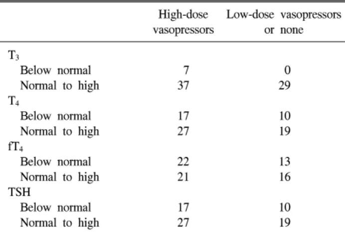
Table 4. Requirement of Vasopressors during ICU Management Compared with Thyroid Hormone Levels
High-dose Low-dose vasopressors vasopressors or none T3 Below normal 7 0 Normal to high 37 29 T4 Below normal 17 10 Normal to high 27 19 fT4 Below normal 22 13 Normal to high 21 16 TSH Below normal 17 10 Normal to high 27 19
Values are number of data sets. T3: triiodothyronine; T4: thyro-xine; fT4: free thyroxine; TSH: thyroid-stimulating hormone.
or ≤0.05μg/kg/min), patients requiring >0.05μg/kg/min of norepinephrine had T3 level below the normal range at sig-nificantly more time points of measurement. In addition, at all time points when the T3 levels were below the normal range, all of the donors required high dose vasopressors infusion (Table 4). The number of patients requiring high-dose vaso-pressor was 11 (58%), 10 (53%), 12 (63%) and 12 at T1, T2, T3, and T4, respectively. There were no differences in the re-quirements of dopamine between low T3 donors and normal T3 donors.
There were 16 kidney and liver donors, and 3 kidney only donors. Also, 5 of kidney and liver donors underwent heart harvest; 1 underwent heart and lung harvest; 3 underwent pan-creas harvest. The lengths of stay in the ICU and hospital were 2.1±0.5 days and 10.8±12.2 days, respectively. The brain death associated complications were coagulopathy (2 patients), thrombocytopenia (2 patients), cardiac ischemia (6 patients), acidosis (1 patient), neurogenic pulmonary edema (3 patients), renal failure (3 patients) and diabetes insipidus (8 patients). The patients used vasopressin due to diabetes insipidus were 2, 2, 2 and 2 at T1, T2, T3, and T4, respectively.
DISCUSSION
In the present study, we have shown that plasma T3 level correlated with requirements of vasopressor during brain-dead donor management in ICU. Although the basal concentration of the anterior pituitary hormones varied considerably relative to the normal range, all were above the sensitivity of the assay
in almost all cases. The concentrations of cortisol, ACTH and testosterone were below normal in most of our patients; the concentration of GH was markedly elevated and was more than 5 ng/ml in 59% of the cases at T2, T3 and T4.
Sequential systemic physiologic changes occur as different areas of the brain stem become ischemic. Pontine ischemia re-sults in mixed vagal and sympathetic stimulation, characterized by bradycardia and hypertension; medulla ischemia results in unopposed sympathetic stimulation as ischemia spreads.2) The function of the pituitary and hypothalamic regulatory system may also become affected as ischemia spreads; a number of hormonal changes occur after brain stem death and reflect an-terior and posan-terior pituitary failure. Not all these hormonal dysfunction are seen in every potential organ donor. The in-cidence and severity of the derangement depends upon the eti-ology and time course of brain stem death, and also increases with time after the onset of brain stem death.2) The basal hor-mone concentrations changed insignificantly during ICU man-agement after the clinical brain death in this study. Since the half-life of the anterior pituitary hormones is less than 1 hour,13) these hormones, therefore, was undoubtedly being re-leased until operation for harvest. The excess increase of GH may be related to a stress reaction or triggered by the in-sulin-induced drop in serum glucose.14) Although anti-diuretic hormone (ADH) was not included in this study, brain death is typically accompanied by diabetes insipidus (DI) reflecting pos-terior pituitary insufficiency. There was early depletion of ADH and development of DI in almost 80% of brain stem dead organ donors.15) In this study, there were 8 patients (42%) with DI and treated with continuous IV vasopressin infusion.
Several studies have highlighted hemodynamic instability, myocardial injury, and impairment in cardiac function after brain death.16-19) This cardiovascular deterioration is associated with impaired cellular oxygen utilization, a shift from aerobic to anaerobic metabolism, depletion of glycogen and myocardial high-energy stores, and the accumulation of lactate.20,21) This ir-regular metabolism has been associated with low levels of T3, T4, and to a lesser extent cortisol and insulin.22) The etiology of this hypothyroid state may be a result of lower than normal TSH levels caused by irreversible damage to the hypothalamus and pituitary, as well as decreased peripheral conversion of T4 to more potent T3. In this study, low incidence of low serum T3 concentration was documented in our brain-dead patients. Some of our patients had a decrease in circulating T4 and TSH concentration, whereas the serum T3 concentrations were
in the normal ranges in most of them. There were no differ-ences in hemodynamics between low T3 donors and normal T3 donors; however, we found significant correlation between low T3 levels and requirements of high-dose vasopressor. These re-sults suggest that serum T3 concentration was a major determi-nant of cardiac function in our brain-dead patients.
The effects of T3 administration in brain-dead patients sparked debate considerable and led to several studies. Several studies documented that no correlation with their hemodynamic status and lower levels of T3 in brain-dead patient.23,24) Other studies showed a benefit only in hemodynamically unstable donors.25,26) Other studies showed that T3 or T4 administration improved cardiovascular status, and reduced inotropic support and the number of donors lost cardiac instability in human brain-dead patients.7,27) Since most of the beneficial effects of T3 is cardiac function,28,29) hormonal replacement therapy in-cluding T3 was recently recommended for donors with a left ventricular ejection fraction less than 45% and/or with unstable hemodynamics.30) Recently, Rosendale et al8) showed a sub-stantial increase in the number of organs transplanted from do-nors receiving three-drug (T3/T4, methylprednisolone, arginine vasopressin) hormone-replacement therapy in the large retro-spective analysis of 10,292 donors. In this study, we observed substantial decrease in plasma hormone including cortisol, ACTH, T4, fT4 and TSH, and DI, reflecting depletion of ADH. We think that these observations will provide the foun-dations for the efficacy and optimal timing of hormonal re-placement therapy in our hospital. The limitation of this study is that the number of donors was not enough to analyze stat-istically the correlation between low T3 donors and requirement of high-dose vasopressor.
In conclusion, we showed substantial decrease in cortisol, ACTH, T4, fT4 and TSH, and significant correlation between T3 concentrations and vasopressor support in brain-dead patients. The addition of hormone therapy in association with an aggressive donor management protocol may help reduce vasopressor support, moreover, maximize the number of organs retrieved from hemodynamically unstable donors. In a time when the transplant waiting list is increasing more than the number of donors, hormonal replacement therapy may be in-tegrated in the management of brain-dead organ donors in ICU.
REFERENCES
1) Copeland JG: Advanced medical therapy does not render heart transplantation obsolete for ambulatory end-stage heart failure
patients: a debate. J Heart Lung Transplant 2001; 20: 725-8. 2) Smith M: Physiologic changes during brain stem death-lessons
for management of the organ donor. J Heart Lung Transplant 2004; 23(9 Suppl): S217-22.
3) Novitzky D, Cooper DK, Morrell D, Isaacs S: Change from aerobic to anaerobic metabolism after brain death, and reversal following triiodothyronine therapy. Transplantion 1988; 45: 32-6.
4) Novitzky D, Cooper DK, Morrell D, Isaacs S: Brain death, triiodothyronine depletion, and inhibition of oxidative phos-phorylation: Relevance to management of organ donors. Transplant Proc 1987; 19: 4110-1.
5) Chen JM, Cullinane S, Spanier TB, Artrip JH, John R, Ed-wards NM, et al: Vasopressin deficiency and pressor hypersensitivity in hemodynamically unstable organ donors. Circulation 1999; 100(19 Suppl): II244-6.
6) Novitzky D, Cooper DK, Rosendale JD, Kauffman HM: Hormonal therapy of the brain-dead organ donor: experimental and clinical studies. Transplantion 2006; 82: 1396-401. 7) Salim A, Martin M, Brown C, Inaba K, Roth B, Hadjizacharia
P, et al: Using thyroid hormone in brain-dead donors to maximize the number of organs available for transplantation. Clin Transplant 2007; 21: 405-9.
8) Rosendale JD, Kauffman HM, McBride MA, Chabalewski FL, Zaroff JG, Garrity ER, et al: Aggressive pharmacologic donor management results in more transplanted organs. Trans-plantation 2003; 75: 482-7.
9) Randell TT, Höckerstedt KA: Triiodothyronine treatment is not indicated in brain-dead multiorgan donors: a controlled study. Transplant Proc 1993; 25: 1552-3.
10) Goarin JP, Cohen S, Riou B, Jacquens Y, Guesde R, Le Bret F, et al: The effects of triiodothyronine on hemodynamic status and cardiac function in potential heart donors. Anesth Analg 1996; 83: 41-7.
11) Wijdicks EF: The diagnosis of brain death. N Engl J Med 2001; 344: 1215-21.
12) Bang EC, Koh SO, Han S, Kim JH, Nam SH: Management of the brain-dead organs donors. Korean J Crit Care Med 1996; 11: 171-7.
13) Barreca T, Perria C, Sannia A, Magnani G, Rolandi E: Evaluation of anterior pituitary function in patients with posttraumatic diabetes insipidus. J Clin Endocrinol Metab 1980; 51: 1279-82.
14) Schrader H, Krogness K, Aakvaag A, Sortland O, Purvis K: Changes of pituitary hormones in brain death. Acta Neurochir 1980; 52: 239-48.
15) Chen EP, Bittner HB, Kendall SW, Van Trigt P: Hormonal and hemodynamic changes in a validated animal model of brain death. Crit Care Med 1996; 24: 1352-9.
16) Shivalkar B, Van Loon J, Wieland W, Tjandra-Maga TB, Borgers M, Plets C, et al: Variable effect of explosive or gradual increase in intracranial pressure on myocardial structure and function. Circulation 1993; 87: 230-9. 17) Riou B, Kalfon P, Arock M, Goarin JP, Saada M, Viars P:
Cardiovascular consequences of severe hypophosphataemia in brain-dead patients. Br J Anaesth 1995; 74: 424-9. 18) Riou B, Dreux S, Roche S, Arthaud M, Goarin JP, Léger P,
et al: Circulating cardiac troponin T in potential heart transplant donors. Circulation 1995; 92: 409-14.
19) Novitsky D, Rose AG, Cooper DK: Injury of myocardial conduction tissue and coronary artery smooth muscle follow-ing brain death in the baboon. Transplantation 1988; 45: 964-6.
20) Cooper DK, Novitzky D, Wicomb WN: The pathophy-siological effects of brain death on potential donor organs, with particular reference to the heart. Ann R Coll Surg Engl 1989; 71: 261-6.
21) Salter DR, Dyke CM, Wechsler AS: Triiodothyronine (T3) and cardiovascular therapeutics: a review. J Card Surg 1992; 7: 363-74.
22) Novitzky D, Wicomb WN, Cooper DK, Tjaalgard MA: Improved cardiac function following hormonal therapy in brain dead pigs: relevance to organ donation. Cryobiology 1987; 24: 1-10.
23) Masson F, Thicoïpe M, Latapie MJ, Maurette P: Thyroid function in brain-dead donors. Transplant Int 1990; 3: 226-33. 24) Robertson KM, Hramiak IM, Gelb AW: Endocrine changes
and hemodynamic stability after brain death. Transplant Proc 1989; 21: 1197-8.
25) Orlowski JP: Evidence that thyroxine (T4) is effective as a hemodynamic rescue agent in management of organ donors. Transplantation 1993; 55: 959-60.
26) Salim A, Vassiliu P, Velmahos GC, Sava J, Murray JA, Belzberg H, et al: The role of thyroid hormone administration in potential organ donors. Arch Surg 2001; 136: 1377-80. 27) Novitzky D, Cooper DK, Reichart B: Hemodynamic and
metabolic responses to hormonal therapy in brain dead potential organ donors. Transplantation 1987; 43: 852-4. 28) Novitzky D, Cooper DK, Swanepoel A: Inotropic effect of
triiodothyronine (T3) in low cardiac output following cardio-plegic arrest and cardiopulmonary bypass: an initial experience in patients undergoing open-heart surgery. Eur J Cardiothorac Surg 1989; 3: 140-5.
29) Novitzky D, Cooper DK, Barton CI, Greer A, Chaffin J, Grim J, et al: Triiodothyronine as an inotropic agent after open-heart surgery. J Thorac Cardiovasc Surg 1989; 98: 972-7. 30) Zaroff JG, Rosengard BR, Armstrong WF, Babcock WD,
D'Alessandro A, Dec GW, et al: Consensus conference report: maximizing use of organs recovered from the cadaver donor: cardiac recommendations. Circulation 2002; 106: 836-41.