Subregional Shape Alterations in the
Amygdala in Patients with Panic Disorder
Sujung Yoon1,2☯, Jieun E. Kim1,2☯, Geon Ha Kim1,3, Hee Jin Kang1,2, Bori R. Kim1,2, Saerom Jeon1,2, Jooyeon Jamie Im1,4, Heejung Hyun1,2, Sohyeon Moon1,5, Soo Mee Lim6,
In Kyoon Lyoo1,2,5*
1 Ewha Brain Institute, Ewha Womans University, Seoul, South Korea, 2 Department of Brain and Cognitive Sciences, Ewha Womans University, Seoul, South Korea, 3 Department of Neurology, Ewha Womans University Mokdong Hospital, Ewha Womans University School of Medicine, Seoul, South Korea,
4 Interdisciplinary Program in Neuroscience, College of Natural Sciences, Seoul National University, Seoul, South Korea, 5 College of Pharmacy, Graduate School of Pharmaceutical Sciences, Ewha Womans University, Seoul, South Korea, 6 Department of Radiology, Ewha Womans University College of Medicine, Seoul, South Korea
☯ These authors contributed equally to this work. *[email protected]
Abstract
Background
The amygdala has been known to play a pivotal role in mediating fear-related responses including panic attacks. Given the functionally distinct role of the amygdalar subregions, morphometric measurements of the amygdala may point to the pathophysiological mecha-nisms underlying panic disorder. The current study aimed to determine the global and local morphometric alterations of the amygdala related to panic disorder.
Methods
Volumetric and surface-based morphometric approach to high-resolution three-dimensional T1-weighted images was used to examine the structural variations of the amygdala, with respect to extent and location, in 23 patients with panic disorder and 31 matched healthy individuals.
Results
There were no significant differences in bilateral amygdalar volumes between patients with panic disorder and healthy individuals despite a trend-level right amygdalar volume reduc-tion related to panic disorder (right,β = -0.23, p = 0.09, Cohen's d = 0.51; left, β = -0.18, p = 0.19, Cohen's d = 0.45). Amygdalar subregions were localized into three groups including the superficial, centromedial, and laterobasal groups based on the cytoarchitectonically defined probability map. Surface-based morphometric analysis revealed shape alterations in the laterobasal and centromedial groups of the right amygdala in patients with panic dis-order (false discovery rate corrected p < 0.05).
a11111
OPEN ACCESS
Citation: Yoon S, Kim JE, Kim GH, Kang HJ, Kim BR, Jeon S, et al. (2016) Subregional Shape Alterations in the Amygdala in Patients with Panic Disorder. PLoS ONE 11(6): e0157856. doi:10.1371/ journal.pone.0157856
Editor: Allan Siegel, University of Medicine & Dentistry of NJ—New Jersey Medical School, UNITED STATES
Received: March 4, 2016 Accepted: June 6, 2016 Published: June 23, 2016
Copyright: © 2016 Yoon et al. This is an open access article distributed under the terms of the Creative Commons Attribution License, which permits unrestricted use, distribution, and reproduction in any medium, provided the original author and source are credited.
Data Availability Statement: Data are subject to an ethical restriction to protect patient data and therefore available from the corresponding author who may be contacted at ([email protected]). Data are available for researchers who meet the criteria for access to confidential data.
Funding: This project was supported by grants of 2015M3C7A1028373 (IKL), 2015M3C7A1028376 (JEK), and 2013R1A2A2A01010309 (SY) from the National Research Foundation of Korea (http://www. nrf.re.kr/nrf_eng_cms/) and by the Ewha Womans University Research Grant of 2015 (SY) from Ewha
Conclusions
The current findings suggest that subregion-specific shape alterations in the right amygdala may be involved in the development and maintenance of panic disorder, which may be attributed to the cause or effects of amygdalar hyperactivation.
Introduction
Patients with panic disorder demonstrate a series of physiological and cognitive symptom clus-ters that are associated with recurrent and unexpected panic attacks, anticipatory anxiety, and phobic avoidance [1–4]. For instance, cardiorespiratory distress and dizziness-related symp-toms were linked to the physiological domain of panic sympsymp-toms, while fear of dying and feel-ings of helplessness were more associated with the cognitive component [2]. Although the neurobiological underpinnings of panic disorder have not yet been established, the amygdala has received much attention as a critical brain structure involved in the pathophysiology of panic disorder [5–7]. Specifically, the amygdala may convey viscerosensory information from the thalamus to the locus coeruleus, periaqueductal gray region, hypothalamus, and parabra-chial nucleus that may contribute to autonomic responses related to the physiological and behavioral arousal during panic attacks [6,7]. Furthermore, the prefrontal cortex in combina-tion with the hippocampus may play an important role in higher-level cognitive control over the amygdala, manifesting the cognitive component of panic symptoms [6,7].
The model of fear conditioning and avoidance responses has been proposed to explain the pathophysiology of several anxiety disorders such as posttraumatic stress disorder, as well as panic disorder. Previous studies have suggested that the amygdala in connections with the pre-frontal cortex, insula, and hippocampus plays an important organizing role in conditioned fear learning [8–10]. However, despite the analogy between panic disorder and fear conditioning and avoidance response model with regard to amygdalar involvement in core symptom mani-festations, biological differences between these conditions should also be considered [6,11]. For instance, panic attacks can spontaneously occur without prior exposure to any aversive sti-muli [6,11].
A series of clinical studies at rest as well as with provocation paradigms have been per-formed to examine the role of the amygdala-centered fear network consisting of the prefrontal cortex, hippocampus, hypothalamus, and brainstem in the development of physiological and cognitive symptom clusters of panic disorder. Earlier functional neuroimaging studies using posi-tron emission tomography or single photon emission computed tomography at resting stage have demonstrated inconsistent findings regarding amygdalar activity in panic disorder [12,13]. More recent resting functional magnetic resonance imaging studies have reported increased amygdalar activation during spontaneous panic attacks [14,15]. However, findings on amygdala activation during provocation states with panicogenic challenges have been inconsistent. The amygdala activity during provocation was found to be greater [16], lower [17], or unchanged [18]. Decreased regional cerebral blood flow in amygdalar activity was suggested to be related to the cognitive component of panic symptoms such as anticipatory anxiety [17].
Amygdalar activation was rather consistently observed in response to emotional stimuli during fear conditioning [8], although some studies with methodological heterogeneity did not replicate this finding [8]. Interestingly, this amygdalar hyperactivation observed during fear conditioning may rapidly become habituated [19].
In contrast to seemingly inconsistent findings on functional imaging of altered amygdalar activity, structural imaging studies have demonstrated volume reductions in the
temporo-Womans University (http://www.ewha.ac.kr/mbs/ ewhaen). The funders had no role in study design, data collection and analysis, decision to publish, or preparation of the manuscript.
Competing Interests: The authors have declared that no competing interests exist.
limbic structures including the amygdala in patients with panic disorder as compared to healthy individuals [20–22]. Similarly, studies using the volumetric approach with manual seg-mentation have demonstrated amygdalar volume reductions in patients with panic disorder [23–25]. Although functional implications of smaller amygdala in panic disorder remain to be elucidated, smaller amygdalar volume is likely to be associated with higher anxiety levels [23].
Lesion studies have been performed to examine the role of the amygdala in fear condition-ing. Patients with unilateral temporal lobectomy [26] or bilateral amygdalar damage [27,28] showed impaired fear conditioning as compared with control subjects, implying the require-ment of an intact amygdala for normal responses to aversive stimuli and then the acquisition of fear memory.
The amygdala is divided into a number of nuclei, which can be categorized to the lateroba-sal, centromedial, and superficial groups in human based on the cytroarchitectonically defined probability map of amygdalar nuclei [29,30]. Amygdalar nuclei have differential connections with various cortical and subcortical structures and are known to have distinct roles in fear and anxiety [29,30]. In this regard, subregion-specific deficits in the amygdala can be assumed to play a role in the development and maintenance of panic disorder [5]. It is important to note that amygdalar volume alterations related to panic disorder do not necessarily reflect equal def-icits across the structure, but rather region-specific changes.
Notwithstanding the value of distinct function of amygdalar subregions, there have been surprisingly little research that has examined subregion-specific information on amygdalar alterations related to panic disorder. Only one study using voxel-based morphometry (VBM) has reported that smaller amygdalar regions that are associated with panic disorder may corre-spond to the central nucleus in the amygdala [23].
Recently, three-dimensional surface-based morphometric analysis of the amygdala has been employed in evaluating more subtle changes in shape composition, which would have been dif-ficult to detect with traditional volumetric analysis [31–33]. Furthermore, local shape and size of the amygdala could be determined using the surface-based morphometric analysis, which can provide subregion-specific information [34]. However, to the best of our knowledge, struc-tural alterations of the amygdala have not been evaluated in patients with panic disorder in terms of shape deformation.
In the current study, we examined the amygdalar volume differences between patients with panic disorder and carefully matched healthy individuals. In order to elucidate a specific role of the amygdalar subregions in panic disorder, three-dimensional surface-based morphometric analysis was also employed and used to determine the location and extent of the panic disor-der-related amygdalar alterations. Furthermore, we examined whether potential subregion-specific alterations may be associated with panic symptom severity.
Material and Methods
Participants
Twenty-three patients with panic disorder and 31 age- and sex-matched healthy individuals participated in the study. Psychiatric diagnoses including panic disorder with or without agora-phobia were assessed using the Structured Clinical Interview for DSM-IV [1]. Severity of panic disorder was assessed using the Panic Disorder Severity Scale (PDSS)[35] and the Zung Self-Rating Anxiety Scale (Z-SAS)[36] only in patients with panic disorder to examine a relation-ship between the extent of amygdalar structural alteration and panic symptom severity. The Hamilton Depression Rating Scale (HDRS)[37] was administered to assess the presence of potential comorbid subclinical depression in both panic and control groups. Exclusion criteria were the presence of current or lifetime history of Axis I or Axis II psychiatric disorders other
than panic disorder in the patient group, major medical or neurological disorders, pregnancy, or contraindication to magnetic resonance imaging including the presence of metal implants or claustrophobia.
All participants in the patient group have a history of taking psychotropic medications including antidepressants and anxiolytics. Participants in this study have been previously reported in other studies [38]. Demographic and clinical information regarding the study par-ticipants is presented inTable 1. This study was approved by the Institutional Review Board (IRB) at the Seoul National University Hospital and all participants provided written informed consent.
Image Acquisition and Amygdala Segmentation
All participants were scanned on a 3T GE whole body imaging system (GE VH/i, General Elec-tric, Milwaukee, WI, USA). High-resolution three-dimensional T1-weighted images were acquired using a three-dimensional spoiled gradient echo pulse sequence with the following parameters: repetition time (TR) = 5.7 ms, echo time (TE) = 1.4 ms, inversion time (TI) = 400 ms, field of view (FOV) = 22 cm, matrix size = 256 x 256, flip angle (FA) = 20°. Axial T2-weighted images (TR = 3500 ms, TE = 118 ms, matrix size = 256 × 192, FOV = 22 cm, FA = 90°, slice thick-ness = 5 mm, 1.5 mm skip) and fluid-attenuated inversion recovery axial images (TR = 9900 ms, TE = 145 ms, TI = 2250 ms, matrix size = 256 × 192, FOV = 22 cm, FA = 90°, slice thickness = 5 mm, 1.5 mm skip) were also obtained and used to screen for possible gross structural abnor-malities. A board-certified neuroradiologist who was blind to the subjects' condition examined all images.
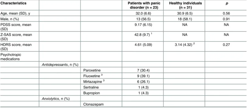
Table 1. Characteristics of Study Participants.
Characteristics Patients with panic
disorder (n = 23) Healthy individuals (n = 31) p Age, mean (SD), y 32.0 (6.6) 30.9 (6.5) 0.56 Male, n (%) 13 (56.5) 18 (58.1) 0.91 PDSS score, mean (SD) 9.17 (6.15) NA NA
Z-SAS score, mean (SD) 42.8 (9.7)1 NA NA HDRS score, mean (SD) 4.61 (5.09) 3.14 (4.32)2 0.27 Psychotropic medications Antidepressants, n (%) Paroxetine 7 (30.4) Fluoxetine3 9 (39.1) Mirtazapine3 6 (26.1) Sertraline 1 (4.3) Bupropion 1 (4.3) Anxiolytics, n (%) Clonazepam
SD, standard deviation; NA, not available; PDSS, Panic Disorder Severity Scale; Z-SAS, Zung Self-Rating Anxiety Scale; HDRS, Hamilton Depression Rating Scale
1Data from one patient with panic disorder was not available. 2Data from 3 healthy individuals were not available.
3One patient was treated with bothfluoxetine and mirtazapine. doi:10.1371/journal.pone.0157856.t001
The amygdalae were segmented from the T1-weighted images using a rater-independent well-validated automatic segmentation method implemented in the FreeSurfer software (http://surfer.nmr.mgh.harvard.edu)[39,40]. This segmentation method consists of several steps, in which T1-weighted images are first processed by performing motion correction and intensity inhomogeneity correction. After removing non-brain tissue, gray matter and white matter are segmented. Then, segmentation of subcortical structures including the amygdalae was performed using an atlas-based approach by assigning each voxel of the preprocessed vol-ume to the corresponding probabilistic information on the amygdalae based on manually labelled training sets [41,42]. An expert visually inspected the final segmented images to assure the appropriateness of the results. Manual editing was performed to correct mislabeled voxels
Fig 1. Anatomical location for the amgydala and hippocampus overlaid on the standard cortical surface and three-dimensional rendering of the amgydalar template. Abbreviations: R, right; L, left.
or prominent anatomical errors if necessary. Anatomical location for the bilateral amygdalae are presented inFig 1.
Shape Analysis
The shape analysis of the amygdala was performed using the University of North Carolina spherical harmonic (SPHARM) shape analysis toolkit ( http://www.nitrc.org/projects/spharm-pdm/)[43,44]. In summary, the segmented bilateral amygdalar images were initially processed to fill any interior holes and minimally smoothed. These processed images were then converted into spherical coordinates, transformed into surface meshes, and then aligned. Spherical har-monic coordinates between corresponding vertices of surface meshes were computed by parameter-based rotation applying the first order ellipsoid from the spherical harmonic coeffi-cients, which could eliminate the effects from the rotation and translation. Then, the spherical harmonic description was sampled into 1,002 triangulated surface points. Finally, the surfaces of the amygdala were spatially aligned to an averaged template using a rigid-body transforma-tion [45]. A detailed description of the method is described elsewhere [32,43,44].
Statistical Analysis
Demographic and clinical characteristics were compared between groups using independent t-tests or chi-square t-tests.
Multiple linear regression analysis was used to examine the effects of diagnosis on amygda-lar volumes adjusting for age, sex, and intracranial volume. Effect sizes (Cohen's d) for group differences in bilateral amygdalar volume and shape were calculated.
Since the amygdala consists of structurally and functionally distinct subregions [5], local shape of the amygdala was investigated to determine the topological preference for potential diagnostic group effects on volumes. Euclidian surface distance was computed at every surface point and used as a dependent measurement for surface contour analysis. Multiple linear regression analysis was applied to estimate the diagnostic group effects on local surface distance at each triangulated point in the amygdala. Localization of the significant group effects on spe-cific amygdalar subregions was performed using cytoarchitectonically defined probability map of amygdalar nuclei group [29]. In this cytoarchitectonically defined probability map, the amygdalar nuclei were divided into 3 groups including the superficial, centromedial, and later-obasal groups [29]. The superficial group included the anterior amygdaloid area, amygdalo-pyriform transition area, amygdaloid-hippocampal area, and ventral and posterior cortical nuclei. The centromedial group included the central nucleus and medial nucleus [29]. The laterobasal group consists of the lateral nucleus, basolateral, basomedial, and paralaminar nuclei. Surface rendering of amygdalar nuclei groups with 40% probability was performed using Slicer (http://www.slicer.org/) and each nuclei group was manually transposed onto the amygdalar template (Fig 2B)[46]. Individual significant vertices were localized to a specific nuclei group of the amygdala based on the highest probability of belonging to the laterobasal, superficial, or centromeidal groups. False discovery rate (FDR) was used to correct for the mul-tiple comparisons.
To investigate the relationship between panic symptom severity and magnitude of surface alteration, mean Euclidian distance of individual surface points that belonged to the clusters showing significant diagnostic effects were calculated. Pearson correlation analysis was used to examine whether the severity of panic symptoms or anxiety symptoms was associated with the mean scaled distance of surface coordinates in each cluster in patients with panic dis-order. All statistical analyses were performed using Stata SE, v11.0 (Stata Corp., College Station, TX).
Results
Demographic and clinical characteristics are presented inTable 1. Patients with panic disorder and healthy individuals were well matched for age and sex. Among 23 patients, 7 (30.4%) patients had comorbid agoraphobia. All patients with panic disorder were treated with a com-bination of antidepressants and/or anxiolytics prior to the study enrollment. Detailed informa-tion on the medicainforma-tion history is shown inTable 1.
Volume Analysis of the Amygdala
There were no differences in left amygdalar volume between patients with panic disorder and healthy individuals (β = -0.18, p = 0.19, Cohen's d = 0.45). However, although the difference did not reach statistical significance, patients with panic disorder showed a trend toward smaller right amygdala in comparison with healthy individuals after adjusting for age, sex, and intracranial volume (β = -0.23, p = 0.09, Cohen's d = 0.51). There were no differences in bilat-eral amygdalar volumes between panic disorder patients with agoraphobia and those without
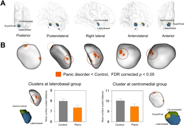
Fig 2. Clusters showing significant surface inward deformation in patients with panic disorder relative to healthy controls. (A) Surface rendering of cytoarchitectonically defined probabilistic maps of the superficial, centromedial, and laterobasal groups of the amygdala transposed onto the amygdalar template is presented. (B) Four clusters of inward deformation related to panic disorder at FDR corrected p < 0.05 are overlaid on the amygdalar template. Bar graphs show that mean surface scaled distance of clusters in the laterobasal and centromedial groups of the amygdala was lower in patients with panic disorder than in healthy individuals. Error bars indicate 95% confidence intervals. Abbreviations: FDR, false discovery rate.
agoraphobia after adjusting for age, sex, and intracranial volume (right,β = 0.17, p = 0.44; left, β = -0.01, p = 0.95).
Bilateral amygdalar volumes were not associated with panic symptom severity measured using the PDSS (right, r = 0.06, p = 0.79; left, r = -0.01, p = 0.95) in patients with panic disorder. There was also no relationship between bilateral amygdalar volumes and scores of the Z-SAS (right, r = -0.11, p = 0.62; left, r = -0.08, p = 0.73) in patients with panic disorder.
Shape Analysis of the Amygdala
Statistical maps for the diagnostic group effects on bilateral amygdalar shape are presented in
Fig 3.
Inward deformation of the right amygdala shape was found in patients with panic disorder relative to healthy individuals. After correcting for multiple comparisons, the diagnostic group effects were significant in 4 clusters within the right amygdala, which were located in the latero-basal and centromedial groups (Fig 2). The effect sizes for inward deformation of the clusters located in the laterobasal and centromedial groups were 0.99 and 0.91, respectively (Fig 2B). Localized shape alterations were found in the left amygdala in patients with panic disorder compared to healthy individuals, although the difference was not statically significant after the correction for multiple comparisons (Fig 3). There were no differences in the mean scaled dis-tances of clusters in the laterobasal (β = 0.10, p = 0.67) and centromedial (r = 0.24, p = 0.32) groups of the amygdala between panic disorder patients with agoraphobia and those without agoraphobia after adjusting for age and sex.
There were no significant relationships between panic symptom severity measured using the PDSS and mean scaled distance of clusters at both laterobasal (r = 0.16, p = 0.46) and cen-tromedial (r = 0.33, p = 0.12) groups of the amygdala, respectively, in patients with panic disor-der. In addition, anxiety symptom severity measured using the Z-SAS was not associated with mean scaled distance of clusters in patients with panic disorder (laterobasal group of the amyg-dala, r = -0.20, p = 0.38; centromedial of the amygamyg-dala, r = 0.40, p = 0.06).
Fig 3. Statistical maps showing the location of shape difference in the amygdala between patients with panic disorder and healthy individuals. The t statistics (A) and probability (B) maps show the results of multiple linear regression analysis for estimating group effects on surface scaled distances of individual triangulated surface points after adjusting for age and sex. Abbreviations: FDR, false discovery rate; R, right; L, left.
Discussion
Using three-dimensional surface-based morphometric analysis, we found surface deformation of the right amygdala related to panic disorder, specifically in the laterobasal and centromedial groups of the amygdala. In human, the laterobasal group of the amygdala consisting of the lat-eral nucleus, basolatlat-eral, basomedial, and paralaminar nuclei is anatomically located in the anterior or ventral segment of the amygdala, while the centromedial group of the amygdala including the central nucleus and medial nucleus corresponds to the posterior or dorsal seg-ment [47]. This is the first in vivo evidence, to our knowledge, demonstrating that panic disor-der-associated structural alterations in the amygdala may be localized and can occur in a subregion-specific manner. Shape alterations of the laterobasal and centromedial groups of the right amygdala were not associated with panic symptom severity.
Amygdalar volume reduction is one of the most replicated findings in patients with panic disorder in previous neuroimaging studies [5,21,24,48]. In line with these findings, we found a trend-level volume reduction in the right amygdala in patients with panic disorder as com-pared with healthy individuals. Through its connections with a variety of other cortical and subcortical structures [49], the amygdala is involved in associative fear learning, processing emotional memory, and regulating autonomic responses to emotion, which are central to the pathophysiology of panic disorder [6]. Amygdalar subregions composed of a number of nuclei may have projections to different brain structures and have known to play distinct roles in emotional regulation [50]. While the role of specific amygdalar subregions have been exten-sively studied in animal models, there is emerging evidence from human studies suggesting subregional specificity of the amygdalar involvement in several anxiety disorders [51,52].
In the current study, we found that panic disorder-related amygdalar structural alterations were prominent in the centromedial group including the central and medical nuclei, which can orchestrate fear responses and convey the viscerosensory information from the laterobasal group of the amygdala to the hypothalamus and brainstem [50,53]. Consistent with this find-ing, one previous study that performed VBM analysis with a small volume correction using bilateral amygdalar masks has revealed panic disorder-related volume deficits in the right amygdalar subregion including central and medial nuclei [23]. Moreover, during panic attack, input information is recognized as fear, triggering the amygdalar nuclei of the centromedial group to evoke autonomic arousal and hypothalamic-pituitary-adrenal axis activation [6]. The amygdalar nuclei of the centromedial group, as the 'controller of the brainstem' [54], appears to be responsible for expression of fear and defensive behaviors [2,55]. In the animal model, stimulation of the nuclei of the centromedial group may lead to a series of symptoms very simi-lar to those during panic attacks [56]. Thus, structural abnormalities in the centromedial group of the amygdala may underlie increased and maladaptive behavioral responses to emotional stimuli in patients with panic disorder. Likewise, increased amygdalar hyperactivation in the safe condition relative to threat condition was reported in patients with panic disorder [57].
Another notable finding was that the areas of reduced amygdalar size in panic disorder patients relative to healthy individuals were primarily located in the laterobasal group of the right amygdala. The laterobasal group of the amygdala, as the 'sensory amygdala', includes lat-eral, basal, and paralaminar nuclei [30] and is responsible for emotional memory formation, fear conditioning, and representation of values [58]. Moreover, the primary function of the laterobasal group of the amygdala includes integration and higher level of neurocognitive pro-cessing of viscerosensory information through reciprocal connections with the cortical and subcortical regions, which were likely to be aberrant in panic disorder [5,6]. Inferred from the animal model, the nuclei from the laterobasal group of the amygdala are expected to be involved in the pathophysiology of panic disorder. Likewise, repeated activation of the
laterobasal group of the amygdala by blocking GABAergic inhibition in the animal model has been reported to lead to a constellation of symptoms frequently observed in panic responses [59]. However, this is the first human study, to our knowledge, to report structural deficits in the laterobasal group of the amygdala in patients with panic disorder.
It remains unresolved whether structural deficits in specific subregions including the latero-basal and centromedial group of the amygdala may be due to the result of repetitive activation during panic attacks or may cause repeated hyperactivation and resultant panic attacks [5]. It is important to note that subregion-specific shape alterations in panic disorder were likely to be independent of panic symptom severity. Similar to our findings, previous volumetric studies did not find relationships between amygdalar volume reductions and clinical measures includ-ing symptom severity [20,24,25]. Further longitudinal studies with a lager sample would be necessary to resolve this cause or effect issue of amygdalar deficits related to panic disorder.
Case studies of bilateral amygdalar damage which is caused by Urbach–Wiethe disease may provide important clues to the amygdalar role in panic disorder. Previous studies have demon-strated that a patient with bilateral amygdalar damage could not acquire conditioned responses to aversive stimuli [28] or recognize fearful stimuli [27]. These findings may suggest the essen-tial role of the amygdala in the formation of fear memory. For instance, the presence of an intact amygdala may be required for normal fear conditioning. However, patients with bilateral amygdalar damage may experience not only spontaneous panic attacks [60] but also evoked ones by challenges with CO2inhalation [61], implying that the structural loss of the amygdala
may contribute to the development of panic attacks [61]. This could be further corroborated by our findings in combination with previous volumetric studies on the amygdalar structural defi-cits in panic disorder. Furthermore, considering the higher occurrence rate of panic attacks led by panicogenic challenges in patients with bilateral amygdalar damage, it can be assumed that an intact amygdala may contribute to the inhibition of panic attack [61].
In the current study, right amygdalar involvement was found in patients with panic disor-der. This is concordant with previous volumetric studies reporting a greater effect size of right rather than left amygdalar deficits in panic disorder [20,23–25]. Hemispheric dominance for fear processing of the amygdala may explain this finding of lateralized shape alterations in the right amygdala. Right hemispheric dominance for mediating emotional behavior and particu-larly for processing negative emotion has been reported [62,63]. In addition, right and left hemispheres have been known to be involved in different aspects of emotion. For instance, the right and left amygdala may be responsible for modulation of acquired and innate fear, respec-tively [64].
There are several limitations to be considered in interpreting the current findings. The three-dimensional surface-based morphometric analysis may detect more subtle changes in shape that could otherwise be overlooked in volumetric analysis across the amygdala, as shown in the results of our current study. Our primary aim was to detect subregion-specific alterations related to panic disorder, but it should be noted the current sample size is relatively small to have sufficient power for detecting moderate effect size of volume difference in bilateral amyg-dalae (right, Cohen's d = 0.51; left, Cohen's d = 0.45) between patients with panic disorder and healthy individuals.
Since it is not possible to distinguish and segment amygdalar nuclei with structural MRI, allocation of shape alterations to specific subregions was performed provisionally based on cytoarchitectonically defined probability map of amygdalar subregions. Such indirect localiza-tion could not account for inter-individual variability of subregional morphometry.
Cellular and molecular mechanisms underlying amygdalar shape alterations could not be determined in this study. Furthermore, the current results could not account for a causal rela-tionship between amygdalar atrophy and panic disorder. Our findings, however, imply that
patients with panic disorder may exhibit subregion-specific shape alterations in the amygdala, which is, to the best of our knowledge, the first report in the human brain in vivo.
Although healthy individuals did not have any kinds of anxiety disorders based on the Structured Clinical Interview for DSM-IV, it should be noted that the PDSS and Z-SAS were not administered in healthy individuals to assess the presence of potential subclinical panic and anxiety symptoms.
In sum, three-dimensional surface-based morphometric analysis was first implemented in the present study to identify the specific location of focal changes in the amygdala related to panic disorder. Panic disorder-related structural alterations were found in the vicinity of the laterobasal and centromedial groups of the right amygdala, both of which together are respon-sible for the formation and modulation of emotional memory and fear responses. The current findings suggest subregion-specific involvements of the right amygdala in panic disorder. Future studies should address the issue of whether amygdalar subregional deficits reflect con-stitutional vulnerability to panic disorder or tear and wear caused by repeated activations dur-ing panic attacks.
Acknowledgments
We thank Eun Namgung for her technical support and also gratefully acknowledge all study subjects for their participation.
Author Contributions
Conceived and designed the experiments: SY JEK IKL. Performed the experiments: SY JEK GHK HJK BRK SML IKL. Analyzed the data: SY JEK SJ JJI HH SM SML IKL. Contributed reagents/materials/analysis tools: SY IKL. Wrote the paper: SY JEK GHK HJK BRK SJ JJI HH SM SML IKL.
References
1. American Psychiatric Association. Diagnostic criteria from DSM-IV-TR. Washington, D.C: American Psychiatric Association; 2000.
2. Cox BJ, Swinson RP, Endler NS, Norton GR. The symptom structure of panic attacks. Compr Psychia-try. 1994; 35(5):349–53. PMID:7995026.
3. Margraf J, Taylor B, Ehlers A, Roth WT, Agras WS. Panic attacks in the natural environment. J Nerv Ment Dis. 1987; 175(9):558–65. PMID:3655782.
4. Craske MG, Kircanski K, Epstein A, Wittchen HU, Pine DS, Lewis-Fernandez R, et al. Panic disorder: a review of DSM-IV panic disorder and proposals for DSM-V. Depress Anxiety. 2010; 27(2):93–112. PMID:20099270. doi:10.1002/da.20654
5. Kim JE, Dager SR, Lyoo IK. The role of the amygdala in the pathophysiology of panic disorder: evi-dence from neuroimaging studies. Biol Mood Anxiety Disord. 2012; 2:20. PMID:23168129. doi:10. 1186/2045-5380-2-20
6. Gorman JM, Kent JM, Sullivan GM, Coplan JD. Neuroanatomical hypothesis of panic disorder, revised. Am J Psychiatry. 2000; 157(4):493–505. PMID:10739407.
7. Dresler T, Guhn A, Tupak SV, Ehlis AC, Herrmann MJ, Fallgatter AJ, et al. Revise the revised? New dimensions of the neuroanatomical hypothesis of panic disorder. J Neural Transm (Vienna). 2013; 120 (1):3–29. PMID:22692647.
8. Sehlmeyer C, Schoning S, Zwitserlood P, Pfleiderer B, Kircher T, Arolt V, et al. Human fear conditioning and extinction in neuroimaging: a systematic review. PLoS One. 2009; 4(6):e5865. PMID:19517024. doi:10.1371/journal.pone.0005865
9. Knight DC, Nguyen HT, Bandettini PA. The role of the human amygdala in the production of conditioned fear responses. Neuroimage. 2005; 26(4):1193–200. PMID:15961053.
10. MacNamara A, Rabinak CA, Fitzgerald DA, Zhou XJ, Shankman SA, Milad MR, et al. Neural correlates of individual differences in fear learning. Behav Brain Res. 2015; 287:34–41. PMID:25819422. doi:10. 1016/j.bbr.2015.03.035
11. Klein DF. False suffocation alarms, spontaneous panics, and related conditions. An integrative hypoth-esis. Arch Gen Psychiatry. 1993; 50(4):306–17. PMID:8466392.
12. Bisaga A, Katz JL, Antonini A, Wright CE, Margouleff C, Gorman JM, et al. Cerebral glucose metabo-lism in women with panic disorder. Am J Psychiatry. 1998; 155(9):1178–83. PMID:9734539. 13. Sakai Y, Kumano H, Nishikawa M, Sakano Y, Kaiya H, Imabayashi E, et al. Cerebral glucose
metabo-lism associated with a fear network in panic disorder. Neuroreport. 2005; 16(9):927–31. PMID: 15931063.
14. Dresler T, Hahn T, Plichta MM, Ernst LH, Tupak SV, Ehlis AC, et al. Neural correlates of spontaneous panic attacks. J Neural Transm (Vienna). 2011; 118(2):263–9. PMID:21203784.
15. Pfleiderer B, Zinkirciran S, Arolt V, Heindel W, Deckert J, Domschke K. fMRI amygdala activation during a spontaneous panic attack in a patient with panic disorder. World J Biol Psychiatry. 2007; 8(4):269– 72. PMID:17853295.
16. Garakani A, Buchsbaum MS, Newmark RE, Goodman C, Aaronson CJ, Martinez JM, et al. The effect of doxapram on brain imaging in patients with panic disorder. Eur Neuropsychopharmacol. 2007; 17 (10):672–86. PMID:17560768.
17. Boshuisen ML, Ter Horst GJ, Paans AM, Reinders AA, den Boer JA. rCBF differences between panic disorder patients and control subjects during anticipatory anxiety and rest. Biol Psychiatry. 2002; 52 (2):126–35. PMID:12114004.
18. Kent JM, Coplan JD, Mawlawi O, Martinez JM, Browne ST, Slifstein M, et al. Prediction of panic response to a respiratory stimulant by reduced orbitofrontal cerebral blood flow in panic disorder. Am J Psychiatry. 2005; 162(7):1379–81. PMID:15994724.
19. Carter RM, O'Doherty JP, Seymour B, Koch C, Dolan RJ. Contingency awareness in human aversive conditioning involves the middle frontal gyrus. Neuroimage. 2006; 29(3):1007–12. PMID:16246595. 20. Asami T, Yamasue H, Hayano F, Nakamura M, Uehara K, Otsuka T, et al. Sexually dimorphic gray
mat-ter volume reduction in patients with panic disorder. Psychiatry Res. 2009; 173(2):128–34. PMID: 19560907. doi:10.1016/j.pscychresns.2008.10.004
21. Lai CH, Hsu YY, Wu YT. First episode drug-naive major depressive disorder with panic disorder: gray matter deficits in limbic and default network structures. Eur Neuropsychopharmacol. 2010; 20(10):676– 82. PMID:20599363. doi:10.1016/j.euroneuro.2010.06.002
22. Sobanski T, Wagner G, Peikert G, Gruhn U, Schluttig K, Sauer H, et al. Temporal and right frontal lobe alterations in panic disorder: a quantitative volumetric and voxel-based morphometric MRI study. Psy-chol Med. 2010; 40(11):1879–86. PMID:20056020. doi:10.1017/S0033291709991930
23. Hayano F, Nakamura M, Asami T, Uehara K, Yoshida T, Roppongi T, et al. Smaller amygdala is associ-ated with anxiety in patients with panic disorder. Psychiatry Clin Neurosci. 2009; 63(3):266–76. PMID: 19566756. doi:10.1111/j.1440-1819.2009.01960.x
24. Massana G, Serra-Grabulosa JM, Salgado-Pineda P, Gasto C, Junque C, Massana J, et al. Amygdalar atrophy in panic disorder patients detected by volumetric magnetic resonance imaging. Neuroimage. 2003; 19(1):80–90. PMID:12781728.
25. Uchida RR, Del-Ben CM, Santos AC, Araujo D, Crippa JA, Guimaraes FS, et al. Decreased left tempo-ral lobe volume of panic patients measured by magnetic resonance imaging. Braz J Med Biol Res. 2003; 36(7):925–9. PMID:12845380.
26. LaBar KS, LeDoux JE, Spencer DD, Phelps EA. Impaired fear conditioning following unilateral temporal lobectomy in humans. J Neurosci. 1995; 15(10):6846–55. PMID:7472442.
27. Adolphs R, Tranel D. Emotion, recognition, and the human amygdala. In: Aggleton JP (Ed.) The amyg-dala. New York: Oxford University Press; 2000:587–630.
28. Bechara A, Tranel D, Damasio H, Adolphs R, Rockland C, Damasio AR. Double dissociation of condi-tioning and declarative knowledge relative to the amygdala and hippocampus in humans. Science. 1995; 269(5227):1115–8. PMID:7652558.
29. Amunts K, Kedo O, Kindler M, Pieperhoff P, Mohlberg H, Shah NJ, et al. Cytoarchitectonic mapping of the human amygdala, hippocampal region and entorhinal cortex: intersubject variability and probability maps. Anat Embryol (Berl). 2005; 210(5–6):343–52. PMID:16208455.
30. Whalen PJ, Phelps EA. The human amygdala. New York: Guilford Press; 2009.
31. Kim JE, Lyoo IK, Estes AM, Renshaw PF, Shaw DW, Friedman SD, et al. Laterobasal amygdalar enlargement in 6- to 7-year-old children with autism spectrum disorder. Arch Gen Psychiatry. 2010; 67 (11):1187–97. PMID:21041620. doi:10.1001/archgenpsychiatry.2010.148
32. Lee JH, Yoon S, Renshaw PF, Kim TS, Jung JJ, Choi Y, et al. Morphometric changes in lateral ventri-cles of patients with recent-onset type 2 diabetes mellitus. PLoS One. 2013; 8(4):e60515. PMID: 23593231. doi:10.1371/journal.pone.0060515
33. Joshi SH, Espinoza RT, Pirnia T, Shi J, Wang Y, Ayers B, et al. Structural Plasticity of the Hippocampus and Amygdala Induced by Electroconvulsive Therapy in Major Depression. Biol Psychiatry. 2016; 79 (4):282–92. PMID:25842202. doi:10.1016/j.biopsych.2015.02.029
34. Veer IM, Oei NY, van Buchem MA, Spinhoven P, Elzinga BM, Rombouts SA. Evidence for smaller right amygdala volumes in posttraumatic stress disorder following childhood trauma. Psychiatry Res. 2015; 233(3):436–42. PMID:26211620. doi:10.1016/j.pscychresns.2015.07.016
35. Shear MK, Maser JD. Standardized assessment for panic disorder research. A conference report. Arch Gen Psychiatry. 1994; 51(5):346–54. PMID:8179458.
36. Zung WW. A rating instrument for anxiety disorders. Psychosomatics. 1971; 12(6):371–9. PMID: 5172928.
37. Hamilton M. A rating scale for depression. J Neurol Neurosurg Psychiatry. 1960; 23:56–62. PMID: 14399272.
38. Yoon S, Jun CS, Jeong HS, Lee S, Lim SM, Ma J, et al. Altered cortical gyrification patterns in panic dis-order: deficits and potential compensation. J Psychiatr Res. 2013; 47(10):1446–54. PMID:23871448. doi:10.1016/j.jpsychires.2013.06.015
39. Fischl B, Sereno MI, Dale AM. Cortical based analysis. II: Inflation, flattening, and a surface-based coordinate system. Neuroimage. 1999; 9(2):195–207. PMID:9931269.
40. Lyoo IK, Kim JE, Yoon SJ, Hwang J, Bae S, Kim DJ. The neurobiological role of the dorsolateral pre-frontal cortex in recovery from trauma. Longitudinal brain imaging study among survivors of the South Korean subway disaster. Arch Gen Psychiatry. 2011; 68(7):701–13. PMID:21727254. doi:10.1001/ archgenpsychiatry.2011.70
41. Fischl B, Salat DH, Busa E, Albert M, Dieterich M, Haselgrove C, et al. Whole brain segmentation: auto-mated labeling of neuroanatomical structures in the human brain. Neuron. 2002; 33(3):341–55. PMID: 11832223.
42. Fischl B, Salat DH, van der Kouwe AJ, Makris N, Segonne F, Quinn BT, et al. Sequence-independent segmentation of magnetic resonance images. Neuroimage. 2004; 23 Suppl 1:S69–84. PMID: 15501102.
43. Styner M, Gerig G, Lieberman J, Jones D, Weinberger D. Statistical shape analysis of neuroanatomical structures based on medial models. Med Image Anal. 2003; 7(3):207–20. PMID:12946464.
44. Styner M, Lieberman JA, McClure RK, Weinberger DR, Jones DW, Gerig G. Morphometric analysis of lateral ventricles in schizophrenia and healthy controls regarding genetic and disease-specific factors. Proc Natl Acad Sci U S A. 2005; 102(13):4872–7. PMID:15772166.
45. Bookstein F. Morphometric tools for landmark data: geometry and biology. New York: Cambridge Uni-versity Press; 1991.
46. Kelley R, Chang KD, Garrett A, Alegria D, Thompson P, Howe M, et al. Deformations of amygdala mor-phology in familial pediatric bipolar disorder. Bipolar Disord. 2013; 15(7):795–802. PMID:24034354. doi:10.1111/bdi.12114
47. Whalen PJ, Shin LM, McInerney SC, Fischer H, Wright CI, Rauch SL. A functional MRI study of human amygdala responses to facial expressions of fear versus anger. Emotion. 2001; 1(1):70–83. PMID: 12894812.
48. Del Casale A, Serata D, Rapinesi C, Kotzalidis GD, Angeletti G, Tatarelli R, et al. Structural neuroimag-ing in patients with panic disorder: findneuroimag-ings and limitations of recent studies. Psychiatria Danubina. 2013; 25(2):108–14. PMID:23793273.
49. LeDoux JE. Emotion circuits in the brain. Annu Rev Neurosci. 2000; 23:155–84. PMID:10845062. 50. Phelps EA, LeDoux JE. Contributions of the amygdala to emotion processing: from animal models to
human behavior. Neuron. 2005; 48(2):175–87. PMID:16242399.
51. Brown VM, LaBar KS, Haswell CC, Gold AL, McCarthy G, Morey RA. Altered resting-state functional connectivity of basolateral and centromedial amygdala complexes in posttraumatic stress disorder. Neuropsychopharmacology. 2014; 39(2):351–9. PMID:23929546. doi:10.1038/npp.2013.197 52. Nicholson AA, Densmore M, Frewen PA, Theberge J, Neufeld RW, McKinnon MC, et al. The
dissocia-tive subtype of posttraumatic stress disorder: unique resting-state functional connectivity of basolateral and centromedial amygdala complexes. Neuropsychopharmacology. 2015; 40(10):2317–26. PMID: 25790021. doi:10.1038/npp.2015.79
53. LeDoux J. Fear and the brain: where have we been, and where are we going? Biol Psychiatry. 1998; 44 (12):1229–38. PMID:9861466.
54. Cardinal RN, Parkinson JA, Hall J, Everitt BJ. Emotion and motivation: the role of the amygdala, ventral striatum, and prefrontal cortex. Neurosci Biobehav Rev. 2002; 26(3):321–52. PMID:12034134.
55. Davis M, Whalen PJ. The amygdala: vigilance and emotion. Mol Psychiatry. 2001; 6(1):13–34. PMID: 11244481.
56. Davis M. The role of the amygdala in fear and anxiety. Ann Rev Neurosci. 1992; 15:353–75. PMID: 1575447.
57. Tuescher O, Protopopescu X, Pan H, Cloitre M, Butler T, Goldstein M, et al. Differential activity of sub-genual cingulate and brainstem in panic disorder and PTSD. J Anxiety Disord. 2011; 25(2):251–7. PMID:21075593. doi:10.1016/j.janxdis.2010.09.010
58. Pessoa L. Emotion and cognition and the amygdala: from "what is it?" to "what's to be done?". Neurop-sychologia. 2010; 48(12):3416–29. PMID:20619280. doi:10.1016/j.neuropsychologia.2010.06.038 59. Shekhar A, Sajdyk TS, Keim SR, Yoder KK, Sanders SK. Role of the basolateral amygdala in panic
dis-order. Ann N Y Acad Sci. 1999; 877:747–50. PMID:10415697.
60. Wiest G, Lehner-Baumgartner E, Baumgartner C. Panic attacks in an individual with bilateral selective lesions of the amygdala. Arch Neurol. 2006; 63(12):1798–801. PMID:17172622.
61. Feinstein JS, Buzza C, Hurlemann R, Follmer RL, Dahdaleh NS, Coryell WH, et al. Fear and panic in humans with bilateral amygdala damage. Nat Neurosci. 2013; 16(3):270–2. PMID:23377128. doi:10. 1038/nn.3323
62. Ley RG, Bryden MP. Hemispheric differences in processing emotions and faces. Brain Lang. 1979; 7 (1):127–38. PMID:435972.
63. Sackeim HA, Greenberg MS, Weiman AL, Gur RC, Hungerbuhler JP, Geschwind N. Hemispheric asymmetry in the expression of positive and negative emotions. Neurologic evidence. Arch Neurol. 1982; 39(4):210–8. PMID:7041863.
64. Lane RD, Nadel L, Ahern G. Cognitive neuroscience of emotion. New York: Oxford University Press; 2000.