Therapeutic use of self-assembled poly
PLGA and PEI core shell nanoparticles
for simultaneous delivery of anti cancer
drug and DNA toward cancer cells.
Jeonghee Kim
The Graduate School
Yonsei University
Therapeutic use of self-assembled poly
PLGA and PEI core shell nanoparticles
for simultaneous delivery of anti cancer
drug and DNA toward cancer cells.
A Dissertation
submitted to the Department of Nanomedical Science
and the Graduate School of Yonsei University
in partial fulfillment of the
requirements for the degree of
Master of Science
Jeonghee Kim
June 2006
This certifies that the master's thesis of Jeonghee Kim is approved
Thesis Supervisor: [Kunhong Kim]
___________________________
[Seung-Joo Haam: Thesis Committee Member #1]
___________________________
[Chae-Ok Yun: Thesis Committee Member #2]
The Graduate School
Yonsei University
Acknowledgements
I would like to express my gratitude to my supervisor prof. Kun-Hong Kim for his excellent guidance and tireless support during my course. I would like to express my appreciation to Dr. Yong-Ho Ahn, Dr. Kyung-Sup Kim, Dr. Man-Wook Hur, Dr. Ho-Geun Yoon, Dr. Jae-Woo Kim, Dr. Sang-Wook Park, and Dr. Woo-Chan Hyung for their encouragement and helpful advice. I especially wish to thank Prof.
Seung-Joo
Haam, and Chae-Ok Yun for their criticisms and thoughtful suggestions. Also I
want to thank Mr. Doo-Wung Park, Mr. Seok-Cheol Kwon, and Mrs.
Hae-Kyung Kim for their help in my laboratory works. I wish to express my
appreciation to the members of Department of Biochemistry and Molecular
Biology and
Department of Nanomedical Science. It was a great pleasure to work
with them and many inspring discussions with them were encouraging and
helpful to my research. I would like to thank Hyeon-Seok Ko, Yoon-Mi Lee,
Hwa-Jin Kim, and Hyang-Tae Choi for encouragement and for encouragement
and helpful advice. I especially wish to thank Mi-Hwa Lim, Sang-Eun Park,
and Dong-Ik Shin for being great friends during my courses. Finally, I would
like to express special thanks to my family and my friends for their love and
support during the days of intensive work. This work may not have been
completed without their support and encouragement.
i
Contents
Contents ···i
List of Figures ···iii
List of Tables ···iii
Abstract ···iv
I. Introduction ··· 1
II. Materials and Methods ··· 6
1. Polymers, plasmid DNA , and drugs ··· 6
2. Preparation of core-shell nanoparticles ··· 7
2.1. PLGA-DOX conjugation··· 7
2.2. Particle precipitation and PEI coating ··· 7
2.3. Folate conjugation ··· 9
2.4. DNA loading ··· 9
3. Characterization of Core-shell nanoparticles ··· 11
3.1. Transmission Electron Microscopy (TEM) ··· 11
3.2. Dynamic Light Scattering (DLS) and zeta potential ··· 11
3.3.1H-Nuclear Magnetic Resonance Spectroscopy (1H-NMR)··· 11
3.4.Gel retardation assay ··· 12
4. Cell culture ··· 12
5. Gene transfection··· 12
6. Cell viability assay ··· 12
ii
Ⅲ Ⅲ Ⅲ
Ⅲ. Result ··· 14
1. Preparation of PLGA-PEI-PEI-DNA core-shell nanoparticles ··· 14
2. Chemical structure investigation ··· 18
3. Selective gene delivery using PLGA-PEI-Folate core-shell nanoparticles ···· 21
4. Cell viability assay ··· 21
5. Drug release test ··· 24
Ⅳ Ⅳ Ⅳ Ⅳ. Discussion··· 26 V. Conclusions ··· 28 References ··· 29
iii
List of Figures
Figure 1. Receptor Endocytosis Cycle ··· 4
Figure 2. Conceptual scheme of nanoparticles for drug and gene delivery ··· 5
Figure 3. Synthetic scheme of PLGA-DOX conjugate ··· 8
Figure 4. Synthetic scheme of PLGA-PEI conjugate··· 10
Figure 5. TEM image of activated PLGA and PLGA-PEI nanoparticles··· 15
Figure 6. Particle size comparison according to PEI quantity··· 16
Figure 7. Zeta potential values and Gel retardation assay··· 17
Figure 8. 1H-NMR spectra of PLGA-PEI ··· 19
Figure 9. IR peak change by conjugation··· 20
Figure 10.Fluorescence microscopic visualization of GFP expression according to N/P ratio in A549 and HeLa cells··· 22
Figure 11.MTT assay for cellular viability ··· 23
Figure 12. Doxorubicin release profile··· 25
List of table
Table 1. Particle size comparison according to PEI quantity ··· 16iv
Abstract
Therapeutic use of self-assembled poly PLGA
a n d P E I c o r e s h e l l n a n o p a r t i c l e s f o r
simultaneous delivery of anti cancer drug and
DNA toward cancer cells.
Jeonghee Kim
Dept. of Nanomedical Science
The Graduate School
Yonsei University
Due to recent developments in drug delivery system (DDS), cancer drugs, DNA, and/or protein can be simultaneous delivered to cancer cells efficiently and selectively. Doxorubicin (DOX) is an anthracycline drug commonly used in cancer chemotherapy, but the dosage of the drug is restricted by its cardiotoxicity and also many cancer cells developed a resistance toward the drug. To overcome or circumvent these problems,
v
we produced self-assembled core-shell nano-particles composed of PLGA [poly (D, L-lactide-co-glycolide)] and PEI (Polyethylenimine). The particles were conjugated with low dose of doxorubicin, EGFP-expressing plasmid, and folic acid for enhancing its selective delivery toward folate receptor overexpressing cancer cells. We used HeLa, a human cervical cancer cell line that express moderate level of folate receptor and has high telomerase activity, and used A549, a human lung adenocarcinoma cell line that express low level of receptor and low telomerase activity. When the cells were treated with the nanoparticles conjugated with folate and GFP expressing plasmid, A549 cells did not show any GFP signal, but HeLa cells showed green fluorescence in approximately 50% of the cells at N/P ratio=12.
Taken together, we documented that the core-shell nanoparticles composed of PLGA and PEI are effective for simultaneous delivery of anti-cancer drug and DNA toward selective cancer cells could be a powerful tool for selective cancer therapy.
Key Words: core-shell nanoparticles, poly (D, L-lactide-co-glycolide), polyethylenimine, DNA, folic acid, doxorubicin, cancer therapy.
1
I. Introduction
Apoptosis is a major mode of cell death induced by chemotherapy. Doxorubicin (DOX), an anthracycline antibiotic, is one of the most important cytostatic drugs used in the field of cancer chemotherapy. It works by interfering with the growth of rapidly growing cancer cells where it binds and intercalates into the DNA strand, thus, inhibiting further DNA and RNA biosynthesis, eventually causing cancer cell death. DOX is usually used in the treatment of neoplastic diseases, such as leukemia and various solid tumors [1]. However, its therapeutic potential has been restricted by its dose limited cardiotoxicity and the resistance developed in the portion of cancer cells. Resistance to DOX has become an obstacle in the clinical treatment of human cancers.
DNA delivery systems have been classified as viral vector-mediated systems and nonviral vector-mediated systems. Non-viral gene-delivery systems are safer to use and easier to produce than viral vectors, but their comparatively low transfection efficiency has limited their applications [2]. Anti-cancer drugs have been chemically conjugated to various polymers for the purpose of its efficient passive targeting to solid tumors [1]. Co-delivery of drugs and DNA has been proposed to achieve the synergistic/combined therapeutic effect of anti-cancer drug and anti-cancer killing gene product [3]. The “enhanced permeation and retention (EPR)” effect on the site of tumor capillaries plays a critical role in accumulating the polymer conjugates in the solid tumors [4]. Efficient therapeutic gene delivery toward cancer cells has been a main goal in cancer gene therapy. Recently, various cationic polymers and lipids have been investigated as nonviral gene carriers that facilitate the intracellular delivery of plasmid DNA into cancer cells [5, 6]. Among these, branched PEI is the one widely
2
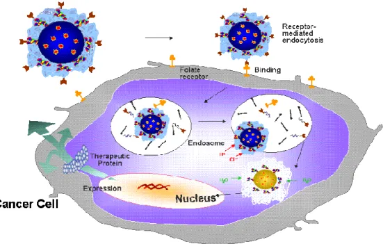
used for gene delivery because of its high transfection efficiency, possibly due to the “proton sponge” effect that destabilizes the endosomal membrane [5, 7] (Figure 1). In this exprement, we produced self-assembled core-shell nano-particles that were composed of PLGA [poly (D, L-lactide-co-glycolide)] and PEI (Polyethylenimine). These nano-particles offer advantages over liposomes, as they are easier to fabricate and are more readily subject to modulation of their size and degree of positive charge. More importantly, high gene transfection efficiency achieved, and thus, it becomes possible to co-deliver anti-cancer drug and cancer killing gene. To achieve simultaneous delivery of cancer drug and DNA to specific cells or tissues wide array of targeting ligands recognized by specific cells has been conjugated to PEI [8, 9]. Folate is the ligands that is most popularly used for target-specific delivery of genes, imaging agents, and anti-cancer agents to folate- receptor over-expressing tumor cells [10, 11]. Accordingly, we conjugated folic acid to the core-shell nanoparticles. DOX, hydrophobic drugs can be incorporated into the PLGA during the particle preparation process. The cationic PEI-shell of the resulting DOX-loaded nano-particles can be used fir deliverying DNA encoding cancer killing protein, as shown schematically in Figure 2. Morphology, size, drug loading efficiency as well as drug release profiles of the nanoparticles were characterized using TEM, DLS, and UV spectrophotometer. The chemical structure of the particles was confirmed at each step by FT-IR and 1 H-NMR. Using GFP as the reporter DNA, a folate-receptor mediated gene transfection efficiency was evaluated against folate receptor over-expressing cancer cells. Taken together, the core-shell nanoaparticles that we produced in this experiment could be useful for simultaneous delivery of anti-cancer drug and DNA encoding cancer killing
3
4
5
Figure 2. Conceptual scheme of core-shell nanoparticels for drug and gene delivery Folate Folate Folate Folate as a as a as a
as a Targeting moiety Targeting moiety Targeting moiety Targeting moiety PLGA polymer matrix
PLGA polymer matrix PLGA polymer matrix PLGA polymer matrix to control rate of to control rate of to control rate of to control rate of drug release drug release drug release drug release
PEI for the gene delivery PEI for the gene deliveryPEI for the gene delivery PEI for the gene delivery Gene (Bax) Gene (Bax) Gene (Bax) Gene (Bax) Anticancer drug Anticancer drug Anticancer drug Anticancer drug (Doxorubicin) (Doxorubicin) (Doxorubicin) (Doxorubicin)
6
II. Materials and Methods
1. Polymers, DNA plasmid, and reagent
Poly(D,L-lactide-co-glycolide) (PLGA) (weight average MW: 5,000) was obtained from Wako (Japan). Polyethylenimine (PEI) with MW 25,000, N-hydroxysuccinimide (NHS), dicyclohexylcarbodiimde (DCC), p-nitrophenyl chloroformate (p-NC), folate, dichloromethane, anhydrous and doxorubicin (DOX) were obtained from Sigma-Aldrich (St. Louis, USA). N,N-dimethylformamide, dimethyl sulfoxide were purchased from Duksan pure chemical Co. (Korea). Double-distilled water was used. EGFP-C1 Plasmid that canexpress GFP protein was obtained from Clontech (Palo Alto, USA).
2. Preparation of core-shell Nanoparticles 2.1. PLGA-DOX conjugation
500 mg of PLGA dissolved in 50 ml of anhydrous dichloromethane was activated by adding 60.5 mg of p-nitrophenyl chloroformate and 39.6 mg of pyridine (PLGA/p-nitrophenyl chloroformate/pyridine stoichiometric molar ratio: 1/3/5) at 0℃. The reaction was carried out for 3 h at room temperature under nitrogen atmosphere. The p-NC activated PLGA (0.05 g) dissolved in 3 ml of dimethylformamide (DMF) was reacted with 5.8 mg of doxorubicin (DOX) in the presence of 4.05 mg of triethylamine (TEA) for 24 h at room temperature under nitrogen atmosphere (stoichiometric molar ratio of activated PLGA/DOX/TEA: 1/1/4) (Figure 3). Unreacted DOX and other chemicals were removed by transfer of the organic phase into deionized water, which was followed by three subsequent dialyzing procedures
7
against deionized water for 4 h (Pierce/Snakeskin Pleated Dialysis Tubing, MWCO 10,000). The purified PLGA-DOX was freeze-dried and stored at -20℃ for further use.
2.2. Particle preparation and PEI coating
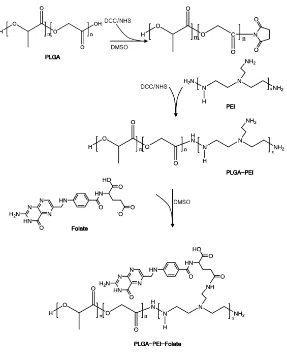
PLGA-DOX (10 mg) in 1ml dimethylsulfoxide directly precipitated in 10 ml of deionized, distilled water. After 8 hours, solution was filtered by filter paper using aspirator. Filtered solution was added to a definite amount of a PEI solution (5 mg) with constant stirring for 1 h. Unactivated PEI and other chemicals were removed by centrifuge at 15,000 rpm for 30 minutes at room temperature (Figure 4).
8
9
2.3. Folate conjugation
In 5 ml of distilled water, 44 mg folic acid was mixed with 184 mg of NHS and 329.6 mg of EDC (Folate/EDC/NHS molar ratio: 1/8/8). The reaction was performed at room temperature for 7 h and then mixed with 50 ml of distilled water and centrifuged at 3,000 rpm. After discarding the pellet, the supernatant was dialyzed and freeze-dried. Folate conjugation was achieved by dissolving activated folate in PLGA-PEI particle solution each molar ratio (Figure 4).
2.4. DNA loading
PEI and DNA solutions were prepared before each experiment at various molar ratios of PEI nitrogen (N) to DNA phosphate (P) up to N/P. The pH of the stock PEI solution was adjusted to the desired pH using HCl. Complex formation always utilized solutions of equal volumes with the least concentrated component being added to the more concentrated one. Samples were continuously stirred during addition and equilibrated at room temperature for 20 min before measurement. Complexes were freshly prepared before each individual measurement. Complexes were formed in 10 mM Tris-HCl (pH 7.4) unless otherwise noted.
10 DCC/NHS DCC/NHS Folate FolateFolate Folate PLGA PLGA PLGA PLGA PEI PEI PEI PEI PLGA PLGA PLGA PLGA---PEI-PEIPEIPEI
O HO HN O HN N O HN H2N N N O -O PLGA PLGA PLGA
PLGA---PEI-PEIPEI-PEI--Folate-FolateFolateFolate
H2N N N NH2 NH2 H x OH O O H O O m n H N N N NH2 NH2 H x O O H O O m n H N N N NH NH2 H x O O H O O m n O HO HN O HN N O HN H2N N N O C O O H O O N O O m n DMSO DMSO DCC/NHS DCC/NHS Folate FolateFolate Folate PLGA PLGA PLGA PLGA PEI PEI PEI PEI PLGA PLGA PLGA PLGA---PEI-PEIPEIPEI
O HO HN O HN N O HN H2N N N O -O PLGA PLGA PLGA
PLGA---PEI-PEIPEI-PEI--Folate-FolateFolateFolate
H2N N N NH2 NH2 H x OH O O H O O m n H N N N NH2 NH2 H x O O H O O m n H N N N NH NH2 H x O O H O O m n O HO HN O HN N O HN H2N N N O C O O H O O N O O m n DMSO DMSO
Figure 4. Synthetic scheme of PLGA-PEI conjugate
11
3. Characterization of Core-shell Nanoparticles 3.1. Transmission Electron Microscopy (TEM)
A concentrated aqueous dispersion of nanoparticles was finely spread over a TEM grid and dried. Then, the surface morphology of the folate conjugated PLGA-PEI nanoparticles was observed using transmission electron microscopy (TEM, LEM-2000, AKASHI).
3.2. Dynamic Light Scattering (DLS) and zeta potential
DLS (Zetasizer Nano ZS, Malvern, UK) analysis of the nanocell and folate attached PLGA-PEI nanoparticles was performed to determine the size and the size distribution. An aircooled argon ion laser was operated at 488nm. Using a digital correlator, the time dependence of the intensity auto-correlation function of the scattered intensity was derived. The size of the nanoparticles was expressed as number-weighted mean diameter in nanometers and was obtained from the measurement of at least three bathes of nanoparticles.
3.3. 1H- nuclear magnetic resonance spectroscopy (1H-NMR) 1
H-NMR spectra were recorded many types of particles for observation of the functional groups using a 500MHz NMR spectrometer (JNM-ECP300, JEOL, UK). In a typical procedure, respective particles were dissolved in DMSO-d6 and directly recorded.
12
3.4. Gel retardation assay
For agarose gel electrophoresis, Core-shell nanoparticle/Folate/DNA complexes mixed with a loading buffer were loaded onto an ethidium bromide containing 1% agarose gel. Gel electrophoresis was performed at room temperature in TEB buffer at 100V for 60 min. DNA bands were visualized using a UV (254 nm) illuminator.
4. Cell culture
Human cervical carcinoma cell line, HeLa cell [folate receptor (+)] and lung carcinoma cell A549 cell [folate receptor (-)] were maintained in Folate-free RPMI1640 (Gibco) supplemented with 10% FBS and 1% penicillin–streptomycin
under conditions of 5% CO2 and 95% humidity.
5. Gene transfection
For in vitro transfection, the cells were split one day prior to transfection and plated in 6-well plates at a density of 4x10⁵ cells/well. Before transfection, the cell culture medium was replaced with Folate-free RPMI1640 containg 1% penicillin– streptomycin (Gibco). The cells were transfected with gene delivery complexes. The transfection medium was then replaced with normal culture medium and the treated cells were cultured for another 24 h.
6. Cell viability assay
For cell viability assay, the cells (10,000 cells/well) were seeded into 96-well plates. The cells were then incubated in culture media containing gene delivery complexes formed at various charge ratios for 24h. Approximately 50 µll of sterile
13
filtered MTT (3-(4,5-dimethyl-thiazol-2-yl)-2,5-diphenyl tetrazolium bromide) (2 mg/ml) stock solution in PBS was added to each well. After 4 h, unreacted dye was removed by aspiration. The formazan crystals were dissolved in 100 µl DMSO per well and measured spectrophotometrically in an ELISA plate reader (Model 550, Bio-Rad) at a wavelength of 595 nm. The cell survival in the presence of complexes was expressed as percentage of cell survival in absence of complexes.
7. Drug release test
Release experiments for controlled release formulation were carried out in an aqueous release medium with the phosphate buffer solution (pH 7.4) at 37.5±0.5 ℃. The beaker was put in a shaking incubator (SI-900, J.O Tech., Korea) at the shaking rate of 150rpm. At predetermined time intervals, 3ml of the aqueous solution were withdrawn and replenished with 3ml of D.D.I. water. The amount of released doxorubicin was monitored by measuring the absorbance using UV spectrophotometer at 480 nm wavelength.
14
III. Result
1. Preparation of PLGA-PEI-PEI-DNA core-shell nanoparticles
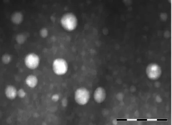
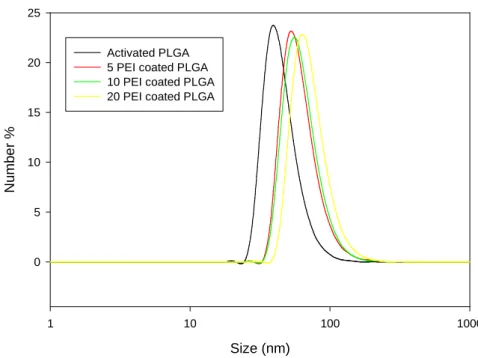
The morphology, average size, and size distribution of PLGA, PLGA-PEI particles were evaluated using TEM and DLS. Figure 5 and 6 showed the photograph and histogram of PLGA and PLGA-PEI particles, respectively.
Based on Dynamic Light Scattering (DLS), the average size of the activated PLGA particles was 63nm. After PEI conjugation, the size of nanoparticles was increased by quantity of conjugating PEI from 83 nm to 92 nm (Table 1). And the PLGA-PEI-FA particles were in the similar size range. This result indicates that the particle size was increased due to the folate molecule.
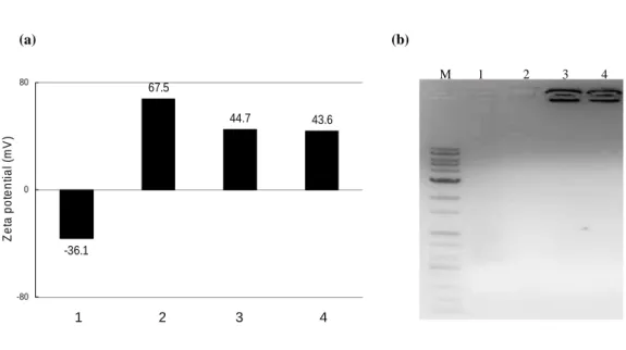
As seen in Figure 7 (a), the zeta potential of PLGA without PEI was -36.1 mV, and became 67.5 mV when PLGA was coated with PEI. When folate and EGFP plasmid were conjugated, zeta potential became 44.7 mV and 43.6 mV at N/P ratio of 8 and 12, respectively. Though folate and EGFP plasmid were conjugated with PLGA-PEI nanoparticles, they maintained positive charge. It was confirmed that gene delivery materials which can load DNA were produced.
Core-shell nanopaticles complexed with EGFP plasmid showed decreased
moility on gel retardation assay (Figure. 7 (b)). Complete retardation of the DNA was achieved at an N/P ratio (molar ratio of nitrogen atom content in the polymer to phosphorous atom content in the DNA) of 8 and 12, respectively. The DNA-binding ability of the blank core–shell nanoparticles was slightly greater than PLGA nanoparticles due to the greater zeta potential of the former.
15
(a) Actvated PLGAnanoparticles
(b) PLGA-PEI nanoparticles
Figure 5. TEM image of (a)a-PLGA and (b) PLGA-PEI nanoparticles
16
Figure 6. Particle size comparison according to PEI quantity
Table 1. Particle size comparison according to PEI quantity
Coated PEI quantity (mg) 0 5 10 20
Size (nm) 63 83 86 92 Size (nm) 1 10 100 1000 N u m b e r % 0 5 10 15 20 25 Activated PLGA 5 PEI coated PLGA 10 PEI coated PLGA 20 PEI coated PLGA
17
Figure 7. Zeta potential values and gel retardation assay of core-shell nanoparticles. ; (a) Zeta potential values (b) Gel retardation assays
1. PLGA; 2. PLGA-PEI-Folate (N/P ratio=0); 3. PLGA-PEI-Folate- EGFP (N/P ratio=8);4. PLGA-PEI-Folate-EGFP ( N/P ratio=12).
-36.1 67.5 44.7 43.6 -80 0 80 Z e ta p o te n ti a l (m V ) 1 2 3 4 (a) (b) M 1 2 3 4
18
2. Chemical structure investigation 1
H-NMR spectrum for PLGA-PEI conjugation was shown in figure 9. In the spectrum, the PLGA peak was observed in 1H NMR spectrum in DMSO-d6 at 1.4 ppm, 4.3 ppm, and 5.2 ppm (for PLGA back bone). NHS group (3.0 ppm) was removed after PEI conjugation process. The characteristic band of PEI was observed at 2.6 ppm and 3.5 ppm in 1H-NMR spectrum in DMSO-d6 at this process (Figure 8).
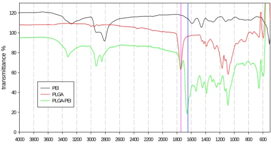
As the conjugation between PLGA and PEI progresses, the FT-IR spectra was changed from NHS succinimidyl ester peak (1780 cm-1) to N-H peak (1640 cm-1) (Figure 9). This indicates that the conjugation between PLGA and PEI is accomplished.
19
Figure 9. 1H-NMR spectra of PEI-PLGA b(1.7) 2.15 a(2.62) 2.62 2.15 2.62 2.15 1.7 1.7 c (3.5) A (5.2) B(4.3) C(1.39) H N N N NH2 NH2 H x O O H O O m n
20 wave number 600 800 1000 1200 1400 1600 1800 2000 2200 2400 2600 2800 3000 3200 3400 3600 3800 4000 tr a n s m it ta n c e % 0 20 40 60 80 100 120 PEI PLGA PLGA-PEI wave number 600 800 1000 1200 1400 1600 1800 2000 2200 2400 2600 2800 3000 3200 3400 3600 3800 4000 tr a n s m it ta n c e % 0 20 40 60 80 100 120 PEI PLGA PLGA-PEI
21
3. Specific gene delivery using PLGA-PEI-Folate core-shell nanoparticles
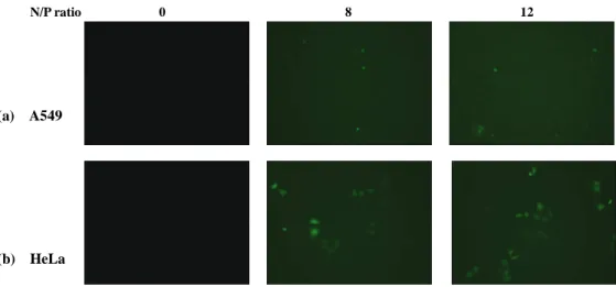
HeLa cells over-express folate receptors, but A549 cells do not. To prove receptor mediated endocytosis of PLGA-PEI-Folate-DNA complexes, transfection in HeLa cells and A549 cells were compared with varying N/P ratios of PLGA-PEI-Folate and DNA. The extent of gene expression steadily increases with the N/P ratio in HeLa, but not in A549 cells (Figure 10). This might be due to folate receptor-mediated endocytosis.
4. Cell viability assay
The efficiency of a gene transfer is dependent on a function of the corresponding toxicity of the polymer and polymer/DNA complex solution. The cell viability was determined in A549 and HeLa cells using PLGA-PEI-Folate, PLGA-PEI-Folate-DNA complexes at N/P ratio=8 and 16, and complex concentrations 0.02μg/μl. Cells with only media and no treatment of polymer or polymer/DNA complexes were considered a negative control. PLGA-PEI-Folate-DNA showed decreased toxicity in comparing without DNA. However, there was an increase in toxicity with increase in N/P ratio and HeLa cells were more sensitive than A549 cells (Figure 11).
22
Figure 10. Fluorescence microscope visualization of GFP expression according to N/P ratio in (a) A549 and (b) HeLa cells
(a) A549
(b) HeLa
23
Figure 11. Cell viability assays using MTT in (a) A549 and (b) HeLa cells at polymer concentrations 0.02μμμg/μμ μμμl. 1. None; 2. PLGA-PEI-Folate; 3. PLGA- PEI-Folate-EGFP(N/P ratio=8); 4. PLGA-PEI-Folate (N/P ratio=12)
0 50 100 150 1 2 3 4 PLGA-PEI-Folate % C e ll V ia b il it y 0 50 100 150 1 2 3 4 PLGA-PEI-Folate % C e ll V ia b il it y (a) (b)
24
5. Drug release test
In vitro release of doxorubicin from PLGA (Figure 12 (a)) and PLGA-PEI nanoparticles (Figure 12 (b)) was performed to investigate the effect of the initial burst and the sustained drug release. Also we measured drug loading efficiency in the nanoparticles using UV spectrophotometer at 480nm wavelength.
In general drug delivery system, the initial burst was suppressed for a few days, but this PLGA-PEI required longer time. As shown in the Figure 12 (b), drug release pattern had more gentle curve than the Figure 12 (a), because of conjugated PEI. Also this result indicated that drug loading efficiency was 7.96 %, approximately.
25 (a) T im e ( D a y ) 0 2 4 6 8 1 0 1 2 1 4 1 6 D ru g r e le a s e d ( % ) 0 2 0 4 0 6 0 8 0 1 0 0 (b) T im e ( D a y ) 0 2 4 6 8 1 0 1 2 1 4 1 6 D ru g r e le a s e d ( % ) 0 2 0 4 0 6 0 8 0 1 0 0
26
IV. Discussion
The purpose of this study is to produce a simultaneous drug delivery system which would deliver anti-cancer drug and DNA to the simultaneous and selective cancer cells. We demonstrated that PLGA-PEI-Folate conjugate could be complexed with DNA and the resultant PLGA-PEI-Folate-DNA complexes showed folate receptor-specific gene transfection. The surface exposed folate ligands on the complexes were recognized by folate receptors on the membrane of cancer cells targeted, resulting in active transport of PLGA-PEI-Folate-DNA complexes into HeLa cells. We used EGFP plasmid, and GFP gene could be effectively delivered into the cells by using PLGA-PEI-Folate conjugate. When the cells were treated with the nanoparticles conjugated with folate and GFP expressing construct, A549 cells did not show any GFP signal, but HeLa cells showed green fluorescence in approximately 50% of the cells at N/P ratio=12. While GFP gene is expressed in HeLa cell, PLGA-PEI-Folate-EGFP plasmid has approximately 50% toxicity. We thought that this toxicity is caused PEI. If we overcome this problem, we will produce powerful tools for all-in-one delivery system of drug and gene. For example, we replace GFP by bax gene. Bax, as a pro-apoptosis gene of the Bcl-2 family, has extensive amino acid homology with Bcl-2, and may form homodimers and heterodimers with Bcl-2 that oppose Bcl-2 function and contribute to cell death. Bax expression is markedly low in esophageal squamous cell carcinoma, breast cancer, hepatocellular carcinoma, and ovarian carcinoma. Recent studies indicate that overexpressions of Bax sensitize human head and gastric cancer cells to various chemotherapeutic agents, and Bax enhances apoptosis in ovarian cancer cell lines. Additionally, we regulated expression
27
of bax gene using human telomerase reverse transcriptase (hTERT) promoter that is active only in cancer cells. Target-specific expression of bax significantly induced apoptosis. The therapeutic effectiveness of bax gene transfection was synergistically enhanced with released DOX in the target cells.
28
V. Conclusions
1. The PLGA-PEI-Folate is used as co-delivery agent of doxorubicin(DOX) and bax gene.
2. The PLGA-PEI-Folate targets folate receptor over-expression cells.
3. Taken together, if the toxicity of nanoparticles is overcome, the nanoparticles have a synergistic effect as the co-delivery of DOX and bax gene using PLGA-PEI-Folate, because the over-expression of the pro-apoptotic bax gene make the cells more sensitive to DOX.
29
References
[1] Rongyi Lin, Biomaterials 26 (2005) 4476–4485 [2] Dan Luo, Nature biotechnology 18 (2000) 33-37
[3] Jian-Yong Zheng , World J Gastroenterol 11 (2005) 3498-3503 [4] Arun K. Iyer, Drug Discovery Today 11(2006) 812-818
[5] Merdan T, Adv Drug Deliv Rev. 54(2002) 715-758 [6] Stefaan C, Pharmaceutical Research. 17(2000) 113-126 [7] Barbara Demeneix , Advances in Genetics, 53(2005) 217-230 [8] O.Boussif, Proc. Natl. Acad. Sci. USA 92(1995)7297-7301
[9] Kyung Chul Cho, Journal of Controlled Release 108 (2005) 121–131 [10] Nikki Parker, Analytical Biochemistry 338 (2005) 284–293
30
국문
국문
국문
국문 요약
요약
요약
요약
암
암
암
암 세포를
세포를
세포를
세포를 향해
향해
향해
향해 동시에
동시에
동시에
동시에 항암제와
항암제와 DNA를
항암제와
항암제와
를
를
를 전달하기
전달하기
전달하기
전달하기 위해
위해
위해
위해
자가
자가
자가
자가 조립되는
조립되는
조립되는 core-shell 구조의
조립되는
구조의
구조의 PLGA-PEI 나노
구조의
나노
나노 입자의
나노
입자의
입자의
입자의
항암
항암
항암
항암 치료의
치료의
치료의 사용
치료의
사용
사용
사용
최근 약물 전달 시스템(drug delivery system, DDS)의 발달로 항암 약물, 핵산(DNA), 단백질은 동시에 선택적, 효율적으로 암 세포로 투여되게 되 었다. Doxorubicin(DOX)은 안트라사이틀린(anthracycline) 계열의 약물로 암 치료를 위한 화학 요법으로 널리 사용되지만, DOX의 투여량은 심장독성 (cardiotoxicity)의 부작용과 많은 암 세포의 내성 때문에 제한을 받는다. 이러한 부작용을 극복하기 위해서 우리는 PLGA(poly (D, L-lactide-co-glycolide))와 PEI(Polyethylenimine)으로 구성된 자가 조립되는 core-shell 구조의 나노 입자를 만들었다. 그리고 적은 양의 doxorubicin, EGF를 발현하는 플라스미드와 folate 수용체가 과발현하는 암세포로 선 택적인 전달을 위해 folate를 융합하였다. 우리는 folate 수용체가 적절히 발현하면서 telomerase 활성도가 큰 자궁경부암 세포인 HeLa 세포와
folate 수용체와 telomerase activity가 작은 폐암 세포인 A549 세포를 사용
31
리하였을 때, A549 세포는 GFP가 발현되지 않았지만 이와 반대로 HeLa 세포는 N/P 비율이 12가 되는 나노 입자에 의해 대략 80% 정도의 비율 로 발현 되었다.
이 모두를 통해, 우리는 PLGA와 PEI로 구성된 core-shell 구조의 나노 입자는 암 세포에 동시 선택적으로 항암제와 DNA 전달체이며, 이는 항 암 세포에 특이적으로 작용하는 항암 치료를 위한 강력한 도구로 사용될 수 있다.
핵심 되는 말:
Core-shell 구조의 나노입자, PLGA, polyethylenimine, folate, DNA, doxorubicin, 항암 치료