Original Article
Expression of DNA methylation-related proteins in
breast phyllodes tumor
Ji Hee Lee, Yu Kyung Lee, Eun-Sol Kim, Ja Seung Koo
Department of Pathology, College of Medicine, Yonsei University, Seoul, South Korea
Received September 19, 2016; Accepted September 27, 2016; Epub May 1, 2017; Published May 15, 2017 Abstract: The purpose of this study is to research the expression of DNA methylation-related proteins in phyllodes tumors of the breast and to study the implication on patient outcomes. We generated tissue microarrays (TMAs) of 196 phyllodes tumors (PT) and performed immunohistochemical staining for 5-meC and the DNA methylation-related proteins DNMT1 and ISL-1. The staining results were analyzed and compared with clinicopathologic param-eters. A total of 196 cases were included in this study, of which 153 were benign, 27 were borderline, and 16 were malignant. The levels of DNMT1, 5 meC, and ISL-1 in the stromal component of tumors increased with increasing grade (P<0.001). Especially, high stromal positivity of DNMT1 and ISL-1 were associated with increased distant metastasis (P=0.001, and P=0.013, respectively). Univariate analysis for factors associated with decreased disease free survival and overall survival identified DNMT1 high positivity (P=0.002 and P<0.001, respectively) and stromal ISL-1 high positivity (P<0.001 and P<0.001, respectively). Among borderline phyllodes tumors, stromal DNMT1 high positivity was associated with decreased OS (P=0.015). In conclusion, DNA methylation and expression of methyl-ation-related proteins in the stromal component increased with increasing histologic grade in phyllodes tumors. In addition, overexpression stromal expression of DNMT1 and ISL-1 was associated with poor prognosis.
Keywords: Breast, methylation, phyllodes tumor Introduction
Phyllodes tumors are a relatively rare tumor type, accounting for 0.3-1.5% all breast tumors, and have histological features similar to fibro-adenoma, a fibroepithelial tumor. Establishing an accurate differential diagnosis including phyllodes tumors is difficult due to their hetero-geneous histological appearance [1, 2]. Clinically, phyllodes tumors are known to be malignant and recurrent and can metastasize to a number of different sites [3]. Although there are differing opinions among researchers regarding histologic classification of phyllodes tumors, the WHO classifies the histologic types of phyllodes tumor as benign, borderline, and malignant [2]. According to this classification scheme, the frequency of phyllodes tumor recurrence and distance metastasis increase with increasing histologic grade and are thus reflective of aggressiveness.
Insensitivity to growth inhibitory signals is an important difference between normal cells and
cancer cells and is meditated primarily by tumor suppressor genes [4]. DNA hypermethylation is an important mechanism of inhibition of tumor suppressor genes and is catalyzed by DNA methyltransferases (DNMTs) [5]. DNMT1,
DNMT2, DNMT3A, and DNMT3B have all been
identified as DNA methyltransferase genes. Among these genes, DNMT1 is both the most common and most important gene involved in maintenance of methyltransferase activity in humans. The DNA residue 5-methylcytosine (5-MeC) is generated by DNMT1 via the addi-tion of a methyl group to the 5’ posiaddi-tion of the cytosine ring in CpG dinucleotides and is a marker of DNMT1 activity. In addition, a previ-ous breast cancer study reported that insulin gene enhancer binding protein-1 (ISL-1) is a direct target of DNMT1 [6].
A number of previous studies have studied the methylation status of genes in phyllodes tumors [7-9], where DNA promoter methylation appears to be more frequent compared to follicular ade-nomas [8]. In addition, one study reported that
the frequency of methylation in phyllodes tumors increases with increasing grade [9]. Given these data, we hypothesized that the expression of DNA methylation-related proteins will increase in phyllodes tumors. Thus, the pur-pose of this study was to investigate the expres-sion and implications of DNA methylation-relat-ed proteins in phyllodes tumor of the breast. Materials and methods
Patient selection
We obtained tissue samples from patients who were diagnosed with phyllodes tumor and who underwent surgery (from 2000 to 2010) in the Department of Pathology in Severance Hos- pital. The study was approved by the Institutional Review Board of Yonsei University Severance Hospital. All tissues were fixed in 10% buffered formalin and embedded in paraffin. All archival hematoxylin and eosin (H&E)-stained slides for each case were reviewed by two pathologists (JS Koo and W Jung). The histologic grade of phyllodes tumors was evaluated using H&E-stained slides according to the WHO blue book criteria [2]. We also obtained associated clini-cal characteristics such as patient age, tumor recurrence, distant metastasis, and survival.
Tissue microarray
A representative area of each H&E-stained tumor slide was selected, and the correspond-ing location on the surface of the paraffin block was marked. The selected area was then punched out, and a 5 mm tissue core was placed in a 5×6 recipient block using a biopsy needle. Two tissue cores were obtained to mini-mize extraction bias. Each separate tissue core was given a unique tissue microarray location number that was linked to a database contain-ing the associated clinical pathology data.
Immunohistochemistry
The antibodies used for immunohistochemistry in this study are shown in Table 1. All
immunos-immunoglobulin and visualized with peroxi-dase-labeled streptavidin using a labeled strep-tavidin biotin kit with 3,3’-diaminobenzidine chromogen as the substrate. As a negative con-trol, the primary antibody incubation step was omitted. Slides were counterstained with Harris hematoxylin. All immunohistochemical markers were analyzed by light microscopy.
Immunohistochemical staining data were eval-uated by multiplying the scores assigned for proportion of stained cells and immunostaining intensity. Specifically, the proportion of stained cells was scored as 0: negative, 1: less than 30% of cells positive, and 2: more than 30% of cells positive. Likewise, immunostaining inten-sity was scored as 0: negative, 1: weak, 2: mod-erate, and 3: strong. A multiplied intensity and proportion score of 0-1 was deemed as a nega-tive staining result, while a score of 2-6 was considered positive [10]. Stromal component positivity was divided into low positive (2-3) and high positive (4-6) categories.
Statistical analysis
Data were analyzed using SPSS for Windows, Version 12.0 (SPSS Inc., Chicago, IL, USA). Student’s t and Fisher’s exact tests were used to determine statistical significance for continu-ous and categorical variables, respectively. The threshold for statistical significance was set as P<0.05. Kaplan-Meier survival curves and log-rank statistics were used to evaluate time to tumor recurrence. Multivariate regression anal-ysis was performed using Cox proportional haz-ards model.
Results
Basal characteristics of phyllodes tumor
The basal characteristics of the 196 phyllodes tumor patients studied in this research are shown in Table 2. Among all cases, 153 were identified as benign, 27 were borderline, and Table 1. Source, clone, and dilution of the antibodies used
Antibody Company Clone Dilution
DNA methylation-related proteins
DNMT1 Abcam, Cambridge, UK 2B5 1:200 5-meC Abcam, Cambridge, UK 33D3 1:200 ISL-1 Abcam, Cambridge, UK Polyclonal 1:200
taining was performed using tissue sections that were formalin-fixed and paraffin-embedded. A microtome was used to obtained 5-μm-thick sections that were transferred to adhesive slides and dried at 62°C for 30 min-utes. After incubation with primary antibodies, immunodetection was per-formed with biotinylated anti-mouse
Table 2. Clincopathological characteristics of phyllodes tumor patients
Parameters N=196 (100%)Total N=153 (100%)PT, Benign PT, BorderlineN=27 (100%) PT, MalignantN=16 (100%) P-value
Age (years, mean ± SD) 40.1±12.3 38.9±12.2 42.3±11.5 47.6±12.9 0.017 Tumor size (cm, mean ± SD) 4.0±2.6 6.7±4.6 4.3±2.5 3.6±2.1 <0.001
Stromal cellularity <0.001 Mild 121 (61.7) 120 (78.4) 1 (3.7) 0 (0.0) Moderate 63 (32.1) 33 (21.6) 23 (85.2) 7 (43.8) Marked 12 (6.1) 0 (0.0) 3 (11.1) 9 (56.2) Stromal atypia <0.001 Mild 156 (79.6) 151 (98.7) 5 (18.5) 0 (0.0) Moderate 30 (15.3) 2 (1.3) 20 (74.1) 8 (50.0) Marked 10 (5.1) 0 (0.0) 2 (7.4) 8 (50.0) Stromal mitosis <0.001 0-4/10 HPFs 154 (78.6) 153 (100.0) 1 (3.7) 0 (0.0) 5-9/10 HPFs 31 (15.8) 0 (0.0) 26 (96.3) 5 (31.2) ≥ 10/10 HPFs 11 (5.6) 0 (0.0) 0 (0.0) 11 (68.8) Stromal overgrowth <0.001 Absent 179 (91.3) 153 (100.0) 24 (88.9) 2 (12.5) Present 17 (8.7) 0 (0.0) 3 (11.1) 14 (87.5) Tumor margin <0.001 Circumscribed 176 (89.8) 150 (98.0) 20 (74.1) 6 (37.5) Infiltrative 20 (10.2) 3 (2.0) 7 (25.9) 10 (62.5) Tumor recurrence 18 (9.2) 5 (3.3) 6 (22.2) 7 (43.8) <0.001 Distant metastasis 8 (4.1) 0 (0.0) 1 (3.7) 7 (43.8) <0.001
PT, phyllodes tumor; HPFs, high-power fields.
Table 3. DNA methylation and expression of proteins related to methylation according to phyllodes tumor grade
Parameters N=196 (100%)Total N=153 (100%)PT, Benign PT, BorderlineN=27 (100%) PT, MalignantN=16 (100%) P-value
DNMT1 (E)* 0.473 Negative 129 (72.1) 107 (70.4) 19 (82.6) 3 (75.0) Positive 50 (27.9) 45 (29.6) 4 (17.4) 1 (25.0) DNMT1 (S) <0.001 Low 183 (93.4) 151 (98.7) 23 (85.2) 9 (56.2) High 13 (6.6) 2 (1.3) 4 (14.8) 7 (43.8)
5 meC (E)* n/a
Negative 179 (100.0) 152 (100.0) 23 (100.0) 4 (100.0) Positive 0 (0.0) 0 (0.0) 0 (0.0) 0 (0.0) 5 meC (S) <0.001 Low 103 (52.6) 94 (61.4) 6 (22.2) 3 (18.8) High 93 (47.4) 59 (38.6) 21 (77.8) 13 (81.2) ISL-1 (E)* 0.194 Negative 149 (83.2) 128 (84.2) 19 (82.6) 2 (50.0) Positive 30 (16.8) 24 (15.8) 4 (17.4) 2 (50.0) ISL-1 (S) <0.001 Low 182 (92.6) 150 (98.0) 22 (81.5) 10 (62.5) High 14 (7.1) 3 (2.0) 5 (18.5) 6 (37.5)
16 were malignant. Age and tumor size increased with increasing histological grade (P=0.017 and P=0.001, respectively). Tumor recurrence and distance metastasis also increased with increasing histological grade (P<0.001). The eight examples of distance metastasis were all lung metastases.
Methylation and expression of methylation-related proteins according to phyllodes tumor grade
In analyzing the methylation status and expres-sion of methylation-related proteins according to histologic type of phyllodes tumor, the expression of methylation-related proteins in
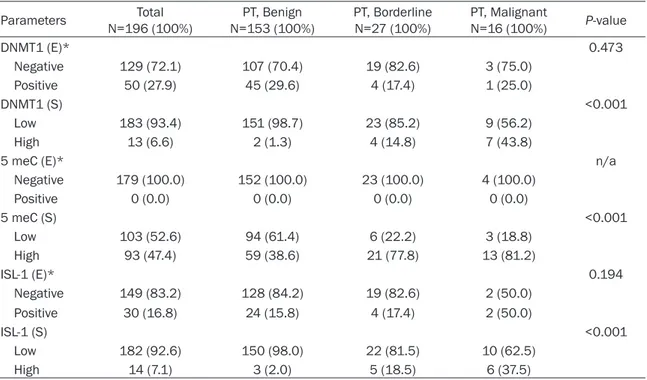
the epithelial component did not appear to be associated with histologic grade. On the other hand, DNMT1, 5 meC, and ISL-1 positivity in the stromal component exhibited significant differ-ences according to histologic grade (Table 3 and Figure 1). Specifically, as histologic grade increased, the ratios of DNMT1, 5 meC, and ISL-1 positivity increased (P<0.001).
Correlations of methylation and the expression of methylation-related proteins with pathologic parameters
Comparison of methylation and expression of methylation-related proteins and pathologic parameters revealed that stromal DNMT1 high Figure 1. DNA methylation and expression of methylation-related proteins according to histologic grade of phyllodes tumor. Levels of DNMT1, 5 meC, and ISL-1 staining in the stromal component increased with increasing histologic grade.
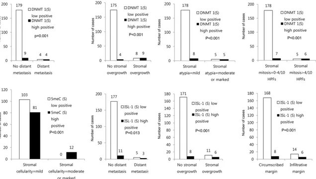
positivity was associated with distant metasta-sis (P=0.001), stromal overgrowth (P<0.001), increased stromal atypia (P<0.001), and increased stromal mitosis (P<0.001). Likewise, high stromal 5 meC positivity was associated
with increased stromal cellularity (P<0.001). Lastly, high stromal ISL-1 positivity was associ-ated with distant metastasis (P=0.013), stro-mal overgrowth (P<0.001), and infiltrative mar-gin (P=0.001, Figure 2).
Figure 2. Correlation of DNA methylation and expression of methylation-related proteins in phyllodes tumor accord-ing to pathologic parameters.
Table 4. Univariate analysis of methylation and methylation-related proteins according to patient prognosis using the log-rank test
Parameters Total/recurrence/ No. of patients metastasis
Disease-free survival Overall survival Median survival
(95% CI) months P-value (95% CI) monthsMedian survival P-value
DNMT1 (E)* 0.242 n/a Negative 129/6/1 174 (167-181) n/a Positive 50/5/0 134 (123-145) n/a DNMT1 (S) 0.002 <0.001 Low 183/14/4 169 (162-176) 179 (175-182) High 13/4/4 96 (65-127) 92 (58-125) 5meC (S) 0.067 n/a Low 103/6/0 172 (164-180) n/a High 93/12/8 155 (143-167) n/a
ISL-1 (E)* 0.633 n/a
Negative 149/9/1 168 (162-175) n/a
Positive 30/2/0 171 (155-186) n/a
ISL-1 (S) <0.001 <0.001
Low 182/13/5 170 (163-176) 178 (173-182)
High 14/5/3 82 (46-119) 106 (78-135)
Impact of methylation and expression of meth-ylation-related proteins on patient prognosis
Univariate analysis to evaluate methylation sta-tus and expression of methylation-related pro-teins on phyllodes tumor patient prognosis showed that decreased DFS and OS were asso-ciated with stromal DNMT1 high positivity (P=0.002 and P<0.001, respectively) and stro-mal ISL-1 high positivity (P<0.001 and P<0.001, respectively, Table 4 and Figure 3). Univariate analysis of phyllodes tumor according to grade showed that high stromal DNMT1 positivity was associated with decreased OS (P=0.015) in borderline phyllodes tumors. According to mul-tivariate Cox analysis, independent factors associated with decreased DFS consisted of histologic grade (hazard ratio: 7.087, 95% CI: 1.857-27.05, P=0.004) and stromal overgrowth (hazard ratio: 10.78, 95% CI: 1.859-62.59, P=0.008), while stromal overgrowth was identi-fied as an independent factor associated with decreased OS (hazard ratio: 59.47, 95% CI: 3.258-1085, P=0.006, Table 5).
Discussion
This research analyzed the relationships between patient outcomes and the methylation and expression of DNA methylation-related pro-teins in phyllodes tumors of the breast. The
results showed that 5 meC staining and the expression of DNMT1 and ISL-1 were increased in the stromal component of phyllodes tumors with increased histologic grade. Consistent with these results, previous studies have reported that TWIST1 [7-9] and RASSF1A [8, 9] are also significantly methylated in phyllodes tumors. Likewise, the methylation frequency of five genes (GSTP1, HIN-1, RAR-beta, RASSF1A, and Twist) has been showed to be increased in phyllodes tumors according to histologic grade [9]. The same study reported that, while the methylation profiles of benign and borderline/ malignant phyllodes tumors are distinguish-able, those of borderline and malignant tumors are not [9]. In contrast to this result, we found that the expression of DNMT1 and ISL-1 was significantly higher in the stromal component of malignant phyllodes tumors compared to bor-derline tumors. A possible explanation for this discrepancy is the use of different experimen-tal methods. Specifically, while previous meth-ylation studies targeting phyllodes tumor evalu-ated the specific methylation status of genes of interest, we focused on the expression of pro-teins involved in DNA methylation (DNMT1) and DNA-methylation target genes (ISL-1). Thus, the increased expression of DNA methylation-relat-ed proteins according to histologic grade that was observed in this study may have been due Figure 3. Disease-free survival (A, B) and overall survival (C, D) according to DNA methylation status and expression of methyla-tion-related proteins in phyllodes tumor. In borderline PTs, high stromal DNMT1 positivity was as-sociated with decreased OS (E).
to carcinogenesis and/or tumor progression fol-lowing epigenetic silencing of various tumor-related genes by DNMT1, especially tumor sup-pressor genes [4]. Thus, as histologic grade of phyllodes tumor increases, the number of genes that undergo methylation-induced silenc-ing increases, which would be consistent with our observation that 5-MeC positivity and expression of DNMT1 were increased in higher grade tumors.
In the present study, expression of DNMT1 and ISL-1 in the stromal component of phyllodes tumors was associated with increased distant metastasis. Consistent with this observation, previous studies have shown that DNMT1 expression is associated with metastasis in both esophageal cancer [11] and stomach can-cer [12], while ISL-1 expression is associated with metastasis in stomach cancer [13]. Increased metastasis following epigenetic silencing by DNA methylation of metastasis-inhibiting genes has been reported [11, 14]. Such metastasis-related genes include proto-cadherin 17, RASSF1A, and DAPK, which were mentioned previously. Among these, RASSF1A has been reported to undergo DNA methylation in phyllodes tumor [8, 9] and should be investi-gated in similar future studies.
Our results showed that high levels of expres-sion of DNMT1 and ISL-1 in the stromal
com-partment of phyllodes tumors were associated with poor prognosis. Consistently, high DNMT1 expression is associated with poor prognosis in malignant lymphoma [15], renal cell carcinoma [16], pancreatic cancer [17], and bladder can-cer [18], while high ISL-1 expression is associ-ated with poor prognosis in gastric cancer [13]. Especially in borderline phyllodes tumors, high stromal DNMT1 expression was significantly associated with decreased OS. Establishing an accurate clinical prognosis for borderline phyl-lodes tumor is difficult because borderline high-grade tumors can be hard to distinguish from benign or malignant tumors [2]. An effec-tive prognostic factor is therefore needed to overcome this problem. Importantly, our results suggest that stromal DNMT1 expression could serve as a prognostic marker of borderline phyl-lodes tumors, a possibility that will require fur-ther study.
A clinically significant outcome of this study is the identification of the epigenetic methylation-related protein DNMT1 as a target in malignant phyllodes tumors. Specifically, by identifying an association between DNMT1 overexpression and increased tumor progression, our study supports the possibility of inhibiting tumorigen-esis by re-expressing tumor suppressor genes that have been inhibited through selective DNMT1 inhibition [19]. Consistent with this possibility, numerous studies have already Table 5. Multivariate analysis of disease-free survival and overall-survival in phyllodes tumor patients Included factor
Disease-free survival Overall survival Hazard
ratio 95% CI P-value Hazard ratio 95% CI P-value
Histologic grade 0.004 n/a
Benign vs. borderline or malignant 7.087 1.857-27.05 n/a n/a
Stromal cellularity 0.317 0.475
Mild vs. moderate or marked 0.336 0.040-2.847 2.805 0.165-47.59
Stromal atypia 0.671 0.866
Mild vs. moderate or marked 0.671 0.106-4.246 0.812 0.072-9.118
Stromal mitosis 0.673 0.426 0-4/10 HPFs vs. >4/10 HPFs 1.874 0.102-34.58 0.209 0.004-9.898 Stromal overgrowth 0.008 0.006 Absent vs. Present 10.78 1.859-62.59 59.47 3.258-1085 Tumor margin 0.158 0.176 Circumscribed vs. Infiltrative 0.395 0.109-1.436 0.333 0.068-1.638 DNMT1 (S) 0.191 0.687 Low vs. High 0.374 0.086-1.631 0.701 0.125-3.934 ISL-1 (S) 0.075 0.432 Low vs. High 2.901 0.899-9.369 1.976 0.362-10.78
attempted to target DNMT1 in various types of cancer [19-22].
In conclusion, the expression of DNA methyla-tion-related proteins in the stromal component of phyllodes tumors of the breast was found to increase with increasing histologic grade, and stromal DNMT1 and ISL-1 overexpression was associated with a poor prognosis.
Acknowledgements
This study was supported by a grant from the National R&D Program for Cancer Control, Ministry of Health & Welfare, Republic of Korea (1420080). This research was supported by Basic Science Research Program through the National Research Foundation of Korea (NRF) funded by the Ministry of Science, ICT and Future Planning (2015R1A1A1A05001209). Disclosure of conflict of interest
None.
Address correspondence to: Dr. Ja Seung Koo, Department of Pathology, College of Medicine, Yonsei University, Severance Hospital, 50 Yonsei-ro, Seodaemun-gu, Seoul 120-752, South Korea. Tel: 82-2-2228-1772; Fax: 82-2-362-0860; E-mail: [email protected]
References
[1] Anderson B, Lawton T, Lehman C and Moe R. Phyllodes tumor. In: Morrow M, Osborne C, edi-tors. Disease of the Breast. Philadelphia: Lippincott & Wilkins; 2004. pp. 991-1006. [2] Tavassoli FA DP. World heath organization
clas-sification of Tumors. Lyon: IARC Press; 2003. [3] Ben Hassouna J, Damak T, Gamoudi A, Chargui
R, Khomsi F, Mahjoub S, Slimene M, Ben Dhiab T, Hechiche M, Boussen H and Rahal K. Phyllodes tumors of the breast: a case series of 106 patients. Am J Surg 2006; 192: 141-147.
[4] Jones PA. DNA methylation and cancer. Oncogene 2002; 21: 5358-5360.
[5] Siedlecki P and Zielenkiewicz P. Mammalian DNA methyltransferases. Acta Biochim Pol 2006; 53: 245-256.
[6] Pathania R, Ramachandran S, Elangovan S, Padia R, Yang P, Cinghu S, Veeranan-Karmegam R, Arjunan P, Gnana-Prakasam JP, Sadanand F, Pei L, Chang CS, Choi JH, Shi H, Manicassamy S, Prasad PD, Sharma S, Ganapathy V, Jothi R and Thangaraju M.
DNMT1 is essential for mammary and cancer stem cell maintenance and tumorigenesis. Nat Commun 2015; 6: 6910.
[7] Do SI, Kim JY, Kang SY, Lee JJ, Lee JE, Nam SJ and Cho EY. Expression of TWIST1, Snail, Slug, and NF-kappaB and methylation of the TWIST1 promoter in mammary phyllodes tumor. Tumour Biol 2013; 34: 445-453.
[8] Huang KT, Dobrovic A, Yan M, Karim RZ, Lee CS, Lakhani SR and Fox SB. DNA methylation profiling of phyllodes and fibroadenoma tu-mours of the breast. Breast Cancer Res Treat 2010; 124: 555-565.
[9] Kim JH, Choi YD, Lee JS, Lee JH, Nam JH, Choi C, Park MH and Yoon JH. Borderline and malig-nant phyllodes tumors display similar promot-er methylation profiles. Virchows Arch 2009; 455: 469-475.
[10] Won KY, Kim GY, Kim YW, Song JY and Lim SJ. Clinicopathologic correlation of beclin-1 and bcl-2 expression in human breast cancer. Hum Pathol 2010; 41: 107-112.
[11] Bai J, Zhang X, Hu K, Liu B, Wang H, Li A, Lin F, Zhang L, Sun X, Du Z and Song J. Silencing DNA methyltransferase 1 (DNMT1) inhibits proliferation, metastasis and invasion in ESCC by suppressing methylation of RASSF1A and DAPK. Oncotarget 2016; [Epub ahead of print]. [12] Qiao F, Zhang K, Gong P, Wang L, Hu J, Lu S.
and Fan H. Decreased miR-30b-5p expression by DNMT1 methylation regulation involved in gastric cancer metastasis. Mol Biol Rep 2014; 41: 5693-5700.
[13] Guo C, Wang W, Shi Q, Chen P and Zhou C. An abnormally high expression of ISL-1 represents a potential prognostic factor in gastric cancer. Hum Pathol 2015; 46: 1282-1289.
[14] Yin X, Xiang T, Mu J, Mao H, Li L, Huang X, Li C, Feng Y, Luo X, Wei Y, Peng W, Ren G and Tao Q. Protocadherin 17 functions as a tumor sup-pressor suppressing Wnt/beta-catenin signal-ing and cell metastasis and is frequently meth-ylated in breast cancer. Oncotarget 2016; [Epub ahead of print].
[15] Zhao H, Zhang LE, Guo S, Yuan T, Xia B, Zhang L and Zhang Y. Overexpression of DNA methyl-transferase 1 as a negative independent prog-nostic factor in primary gastrointestinal diffuse large B-cell lymphoma treated with CHOP-like regimen and rituximab. Oncol Lett 2015; 9: 2307-2312.
[16] Li M, Wang Y, Song Y, Bu R, Yin B, Fei X, Guo Q and Wu B. Aberrant DNA methyltransferase 1 expression in clear cell renal cell carcinoma development and progression. Chin J Cancer Res 2014; 26: 371-381.
[17] Zhang JJ, Zhu Y, Zhu Y, Wu JL, Liang WB, Zhu R, Xu ZK, Du Q and Miao Y. Association of in-creased DNA methyltransferase expression
with carcinogenesis and poor prognosis in pancreatic ductal adenocarcinoma. Clin Transl Oncol 2012; 14: 116-124.
[18] Wu CT, Wu CF, Lu CH, Lin CC, Chen WC, Lin PY and Chen MF. Expression and function role of DNA methyltransferase 1 in human bladder cancer. Cancer 2011; 117: 5221-5233. [19] Subramaniam D, Thombre R, Dhar A and
Anant S. DNA methyltransferases: a novel tar-get for prevention and therapy. Front Oncol 2014; 4: 80.
[20] Amato RJ, Stephenson J, Hotte S, Nemunaitis J, Belanger K, Reid G and Martell RE. MG98, a second-generation DNMT1 inhibitor, in the treatment of advanced renal cell carcinoma. Cancer Invest 2012; 30: 415-421.
[21] Mutze K, Langer R, Schumacher F, Becker K, Ott K, Novotny A, Hapfelmeier A, Hofler H and Keller G. DNA methyltransferase 1 as a predic-tive biomarker and potential therapeutic target for chemotherapy in gastric cancer. Eur J Cancer 2011; 47: 1817-1825.
[22] Thottassery JV, Sambandam V, Allan PW, Maddry JA, Maxuitenko YY, Tiwari K, Hollingshead M and Parker WB. Novel DNA methyltransferase-1 (DNMT1) depleting anti-cancer nucleosides, 4’-thio-2’-deoxycytidine and 5-aza-4’-thio-2’-deoxycytidine. Cancer Chemother Pharmacol 2014; 74: 291-302.