저작자표시-비영리-변경금지 2.0 대한민국 이용자는 아래의 조건을 따르는 경우에 한하여 자유롭게 l 이 저작물을 복제, 배포, 전송, 전시, 공연 및 방송할 수 있습니다. 다음과 같은 조건을 따라야 합니다: l 귀하는, 이 저작물의 재이용이나 배포의 경우, 이 저작물에 적용된 이용허락조건 을 명확하게 나타내어야 합니다. l 저작권자로부터 별도의 허가를 받으면 이러한 조건들은 적용되지 않습니다. 저작권법에 따른 이용자의 권리는 위의 내용에 의하여 영향을 받지 않습니다. 이것은 이용허락규약(Legal Code)을 이해하기 쉽게 요약한 것입니다. Disclaimer 저작자표시. 귀하는 원저작자를 표시하여야 합니다. 비영리. 귀하는 이 저작물을 영리 목적으로 이용할 수 없습니다. 변경금지. 귀하는 이 저작물을 개작, 변형 또는 가공할 수 없습니다.
석사학위논문
RAW 264.7 세포에서 제주왕지네
(
Scolopendra subspinipes mutilans
)
추출물의 염증조절 기전에 관한 연구
제주대학교 대학원
생물학과
박 재 현
RAW 264.7 세포에서 제주왕지네
(
Scolopendra subspinipes mutilans
)
추출물의 염증조절 기전에 관한 연구
지도교수 이 선 령
박 재 현
이 논문을 이학 석사학위 논문으로 제출함
2018년 2월
박재현의 이학 석사학위 논문을 인준함
심사위원장 ㊞
위 원 ㊞
위 원 ㊞
제주대학교 대학원
2018년 2월
Study on the inflammatory mechanisms of the
Scolopendra subspinipes mutilans extract
in RAW 264.7 cells
Jae-Hyeon Park
(Supervised by professor Sun-Ryung Lee)
A thesis submitted in partial fulfillment of the requirement
for the degree of Master of Science
2018. 2.
This dissertation has been examined and approved by
Chairperson of the Committee
(Name and signature)
DEPARTMENT OF BIOLOGY
GRADUATE SCHOOL
JEJU NATIONAL UNIVERSITY
ABSTRACT
Inflammation is the part of the immune response to protect the body from extracellular stimuli or damage. However, excessive inflammation caused by activation of macrophage can lead to inflammatory disorder. Centipede has long been used in korea as traditional medicine, but little is known about its mechanisms. In present study, the anti-inflammatory effect of ethanol extract of Scolopendra subspinipes mutilans was investigated in lipopolysaccharide (LPS)-induced RAW 264.7 cells. Scolopendra subspinipes mutilans ethanol extract (SSE) inhibited nitric oxide (NO) production and inducible NO synthase (iNOS) protein expression in concentration dependent manner (10, 100, 200 ㎍/㎖).
Pro-inflammatory cytokines, such as interleukin-1β (IL-1β) and
interleukin-6 (IL-6) were does dependently attenuated by SSE. SSE inhibited nuclear factor-kappa B (NF-κB) translocation from the cytoplasm to the nucleus, and decreased degradation of inhibitory kappa B-alpha (IκB-α) in LPS-stimulated RAW 264.7 cells. In addition exposure of SSE and pyrrolidine dithiocarbamate (PDTC), an NF-κB specific inhibitor, were decreased NO release and iNOS expression significantly. SSE also attenuated LPS-stimulated phosphorylation of extracellular signal-regulated kinases 1/2 (ERK 1/2). Co-treatment with SSE and U0126, an MEK inhibitor, reduced LPS-induced NO production and iNOS expression compared to that in SSE-treated or U0126-treated cells. These results indicate that SSE down-regulated NO production and iNOS
expression through ERK 1/2 and NF-κB signaling pathway in
LPS-stimulated RAW 264.7 cells. In conclusion, ethanol extract of Scolopendra subspinipes mutilans has significant inhibitory effect on
inflammation and it is might have therapeutic potential to inflammatory diseases.
Key word : Inflammation, Scolopendra subspinipes mutilans, nitric oxide (NO), cytokine, nuclear factor-kappa B (NF-κB), extracellular signal- regulated kinases 1/2 (ERK-1/2).
A B S T R A C T --- 1 목 차 --- 3 L I S T O F T A B L E --- 5 L I S T O F F I G U R E S --- 6 Ⅰ. 서 론 --- 8 Ⅱ. 재 료 및 방 법 --- 12 1. 시료 준비 --- 12 2. 세포 배양 --- 13 3. 세포 독성 측정 --- 13 4. NO 생성량 측정 --- 13 5. 단백질 추출 --- 14 6. Western blot --- 14
7. Reverse transcription polymerase chain reaction --- 15
8. 통계처리 --- 16
Ⅲ. 결 과 --- 18 1. 제주 왕지네 추출물이 염증반응에 미치는 영향 --- 18 (1) 왕지네 추출물의 세포 독성 --- 18 (2) NO 생성 및 iNOS 발현 억제 --- 18 (3) Pro-inflammatory cytokine 발현 억제 --- 19 2. 제주왕지네 추출물이 NF-κB signaling에 미치는 영향 --- 24 3. 제주왕지네 추출물이 ERK 1/2 signaling 에 미치는 영향 --- 29 Ⅳ. 고 찰 --- 34 Ⅴ. 참 고 문 헌 --- 38 Ⅵ. 요 약 --- 46
Table 1. The primer sequences of the genes used in reverse transcription
polymerase chain reaction (RT-PCR) analysis. --- 17
Figure 1. Photo of Scolopendra subspinipes mutilans. --- 12
Figure 2. Effect of Scolopendra subspinipes mutilans extract (SSE) on the viability of RAW 264.7 cells. --- 20
Figure 3. Inhibitory effect of Scolopendra subspinipes mutilans
extract (SSE) on LPS-stimulated NO production. --- 21
Figure 4. Inhibitory effect of Scolopendra subspinipes mutilans extract (SSE) on LPS-stimulated iNOS protein
expression. --- 22
Figure 5. Inhibitory effect of Scolopendra subspinipes mutilans extract (SSE) on LPS-stimulated pro-inflammatory
cytokines (IL-1β, IL-6) expression. --- 23
Figure 6. Effect of Scolopendra subspinipes mutilans extract (SSE) on activation of NF-κB (p65) in LPS-stimulated
RAW 264.7 cells. --- 25
Figure 7. Effect of Scolopendra subspinipes mutilans extract (SSE) on expression of IκB in LPS-stimulated RAW 264.7
cells. --- 26
Figure 8. Involvement of NF-κB (p65) signaling on LPS-induced
NO production. --- 27
Figure 9. Involvement of NF-κB (p65) signaling on LPS-induced
iNOS expression. --- 28
Figure 10. Effect of Scolopendra subspinipes mutilans extract (SSE) on activation of ERK 1/2 in RAW 264.7 cells. --- 30
Figure 11. Inhibitory Effect of Scolopendra subspinipes mutilans extract (SSE) on phosphorylation of ERK 1/2 in
LPS-stimulated RAW 264.7 cells. --- 31
Figure 12. Involvement of ERK 1/2 signaling on LPS-induced NO
production. --- 32
Figure 13. Involvement of ERK 1/2 signaling on LPS-induced iNOS
Ⅰ. 서 론
염증 (Inflammation)은 특정 외부 자극이나 손상으로부터 신체를 보호하기 위해 발생하는 면역반응이다 (Limtrakul et al., 2016). 그러나 염증과정에서 발생하는 오작동으로 인하여 염증이 억제되지 않고 지속적으로 유지되는 만성 염증 상태는 염증성 장 질환, 류마티스성 관절염, 심혈관질환, 신경퇴행성 질 환, 당뇨, 암과 같은 질환의 발병원인이 된다 (Ferrero-Miliani et al., 2007; Kim et al., 2016). 즉, 염증은 조직 손상을 완화하고 우리 몸의 항상성을 유 지하기도 하지만 질병을 유발하는 주요 인자로도 작용한다 (Janssen et al., 2012). 체내에서 염증 반응, 항균 작용과 관련된 기능을 담당하며 항상성과 선천 성 면역을 유지하여 질병의 발생과 연관되어 있는 대식세포 (macrophage)는 lipopolysaccharide (LPS)와 같은 염증성 자극에 의해 활성화 된다 (Saha et al., 2017; Na et al., 2017). LPS는 gram-negative bacteria 외막의 주성분 으로 세포에 노출 되었을 시 강력한 염증을 유도하는 내독소로 알려져 있다 (Alexander et al., 2001). 대식세포에 LPS를 처리하면 세포 표면에 존재하 는 toll-like receptors (TLRs)를 통해 염증 신호가 유도되어 대식세포를 활 성화 시키는 것으로 보고되었다 (Maris et al., 2006; Doyle et al., 2006). 활성화된 대식세포는 nitric oxide (NO), prostaglandin E2 (PGE2), inducibleNO synthase (iNOS), cyclooxygenase-2 (COX-2), interleukin-1β (IL-1β), interleukin-6 (IL-6), tumor necrosis factor-α (TNF-α)와 같은 전염증 인자 (pro-inflammatory factors)의 발현을 유도한다 (Meng et al., 2015).
Nitric oxide (NO)는 세포 내·외부적으로 중요한 신호전달 분자이며 순환 계, 신경계, 면역계에서 생리학적 기전을 조절하는 다양한 역할을 하는 반면 free radical oxygen (NO˙)이 세포독성 요소로 작용하여 병리학적 과정과도 연관되어 있다 (Aktan. 2004). NO는 NO synthase (NOS)에 의해 L-arginine으로부터 합성되며, endothelial NOS (eNOS), neuronal NOS (nNOS), inducible NOS (iNOS) 3종류의 NOS isoform이 존재한다 (Zamora
et al., 2000). nNOS와 eNOS에 의해 소량 생성되는 NO는 시냅스 가소성 (synaptic plasticity), 혈압 조절, 혈관 확장 및 보호 등 항상성 유지에 중요 한 역할을 한다. 이들과 달리 iNOS는 보통의 세포에서는 발현되지 않지만 LPS나 pro-inflammatory cytokine과 같은 염증성 자극에 의해서 유도되어 NO의 생성이 과도하게 증가되고 이에 따라 염증과 부종, 조직 손상 등을 유 발하게 된다 (Förstermann et al., 2012; Chen et al., 2015).
Nuclear factor-kappa B (NF-κB)는 cytokine, chemokine, growth factor, cell adhesion molecule 등의 발현에 영향을 미치는 전사 인자이며 면역과 염증 반응을 조절하는 중요한 역할을 한다 (Beinke et al., 2004). NF-κB는 p65와 p50 단백질이 heterodimer 상태로 존재하며 inhibitory kappa B (IκB)라는 억제 단백질과 complex를 이루어 세포질에 위치하고 있 다 (Li et al., 2002). LPS와 같은 염증 자극이 전해질 경우 IκB kinase (IKK)의 활성화에 의해 IκB를 인산화 시키고, NF-κB complex와 분리되어 NF-κB의 활성화를 유도하며 인산화 된 IκB는 proteasome에 의해 분해된다 (Karin, 1999). 활성화된 NF-κB는 핵 내부로 전이 (translocation)되면서 프 로모터에 부착하여 iNOS, COX-2, IL-1β, IL-6와 같은 전염증 인자를 발현 하게 된다 (Seo and Huh et al., 2012). 따라서 활성화된 NF-κB의 전이를 억제하는 것이 염증 질환 예방 및 치료의 주요 원인 중 하나로 여겨져 대식세 포의 과도한 활성을 억제하는 소재의 연구가 다양하게 진행되고 있다. 식물 한약재인 속단 (Phlomis younghusbandii)의 뿌리에서 분리한 화합물인 임페 라토린 (Imperatorin)과 한의학에서 사용되는 약초인 가미소요산의 물 추출물 은 LPS로 유도된 RAW 264.7 세포에서 NF-κB의 핵 내부로 전이를 억제하 여 염증 인자들의 발현이 저해되는 것을 보여주어 염증질환 치료 및 예방에 있어서 NF-κB의 중요성을 제시하였다 (Zang et al., 2017; Jin et al., 2016).
곤충은 주로 해충을 잡아먹는 천적으로 이용되거나, 화분 매개용, 애완용, 사료용 등으로 이용되고 있다. 뿐만 아니라, 약용 및 식용으로도 이용되기 시 작하면서 생물자원으로써의 가치가 증가하고 있다. 2015년 기준으로 우리나 라 곤충 산업의 시장규모는 3000억원에 이르고 2020년에는 5,600억원으로
성장할 것으로 예측하고 있다 (Kim et al., 2016). 또한 생물자원을 이용하였 을 때 발생되는 이익은 자원을 제공한 나라와 이용한 나라가 공유하도록 규정 한 국제 규범인 나고야의정서 (nagoya protocol)가 채택됨에 따라 생물자원 을 발굴하고 데이터베이스를 구축해 나가는 것이 생물다양성협약을 대비하는 데 중요한 역할을 하고 있다 (Jang et al., 2012). 최근, 곤충의 유용물질에 대한 연구도 다양하게 진행되고 있다. 참콩풍뎅이 (Popillia flavosellata)추출물의 항염증 효과 (Yoon et al., 2015), 무당벌레 (Harmonia axyridis)에서 분리한 HaGF peptide의 항염증 효과 (Kim et al.,
2013), 애기뿔소똥구리 (Copris tripartitus)에서 유래한 CopA3 peptide의 항
염증 효과 (Kim et al., 2013), 벼메뚜기 (Oxya chinensis sinuosa) 추출물의 항염증, 항산화 및 항균 효과 (Yoon et al., 2014; Kim et al., 2015), 미국산 바퀴 (Periplaneta americana L.) 추출물의 항균 효과 (Kim et al., 2015) 등 이 보고되었다. 이처럼 곤충에서 유래한 물질을 바탕으로 진행한 연구는 염증 질환의 치료제와 항균제로써의 이용가능성을 제시하였다.
곤충의 유용물질에 대한 생리활성이 알려지면서 왕지네의 생리활성 연구 또한 활발하게 진행 되고 있다. Ding 등 (Ding et al., 2016)의 연구에 따르 면 왕지네 추출물에서 분리한 화합물이 U87 human glioma cancer cell에서 apoptosis를 유도하였고, Ma 등 (Ma et al., 2014)의 연구에서 왕지네 에탄 올 추출물이 A375 human melanoma cell의 증식을 억제하여 항암효과가 있 음을 보고하였다. Kim 등 (Kim et al., 2014)의 연구에서는 왕지네 에탄올 추출물이 B16/F10 melanoma cell에서 tyrosinase의 활성을 저해하여 멜라 닌 생성을 억제해 미백효과가 있음을 보고하였다. 또한 왕지네에서 분리한 Scolopendrasin Ⅴ와 Scolopendin peptide가 미생물의 세포막을 파괴하여 강력한 항균효과가 있음이 보고되었다 (Lee et al., 2015; Lee et al., 2017).
왕지네 (Scolopendra subspinipes mutilans)는 오래전부터 한국에서 관절
염, 뇌졸중, 경련, 중풍, 파상풍, 림프선염, 암종 등의 질병에 약재로 사용되어 왔다 (Pemberton, 1999). 또한 민간에서는 왕지네를 술에 담가 1년 이상 숙 성시켰을 경우 관절염 및 통증에 효과가 있는 것으로 알려져 왔으나 (Kim et al., 2010) 이에 대한 정확한 연구는 아직 보고된 바 없다. 따라서 본 연구에
서는 제주도에 서식하는 왕지네 에탄올 추출물 (Scolopendra subspinipes mutilans ethanol extract, SSE)의 항염증 활성을 조사하기 위하여 LPS로 유 도된 RAW 264.7 세포에서 왕지네 추출물이 염증인자들의 발현에 미치는 영 향과 이들이 어떠한 신호전달 경로를 통해 조절되는지 분석하였다.
Ⅱ. 재료 및 방법
1. 시료 준비
본 연구에서 사용한 왕지네 (Scolopendra subspinipes mutilans)는 제주
마트 (www.jejumart.co.kr, Korea)에서 구입하였다 (Figure 1). 건조된 상태 의 왕지네 전충에서 독성이 있는 머리, 꼬리, 다리를 제거하여 지네 몸통만 추출에 사용하였다. 건조된 지네몸통을 파쇄기로 분쇄한 후 60% 에탄올 (EtOH)을 이용하여 상온에서 3시간 동안 추출하였다. 추출물은 filter paper (Whatman, No.2)를 이용하여 여과 후 감압 회전 농축기 (Bunchi, R-200, Switzerland)로 농축하였고, 동결 건조를 실시하여 얻은 powder 상태의 시료 는 –20℃에 보관하였다. 실험에 사용한 시료는 EtOH과 PBS (phosphate buffered saline)를 1:1로 혼합한 용매에 녹여 사용하였다.
Figure 1. Photo of Scolopendra subspinipes mutilans. The photographs are shown on the ventral and dorsal side of Scolopendra subspinipes mutilans.
2. 세포 배양
Murine macrophage RAW 264.7 세포는 American Type Culture Collection (ATCC, USA)에서 구입하였다. 세포 배양은 10% fetal bovine serum (FBS, GIBCO, USA)과 1% penicillin streptomycin (P/S, GIBCO, USA)이 첨가된 Dulbecco's modified Eagle's medium (DMEM, GIBCO,
USA)을 이용하여 37℃, 5% CO2 incubator에서 배양하였다. 세포는 2일에
한번 계대 배양하여 유지하였다.
3. 세포 독성 측정
세포의 독성은 MTT assay를 통해 확인하였다 (Stockert et al., 2012). RAW 264.7 세포를 48 well cell culture plate에 1.8 × 10⁵ cells/㎖로 분
주하여 18시간 동안 CO2 incubator에서 배양 한 후 시료 또는 LPS를 24시
간 동안 처리한 다음 3-(4,5-dimethylthiazol-2-yl)-2,5-diphenyl-tetra- zolium bromide (MTT, Amnesco, USA) 시약을 각 well에 처리하였다. 3시 간 동안 반응시킨 후 배양 상층액을 제거하고 dimethyl sulfoxide (DMSO)로 생성된 formazan crystals을 녹여 ELISA reader (molecular devices, USA) 를 이용하여 570 nm에서 흡광도(O.D)를 측정하였다. 세포독성은 시료를 처리 하지 않은 대조군의 O.D값과 비교하여 다음과 같이 계산하였다.
Cell viability (%) = {(sample O.D570 / control O.D570)} × 100
4. NO 생성량 측정
NO의 생성은 1.8 × 10⁵ cells/㎖로 분주한 48 well plate의 세포에 시료
및 LPS (Sigma aldrich, USA)를 24시간 처리하여 배지로 분비되는 NO의 양 을 측정 하여 확인하였다. 배지 상층액과 griess reagent (1% sulfanilamide, 0.1% napthylethylene diamine, 2.5% phosphoric acid)를 1:1로 혼합하여 암실조건에서 10분 동안 반응시킨 후 540 nm에서 흡광도를 측정하였다.
Sodium nitrite (NaNO2)를 이용하여 표준곡선을 작성한 후 측정한 흡광도 값
5. 단백질 추출
시료 및 LPS (100 ng/㎖)가 처리된 세포를 cold-PBS로 1회 수세한 후 protease & phosphatase inhibitor (1 mM PMSF, 1 mM Na3VO4, 1 mM
NaF, 1 ㎍/㎖ aprotinin, 1 ㎍/㎖ pepstatin, 1 ㎍/㎖ leupeptin)가 첨가된 RIPA buffer (50 mM Tris-Hcl pH 7.4, 150 mM NaCl, 2 mM EDTA, 1% NP-40, 0.1% SDS)를 이용하여 30분 동안 lysis시킨 후 4℃, 13,000 rpm에 서 15분간 원심분리하여 단백질을 얻었다.
세포질 단백질과 핵 단백질 분리는 NE-PER Nuclear and Cytoplasmic Extraction kit (Thermo scientific, USA)를 이용하였다. Cold-PBS로 세포 를 회수한 후 Cytoplasmic Extraction Reagent (CER)을 첨가하여 10분 동 안 ice에서 반응하여 원심분리 (4℃, 15,000g, 5분) 후 얻어진 상층액을 세포 질 단백질로 사용하였고 남은 pellet에 Nuclear Extraction Buffer (NER)을 첨가하여 40분 동안 ice에서 반응하고 원심분리 (4℃, 15,000g, 15분)하여 핵 단백질을 추출하였다.
6. Western blot
세포에서 추출한 단백질은 Bio-Rad protein assay kit (Bio-Rad, USA)를 사용하여 정량하였고 동일한 양의 단백질을 sodium dodecyl sulfate (SDS) polyacrylamide gel에서 전기영동 하여 분리 한 후 nitrocellulose (NC) membrane (GE healthcare life sciences, UK)에 전이 시켰다. NC membrane을 3% skim milk (BD Diagnostic, USA)를 함유한 Tris-buffered saline-0.1% Tween 20 (0.1% TBS-T) 용액으로 1시간 동안 blocking 시킨 후 1차 항체와 반응시켰다. 단백질 발현 분석을 위해 1차 항체는 anti-rabbit iNOS (Santacruz biotechnology, USA), anti-mouse β-actin (SIGMA aldrich, USA), anti-rabbit NF-κB (Cell signaling technology, USA), anti-mouse IκB-α (Cell siganling technology, USA), anti-rabbit phospho-ERK 1/2, anti-rabbit ERK 1/2 (Cell signaling technology, USA) 를 사용하였다. 1차 항체 반응이 끝난 NC membrane은 0.1% TBS-T용액으 로 3회 세척 한 후 peroxidase가 conjugated된 2차 항체인 goat anti-
mouse IgG, goat anti-rabbit IgG (Santacruz biotechnology, USA)를 이용 하여 반응시킨 후 TBS-T용액으로 세척하였다. SuperSignal West Pico Chemiluminescent Substrate (Thermo fisher scientific, USA)로 5분간 반 응시킨 후 암실에서 X-ray film (Agfa gevaert N.V., Belgium)을 이용하여 분석하였다.
7. Reverse transcription polymerase chain reaction
세포로부터 RNA분리는 easy-BLUE Total RNA Extraction Kit (iNrRON Biotechnology, Korea)를 사용하였다. RNA extraction buffer를 첨가해 microtube에 수거한 후 chloroform 200 ㎕를 넣고 voltexing하여 세포를 lysis시키고 4℃, 13,000 rpm에서 10분간 원심분리 하였다. 상층액 400 ㎕를 새로운 microtube에 옮기고 동량의 isopropanol을 첨가하여 2~3회 inverting 하여 상온에서 10분간 반응시킨 후 원심분리 (4℃, 13,000 rpm, 5분)하여 RNA를 침전시켰다. 75% EtOH를 이용하여 2~3회 수세하고 RNA를 UltraPureTM DNase/RNase-free distilled water (Thermo fisher scientific,
USA)에 녹여 -70℃에 보관하였다. RNA는 nanodrop 2000 spectrometer (Nanodrop, USA)를 이용하여 정량하였으며, A260 / A280㎚ 값이 1.7-2.0 의 범위를 갖는 RNA를 실험에 사용하였다.
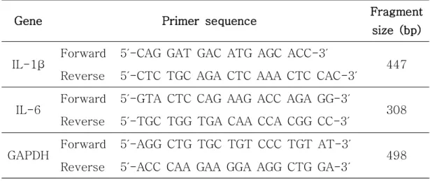
중합효소반응은 Maxime RT-PCR PreMix Kit (iNtRON Biotechnology, Korea)를 이용하였다. 동일한 양의 RNA와 primer를 reaction mixture tube 에 넣고 혼합한 후 중합효소반응을 진행하였으며, 사용된 primer의 염기서열 은 Table 1에 나타내었다. 역전사 중합효소반응의 조건으로는 45℃에서 30 분, 94℃에서 5분 동안 1회 진행하여 cDNA를 합성하였으며, 이후 94℃에서 45초간 denaturation, 57℃에서 45초간 annealing, 72℃에서 1분간 extension 과정을 27회 진행하였다. 최종 PCR 산물을 Red-safe nucleic acid staining solution (iNtRON Biotechnology, Korea)으로 염색한 1.5% agarose gel에서 전기영동한 후 Gelmanager (Prime-tech corparation, Korea)에서 band를 확인하였다.
8. 통계처리
실험 결과는 평균과 표준편차 (mean ± S.D)로 나타내었고 student's t-test 또는 SPSS (statistical package for social science, ver. 18)를 이용 하여Duncan’s multiple range test로 통계학적 유의성을 검증하였다.
Gene Primer sequence Fragment size (bp)
IL-1β Forward 5′-CAG GAT GAC ATG AGC ACC-3′ 447
Reverse 5′-CTC TGC AGA CTC AAA CTC CAC-3′
IL-6 Forward 5′-GTA CTC CAG AAG ACC AGA GG-3′ 308
Reverse 5′-TGC TGG TGA CAA CCA CGG CC-3′
GAPDH Forward 5′-AGG CTG TGC TGT CCC TGT AT-3′ 498
Reverse 5′-ACC CAA GAA GGA AGG CTG GA-3′
Table 1. The primer sequences of the genes used in reverse transcription polymerase chain reaction (RT-PCR) analysis.
Ⅲ. 결 과
1. 제주왕지네 추출물이 염증 반응에 미치는 영향
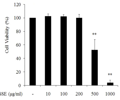
(1) 왕지네 추출물의 세포 독성 왕지네 에탄올 추출물의 세포독성을 확인하기 위하여 MTT assay를 수행 하였다. RAW 264.7 세포에 왕지네 추출물을 농도별로 처리한 후 세포생존율 (cell viability)을 확인한 결과 200 ㎍/㎖ 이하의 농도에서는 대조군과 비슷한 수준의 세포생존율을 나타내었으나 500, 1000 ㎍/㎖의 농도에서 각각 52%, 4%의 생존율을 나타내어 강한 독성을 나타내었다 (Figure 2). 이 결과를 바 탕으로 세포 생장에 영향을 미치지 않는 추출물의 농도인 10~200 ㎍/㎖을 선정하여 세포 실험을 진행하였다. (2) NO 생성 및 iNOS 발현 억제 대식세포에서 LPS를 처리하면 iNOS의 발현에 의해 NO 생성이 증가되고 과도할 경우 염증, 부종, 조직손상을 유발하게 된다 (Förstermann et al., 2012). 따라서 왕지네 추출물의 항염증 활성을 알아보기 위해 추출물이 염증 자극에 의해 합성되는 iNOS 발현 및 NO 생성에 어떠한 영향을 미치는지 분 석하였다. 먼저 NO 생성률(%)을 확인하기 위해 세포독성이 나타나지 않는 농 도의 추출물을 (10~200 ㎍/㎖) 처리하여 griess reagent로 측정한 결과 100 ng/㎖ LPS 처리군에서의 NO 생성률이 대조군 보다 5배 정도 증가하였고 왕 지네 추출물이 10, 100, 200 ㎍/㎖ 처리됨에 따라 각각 1%, 30%, 56%의 NO 생성 억제율을 나타내었다 (Figure 3).또한 NO 분비를 조절하는 단백질인 iNOS의 발현을 western blot을 통하 여 분석한 결과 RAW 264.7 세포에 LPS (100 ng/㎖)의 처리는 iNOS 단백질 발현양을 현저하게 증가시킨 반면 왕지네 추출물의 처리는 농도가 증가함에 따라 iNOS 발현을 강력하게 억제시키는 것을 관찰할 수 있었다 (Figure 4).
(3) Pro-inflammatory cytokine 발현 억제
대식세포에서 자극에 의해 발현되는 염증성 cytokine으로 알려진 IL-1β, IL-6의 분비는 iNOS 발현을 증가시켜 NO 생성에 영향을 미치는 것으로 보 고되어 있다 (Lee et al., 1993; Kaur et al., 2014). 따라서 왕지네 추출물이 LPS에 의해 유도된 IL-1β, IL-6 발현에 어떠한 영향을 미치는지 확인하기 위하여 RT-PCR 방법을 이용하여 RNA 발현양을 분석하였다. 100 ng/㎖ 농 도의 LPS 처리에 의하여 IL-1β와 IL-6의 발현이 현저하게 증가하였으나, 왕 지네 추출물이 처리된 실험군에서는 농도 의존적으로 IL-1β, IL-6의 발현이 저해되는 것을 확인하였다 (Figure 5).
Figure 2. Effect of Scolopendra subspinipes mutilans extract (SSE) on the viability of RAW 264.7 cells. Cells were treated with the indicated concentrations of SSE for 24 hr. Cytotoxicity of SSE was analyzed by MTT assay. The data are represented as mean ± S.D of five independent experiments. ** P < 0.001 compared to control group.
Figure 3. Inhibitory effect of Scolopendra subspinipes mutilans extract (SSE) on LPS-stimulated NO production. Cells were pre-treated with indicated concentration of SSE for 1 hr, and then stimulated with LPS (100 ng/㎖) for 24 hr. Cell viability and production of NO were determined by MTT assay and griess reagent. The data are represented as mean ± S.D of five independent experiments. # P < 0.001 compared to control group, ** P < 0.001 compared to LPS treated group.
Figure 4. Inhibitory effect of Scolopendra subspinipes mutilans extract (SSE) on LPS-stimulated iNOS protein expression. Cells were pre- treated with indicated concentration of SSE for 1 hr, and then stimulated with LPS (100 ng/㎖) for 24 hr. iNOS expression was analyzed by western blot. The relative iNOS expression was quantified by densitometry and normalized to β-actin expression. The data are represented as mean ± S.D of three independent experiments. # P < 0.001 compared to control group, ** P < 0.001 compared to LPS treated group.
Figure 5. Inhibitory effect of Scolopendra subspinipes mutilans extract (SSE) on LPS-stimulated pro-inflammatory cytokines (IL-1β, IL-6) expression. Cells were pre-treated with indicated concentration of SSE for 1 hr, and then stimulated with LPS (100 ng/㎖) for 12 hr. RNA expression of cytokines were analyzed by RT-PCR.
2. 왕지네 추출물이 NF-κB signaling에 미치는 영향
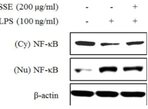
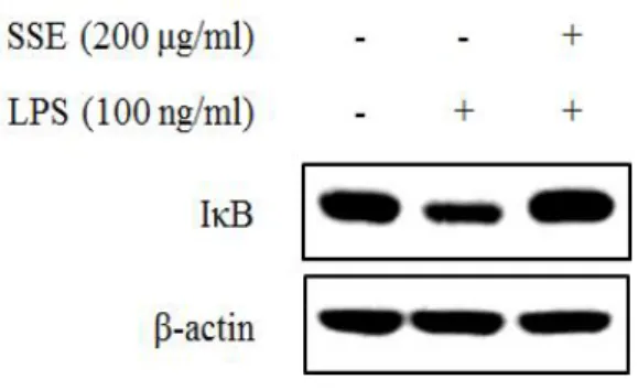
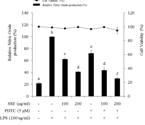
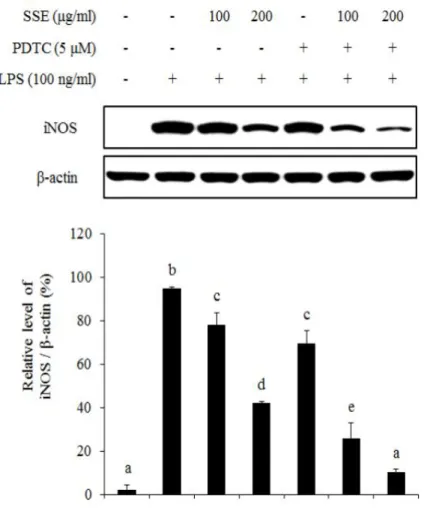
대식세포에서 염증 자극에 의해서 NF-κB는 IκB가 분해됨에 따라 활성화 되어 핵 내부로 전이된다. 핵 내부에서 NF-κB는 전사 인자로 작용하여 여러 가지 염증 인자들의 발현을 조절하게 된다 (Seo and Huh et al., 2012). 따 라서 NO 생성과 iNOS 발현을 조절하는 왕지네 추출물이 NF-κB 신호전달계 와 어떠한 연관성이 있는지 알아보기 위하여 NF-κB와 IκB 단백질 발현정도 를 western blot을 이용하여 분석하였다. 왕지네 추출물이 NF-κB 단백질의 핵 내부로의 전이에 미치는 효과를 확 인하기 위하여 세포질 단백질과 핵 단백질을 각각 분리하여 NF-κB 단백질 발현량을 분석하였다. 그 결과 세포질의 경우 LPS의 처리는 NF-κB의 핵 내 부로의 전이를 증가시켜 NF-κB 발현을 감소시켰고, 왕지네 추출물 처리는 LPS에 의한 NF-κB 단백질의 전이를 억제시켰다. 핵 분획의 경우 LPS 처리 에 의해 NF-κB의 발현이 증가되었고, 왕지네 추출물 처리에 의해 NF-κB 발 현량이 감소되어 세포질에서의 발현 양상과 상반된 결과를 나타내었다 (Figure 6). 또한 NF-κB 신호전달에 중요한 역할을 하는 IκB 발현 양상을 분석한 결과, LPS 처리 75분 후 IκB의 발현이 현저하게 감소하였고, 왕지네 추출물 처리는 LPS에 의해 감소된 IκB 발현을 증가시키는 것을 확인할 수 있 었다 (Figure 7). 왕지네 추출물이 유도하는 NO 생성 저해 기전이 NF-κB signaling을 통해 조절되는지를 검증하기 위해 NF-κB specific inhibitor인 pyrrolidine dithiocarbamate (PDTC)를 처리하여 NO 생성률과 iNOS 발현 양상을 분석 하였다. NO 생성률을 살펴보면 왕지네 추출물과 PDTC가 각각 처리된 실험 군에서는 NO 생성 저해율이 각각 37%, 28% 정도로 유사하게 나타났으며 이 들을 동시처리 했을 때 NO 생성 억제가 더욱 증가하여 56%의 저해율을 나 타내었다 (Figure 8). 이러한 결과는 동일한 농도에서 NO 합성 효소인 iNOS 의 발현과 유사한 양상을 나타내었다. 즉, 왕지네 추출물과 PDTC를 단독으로 처리하였을 경우보다 이들을 동시에 처리하였을 때 iNOS 발현이 더욱 감소하 는 것을 확인하였다 (Figure 9).
Figure 6. Effect of Scolopendra subspinipes mutilans extract (SSE) on activation of NF-κB (p65) in LPS-stimulated RAW 264.7 cells. Cells were pre-treated with SSE (200 ㎍/㎖) for 1 hr, and then stimulated with LPS (100 ng/㎖) for 75 min. The expression of NF-κB protein at cytoplasm (Cy) and nucleus (Nu) was analyzed by western blot.
Figure 7. Effect of Scolopendra subspinipes mutilans extract (SSE) on expression of IκB in LPS-stimulated RAW 264.7 cells. Cells were pre-treated with SSE (200 ㎍/㎖) for 1 hr, and then stimulated with LPS (100 ng/㎖) for 75 min. The expression of IκB was analyzed by western blot.
Figure 8. Involvement of NF-κB (p65) signaling on LPS-induced NO production. Cells were pre-treated with SSE and/or PDTC for 1 hr, and then stimulated with LPS (100 ng/㎖) for 24 hr. Cytotoxicity and production of NO were analyzed by MTT assay and griess reagent. Different letters indicate significant differences among group at P < 0.05 as determined by Duncan’s multiple range test.
Figure 9. Involvement of NF-κB (p65) signaling on LPS-induced iNOS expression. Cells were pre-treated with SSE and/or PDTC for 1 hr, and then stimulated with LPS (100 ng/㎖) for 24 hr. Expression of iNOS was determined by western blot. The relative iNOS expression was quantified by densitometry and normalized to β-actin expression. The data are represented as mean ± S.D of three independent experiments. Different letters indicate significant differences among group at P < 0.05 as determined by Duncan’s multiple range test.
3. 제주왕지네 추출물이 ERK 1/2 signaling에 미치는 영향
왕지네 추출물이 NF-κB signalig pathway를 통해 염증이 조절되는 것을 확인하였고, 또 다른 신호전달경로의 하나인 ERK 1/2와 연관성을 확인하기 위하여 ERK 1/2의 활성화 정도를 분석하였다. LPS 처리군의 경우 30분 이 후부터 ERK 1/2의 인산화를 현저히 증가시켰으나 왕지네 추출물의 처리는 phospho-ERK 1/2의 발현 양상에 유의미한 영향을 미치지 않았다 (Figure 10). 반면 왕지네 추출물을 24시간 동안 처리한 경우 ERK 1/2의 인산화를 농도 의존적 (10, 100, 200 ㎍/㎖)으로 저해하는 것을 확인할 수 있었다 (Figure 11).왕지네 추출물의 염증 억제 조절이 ERK 1/2 signaling pathway를 통해 이루어짐을 확인하기 위하여 왕지네 추출물과 ERK 1/2 상위신호분자인 MEK 의 inhibitor, U0126을 각각 또는 동시 처리하여 NO 생성과 iNOS 발현을 분 석하였다. 그 결과 NO 생성의 경우 왕지네 추출물과 U0125을 단독으로 처리 한 실험군에서는 각각 30%, 32%의 NO 생성억제 효과를 나타내었고, 동시에 처리했을 때 이보다 더 증가한 53%의 억제효과를 나타내었다 (Figure 12). iNOS 발현 또한 왕지네 추출물과 U0126을 각각 처리한 실험군보다 이들을 동시에 처리한 실험군에서 유의성 있게 iNOS 발현이 감소하는 것을 확인하였 다 (Figure 13).
Figure 10. Effect of Scolopendra subspinipes mutilans extract (SSE) on activation of ERK 1/2 in RAW 264.7 cells. Cells were pre-treated with SSE (200 ㎍/㎖) for 1 hr, and then stimulated with LPS (100 ng/㎖) for indicated time. The protein levels were analyzed by western blot.
Figure 11. Inhibitory Effect of Scolopendra subspinipes mutilans extract (SSE) on phosphorylation of ERK 1/2 in LPS-stimulated RAW 264.7 cells. Cells were pre-treated with SSE for 1 hr, and then stimulated with LPS (100 ng/㎖) for 24 hr. The protein levels were analyzed by western blot.
Figure 12. Involvement of ERK 1/2 signaling on LPS-induced NO production. Cells were pre-treated with SSE (100 ㎍/㎖) and/or U0126 (5 μM) for 1 hr, and then stimulated with LPS (100 ng/㎖) for 24 hr. Cell viability and production of NO were analyzed by MTT assay and griess reagent. The data are represented as mean ± S.D of three independent experiments. Different letters indicate significant differences among group at P < 0.05 as determined by Duncan’s multiple range test.
Figure 13. Involvement of ERK 1/2 signaling on LPS-induced iNOS protein expression. Cells were pre-treated with SSE (100 ㎍/㎖) and/or U0126 (5 μM) for 1 hr, and then stimulated with LPS (100 ng/㎖) for 24 hr. Protein levels were analyzed by western blot. The relative iNOS expression was quantified by densitometry and normalized to β-actin expression. The data are represented as mean ± S.D of three independent experiments. Different letters indicate significant differences among group at P < 0.05 as determined by Duncan’s multiple range test.
Ⅳ. 고 찰
유용생물자원에 대한 연구는 주로 식물군을 중심으로 진행이 되어 왔으나 최근에 들어서 곤충에서 유래한 생리활성물질에 대한 연구 또한 활발하게 진 행되면서 (Yoon et al., 2015) 농업적, 산업적, 의학적으로 곤충에 대한 가치 가 증가하고 있다. 또한 생물다양성 협약인 나고야 의정서가 채택됨에 따라 천연물을 활용한 생물자원의 탐색 및 다양성 확보가 더욱 중요시 되고 있다 (Jang et al., 2012). 이에 따라 본 연구에서도 예로부터 민간요법으로 사용되어온 왕지네의 효능을 알아보고자 Scolopendra subspinipes mutilans를 60%
에탄올을 이용하여 추출한 왕지네 추출물이 염증반응에 미치는 효과와 조절 기전을 분석하였다.
왕지네 (Scolopendra subspinipes mutilans)는 분류학적으로 절지동물문
(Arthropoda), 순각강 (Chilopoda), 왕지네목 (Scolopendromorpha), 왕지네 과 (Scolopendridae)에 속하며 오래전부터 왕지네과에 속한 지네류의 전충을 말려 오공(蜈蚣)이라고 하였다. 오공(蜈蚣)은 동물성 한약재로 여러 질병 치료 에 많이 사용되었을 뿐만 아니라 민간요법으로 술에 담가 숙성시킨 후 먹으면 관절염에 효과가 있는 것으로 알려져 있다 (Kim et al., 2008; Kim et al., 2010). 그러나 전통적으로 많이 사용된 것에 비하여 왕지네의 생리활성물질 에 대한 연구는 부족한 실정이다. 최근 곤충의 대한 다양한 생리활성이 주목 받으면서 한국 또는 중국에서 전통적으로 사용되었던 왕지네에 관련된 연구도 활발히 진행되고 있다. 왕지네는 항암효과 (Ding et al., 2016; Ma et al., 2014), 항균효과 (Lee et al., 2015; Lee et al., 2017), 미백효과 (Kim et al., 2014)등을 가지는 것으로 알려져 있으며, 다양한 질병 치료에 지네 독 (venom)을 이용하는 연구 또한 보고되어 있다 (Hakim et al., 2015).
체내에서 iNOS에 의해 생성되는 NO는 항바이러스, 항미생물, 항기생충, 항종양에 효과가 있으나, 과도하게 발현될 경우 천식, 관절염, 다발성 경화증, 대장염, 신경퇴행성 질환, 종양형성 등 다양한 질병 발생의 원인이 된다 (Pautz et al., 2010). 왕지네 추출물은 RAW 264.7 대식세포에서 LPS에 의
해 급격하게 증가한 NO 생성을 농도 의존적으로 저해하였고 (Figure 3), iNOS 발현 또한 추출물의 농도가 증가함에 따라 강력하게 저해하는 것을 관 찰하였다 (Figure 4). NO 생성을 특이적으로 억제하는 물질은 염증성 질환의 치료에 있어서 중요한 역할을 한다는 연구 (Sharma et al., 2007)를 바탕으 로 볼 때 왕지네 추출물이 활성화된 대식세포에서 과도하게 생성되는 NO를 억제하여 염증 질환을 완화할 수 있는 가능성을 보여주는 것으로 사료된다. Cytokine은 극히 적은 농도로 작용하여 면역세포의 증식, 활성화, 항원제 시 등과 연관되어 다양한 면역반응을 조절하는 역할을 하는 반면, 당뇨병, 염 증성 장 질환, 다발성 경화증과 같은 자가면역 질환과도 연관이 있다 (Maes et at., 1998; O’Shea et al., 2008). 대표적인 pro-inflammatory cytokine으 로 IL-1β와 IL-6 등이 알려져 있으며 이들은 iNOS 단백질의 발현을 조절하 여 NO 생성에 영향을 미치는 것으로 연구된 바 있다 (Lee et al., 1993; Kaur et al., 2014). Figure 5에서 보듯이 왕지네 추출물이 LPS에 의해 증가 된 IL-1β와 IL-6 발현을 농도 의존적으로 감소시키는 것을 확인하였다. 이러 한 결과는 왕지네 추출물이 pro-inflammatory cytokine의 발현을 억제함으 로써 iNOS 발현과 NO 생성을 저해하여 항염증 활성을 가지고 있음을 보여주 는 것이다. 대식세포에서 NF-κB:IκB 복합체는 세포질에 존재하며 LPS와 같은 염증 자극에 의해 IκB가 분해되어 활성화된 NF-κB가 핵 내부로 전이된다 (Karin, 1999). 전이된 NF-κB는 cytokine, adhesion molecule 등과 같은 염증반응 관련인자들의 발현에 연관된 유전자의 promoter에 부착하여 발현을 조절하는 전사인자 역할을 한다 (Kan et al., 2010). 본 연구에서 왕지네 추출물이 NF-κB signalig에 미치는 영향을 조사한 결과 세포질에서 NF-κB는 LPS에 의해 핵 내부로 전이됨에 따라 발현 양상이 감소하였고 이와 반대로 핵에서는 NF-κB 발현양이 증가하는 것을 관찰하였다. 그러나 왕지네 추출물 처리는 NF-κB가 핵 내부로 전이되는 것을 억제하였다 (Figure 6). 또한 LPS에 의해 분해가 일어난 IκB 단백질은 왕지네 추출물에 의해 복귀되는 것을 확인하였 다 (Figure 7). 이러한 결과는 임페라토린 (Imperatorin)과 가미소요산 물 추 출물의 처리가 LPS 자극에 의한 NF-κB의 핵 내부로 전이를 억제하여 염증
인자들의 발현이 저해된다는 연구 (Zang et al., 2017; Jin et al., 2016)와 유사한 결과로 염증과정에서 왕지네 추출물이 NF-κB의 활성화를 억제하여 염증인자들을 조절할 것으로 보여진다. 또한 NF-κB inhibitor인 PDTC를 이 용한 실험을 바탕으로 왕지네 추출물이 NO 생성과 iNOS 발현 조절에 있어서 NF-κB signaling pathway를 통해 조절됨을 검증하였다 (Figure 8 & 9). Tergaonkar의 (Tergaonkar, 2006) 연구에서 NF-κB 단백질 조절에 대한 정 확한 이해는 염증관련 질병의 치료에 도움이 될 수 있음을 제시하였다. 따라 서 왕지네 추출물에 의한 NF-κB signaling의 활성 조절은 염증으로 인해 유 발되는 다양한 질병을 효율적으로 제어할 수 있을 것으로 생각된다.
RAW 264.7 세포는 LPS 자극에 대하여 mitogen-activated protein kinases (MAPKs)의 활성화를 통해 염증신호가 조절된다고 연구된 바 있다 (Kang et al., 2006). MAPKs는 extracellular signal-regulated kinases 1/2 (ERK 1/2), c-jun amino-terminal kinases 1/2/3 (JNK 1/2/3), p38 3가지 pathway가 존재하며, 이들은 세포 생장 및 분화, stress response, apoptosis, inflammation 등에 연관되어 있는 것으로 알려져 있다 (Aouadi et al., 2006). JNK 1/2/3는 c-jun을 인산화 시켜 전사인자인 activating protein-1 (AP-1)을 활성화시키는 것으로 알려져 있고 (Karin et al., 1997), p38은 pro-inflammatory cytokine의 생산과 COX-2를 유도하여 손상된 조 직을 회복하는 역할을 한다고 알려져 있다 (Zarubin et al., 2005). ERK 1/2는 NF-κB를 활성화시켜 DNA-binding activity를 증가시키고 pro- inflammatory mediators의 발현에 영향을 미치는 것으로 알려져 있다 (Aga et al., 2004; Hu et al., 2004). 따라서 본 연구에서는 왕지네 추출물이 NF-κB 활성에 상위신호전달 경로로 작용하는 ERK 1/2 활성화에 미치는 영향을 조사하였다. LPS에 의해 phospho-ERK 1/2의 발현은 증가하였으나 왕지네 추출물에 의한 억제 효과는 반응 초기인 30분에서 90분 사이에서는 나타나지 않았다 (Figure 10). 그러나 반응 24시간 후에는 phospho-ERK 1/2의 발현 이 현저히 감소되는 것을 확인하였다 (Figure 11). 또한 ERK 1/2 신호전달계 를 저해하기 위해 MEK inhibitor인 U0126을 이용한 실험에서 왕지네 추출물 의 NO 생성과 iNOS의 발현 조절은 ERK 1/2 signaling pathway를 통해 이
루어짐을 확인하였다 (Figure 12 & 13). 추가적으로 왕지네 추출물이 다른 MAPKs (JNK 1/2/3, p38)의 활성화에 미치는 영향과 이들 MAPKs 간의 상 관관계에 대한 연구가 필요할 것으로 생각된다. 현대사회에서는 여러 가지 원인으로 인해 만성 염증이 유발되어 대사증후 군, 암, 치매 등과 같은 질환의 발병률이 증가하고 있다 (Kang et al., 2014). 염증은 다양한 질병을 유발하는 원인으로 작용하기 때문에 항염증 효능을 가 진 천연 화합물은 치료와 예방에 있어서 중요하게 사용될 수 있다 (Dvořák et al., 2006). 이에 따라 천연물에서 유래한 다양한 생리활성물질들이 기능성 소재로 주목받고 있으며 이에 대한 연구가 증가하고 있다 (Jin et al., 2013). 본 연구에서는 왕지네 추출물의 생리활성 효과를 조사하기 위해 LPS로 유도 된 RAW 264.7 대식세포에서 항염증 작용을 확인하였다. 결론적으로 왕지네 추출물은 NO의 생성과 iNOS 발현 뿐 만 아니라 pro-inflammatory cytokine 의 생성을 효과적으로 억제함으로써 항염증 소재로서의 가능성을 나타내었으 며, 이러한 항염 효능 조절을 ERK 1/2와 NF-κB signaling pathway를 통해 이루어지고 있음을 보여주었다. 하지만 왕지네 추출물의 항염증 소재로서의 개발을 위해 기능성 성분 분석과 추가적인 기전 연구가 필요할 것으로 사료된 다.
Ⅴ. 참고 문헌
Aga M, Watters JJ, Pfeiffer ZA, Wiepz GJ, Sommer JA, Bertics PJ (2004). Evidence for nucleotide receptor modulation of cross talk between MAPkinase and NF-B signaling pathways in murine RAW 264.7 macrophages. Am J Physiol Cell Physiol. 286(4):923–930.
Aktan F (2004). iNOS-mediated nitric oxide production and its regulation. Life Sci. 75(6):639–653.
Alexander C, Rietschel ET (2001). Bacterial lipopolysaccharides and innate immunity. J Endotoxin Res. 7(3):167-202.
Aouadi M, Binetruy B, Caron L, Le Marchand-Brustel Y, Bost F (2006). Role of MAPKs in development and differentiation: Lessons from knockout mice. Biochimie. 88(9):1091-1098.
Chen X, Tang SA, Lee EK, Qiu Y, Wang R, Duan HQ, Dan S, Jin M, Kong D (2015). IVSE, isolated from Inula japonica, suppresses LPS-induced NO production via NF-κB and MAPK inactivation in RAW264.7 cells. Life Sci. 124:8–15.
Ding D, Guo YR, Wu RL, Qi WY, Xu HM (2016). Two new isoquinoline alkaloids from Scolopendra subspinipes mutilans induce cell cycle arrest and apoptosis in human glioma cancer U87 cells. Fitoterapia.
110:103–109.
NFkB to new insights into transcriptional regulations in innate immunity. biochem pharmacol. 72(9):1102–1113.
Ferrero-Miliani L, Nielsen OH, Andersen PS, Girardin SE (2007). Chronic inflammation: Importance of NOD2 and NALP3 in interleukin-1b
generation. Clin Exp Immunol. 147(2):227–235.
Förstermann U, Sessa WC (2012). Nitric oxide synthases: Regulation and function. Eur Heart J. 33(7):829–837.
Hakim MA, Yang S, Lai R (2015). Centipede venoms and their
components: Resources for potential therapeutic applications. Toxins. 7(11):4832-4851.
Hu J, Nakano H, Sakurai H, Colburn NH (2004). Insufficient p65
phosphorylation at S536 specifically contributes to the lack of NF-kB activation and transformation in resistant JB6 cells. Carcinogenesis. 25(10):1991-2003.
Hwang KA, Hwang YJ, Song J (2017). Aster yomena extract ameliorates pro-inflammatory immune response by suppressing NF-kB activation in RAW 264.7 cells. J Chin Med Assoc. 1-9.
Jang HS, Park HJ (2012). Real option analysis of pharmaceutical R&D investment under the nogoya industry. Kor J Heal Eco Pol.
18(3):121-138.
Janssen WJ, Henson PM (2012). Cellular regulation of the inflammatory response. Toxicol Pathol. 40(2):166–173.
Jin KS, Oh YN, Lee JY, Son BY, Choi WB, Lee EW Hyun, Kwon J, Kim BW. Korean J (2013). Anti-oxidative and anti-inflammatory activities of seven medicinal herbs including tetrapanax papyriferus and piper
longum Linne. Microbiol. Biotechnol. 41(2):253–262.
Jin SE, Kim OS, Yoo SR, Seo CS, Kim YJ, Shin HK, Jeong SJ (2016). Anti-inflammatory effect and action mechanisms of traditional herbal formula Gamisoyo-san in RAW 264.7 macrophages. BMC Complement Altern Med. 16:219.
Kan SH, Huang F, Tang J, Gao Y, Yu CL (2010). Role of intrapulmonary expression of inducible nitric oxide synthase gene and nuclear factor κB activation in severe pancreatitis-associated lung injury. Inflammation. 33(5):287-294.
Kang BK, Kim MJ, Kim KBWR, Ahn NK, Choi YU, Bark SW, Pak WM, Kim BR, Park JH, Bae NY, Ahn DH (2015). The anti-inflammatory effect of skipjack tuna (Katsuwonus pelamis) oil in LPS-induced RAW 264.7 cells and mouse models. Microbiol. Biotechnol. Lett. 43(1):45–55.
Kang YJ, Wingerd BA, Arakawa T, Smith WL (2006). Cyclooxygenase-2 gene transcription in a macrophage model of inflammation. J Immunol. 177(11):8111-8122.
Karin M (1999). How NF-kB is activated: The role of the IkB kinase (IKK) complex. Oncogene. 18:6867-6874.
Opinion in Cell Biology. 9(2):240-246.
Kaur J, Syngle A, Krishan P, Vohra K, Garg N, Kaur L, Chhabra M (2014). IL-6 inhibition improves nitric oxide in rheumatoid arthritis. Inte J Rheuma Clin Immun. 2(1) DOI: 10.15305.
Kim DH, Kim HJ, Lee JY, Hwang JS, Kim IW, Lee SG, Jeong HG An BJ (2013). Anti-inflammatory effect of HaGF peptide of Harmonia axyridis. Journal of Life Science. 23(4):495-500.
Kim HJ, Kang SJ, Kim SG, Kim JE, Koo HY (2015). Antioxidant activity and antimicrobial activity of the grasshopper, Oxya chinensis sinuosa. J Seric Entomol Sci. 53(2):130-134.
Kim HJ, Kim DH, Lee JY, Hwang JS, Lee JH, Lee SG, Jeong HG, An BJ (2013). Study of anti-inflammatory effect of CopA3 peptide derived from Copris tripartitus. Journal of Life Science. 23(1):38-43.
Kim IW, Lee JH, Kwon YN, Kim SH, Yun EY, Nam SH, Ahn MY, Hwang JS (2014). Inhibitory effect of melanin synthesis using organic solvent extracts from Scolopendra subspinipes mutilans. J Seric Entomol Sci. 52(1):1-5.
Kim JE, Kim SG, Kang SJ, Kim CS, Choi YS (2015). Effect of antioxidation and antibacterial activity on crude extract and
characterization of american cockroaches (Periplaneta americana L.) in Korea. J Seric Entomol Sci. 53(2):135-142.
apoptosis by Scolopendra subspinipes mutilans in human leukemia HL-60 cells through Bcl-xL regulation. J Korean Soc Food Sci Nutr. 37(11):1408-1414.
Kim KS, Lee DS, Kim DC, Yoon CS, Ko WM, Oh HC, Kim YC (2016). Anti-inflammatory effects and mechanisms of action of coussaric and betulinic acids isolated from Diospyros kaki in lipopolysaccharide- stimulated RAW 264.7 macrophages. Molecules 21(9):1206.
Kim SC,, Seo GY, Lee SW, Park SJ, Kim JH, Ahn SH, Hwang SY (2010). Biological activities of scolopendrid pharmacopuncture. Journal of Pharmacopuncture. 13(3):5-13.
Lee JH, Kim IW, Kim MA, Ahn MY, Yun EY, Hwang JS (2017).
Antimicrobial activity of the Scolopendrasin V peptide identified from the centipede Scolopendra subspinipes mutilans. J Microbiol Biotechnol. 27(1):43–48.
Lee SC, Dickson DW, Liu W, Brosnan CF (1993). Induction of nitric oxide synthase activity in human astrocytes by interleukin-1β and
interferon-γ. J Neuroimmun. 46(1–2):19-24.
Lee WY, Hwang JS, Lee DG (2015). A novel antimicrobial peptide,
Scolopendin, from Scolopendra subspinipes mutilans and its microbicidal mechanism. Biochimie. 118:176-184.
Li X, Stark GR (2002). NFkB-dependent signaling pathways. Experimental Hematology. 30:285–296.
Limtrakul P, Yodkeeree S, Pitchakarn P, Punfa W (2016). Anti- inflammatory effects of proanthocyanidin-rich red rice extract via suppression of MAPK, AP-1 and NF-κB pathways in RAW 264.7 macrophages. Nutr Res Pract. 10(3):251-258.
Ma W, Liu R, Qi J, Zhang Y (2014). Extracts of centipede Scolopendra subspinipes mutilans induce cell cycle arrest and apoptosis in A375 human melanoma cells. Oncol Lett. 8(1):414-420.
Maes M, Song C, Lin A, Jongh RD, Gastel AV, Kenis G, Bosmans E, Meester ID, Benoy I, Neels H, Demedts P, Janca A, Scharpé S, Smith RS (1998). The effects of psychological stress on human: Increased production of pro-inflammatory cytokines and Th1-like response in stress-induced anxiety. Cytokine. 10(4) 313-318.
Maris NA, Dessing MC, de Vos AF, Bresser P, van der Zee JS, Jansen HM, Spek CA, van der Poll T (2006). Toll-like receptor mRNA levels in alveolar macrophages after inhalation of endotoxin. Eur Respir J.
28(3):622-6.
Meng XM, Tang PM, Li J, Lan HY (2105). Macrophage phenotype in kidney injury and repair. Kidney Dis (Basel). 1(2):138–146.
Na YR, Je S, Seok SH (2017). Metabolic features of macrophages in inflammatory diseases and cancer. Cancer Lett. 413:46-58.
O’Shea JJ, Murray PJ (2008). Cytokine signaling modules in inflammatory response. Immunity. 28(4):477-487
Pautz A, Art J, Hahn S, Nowag S, Voss C, Kleinert H (2010). Regulation of the expression of inducible nitric oxide synthase. Nitric Oxide 23(2):75–93.
Pemberton RW (1999). Insects and other arthropods used as drugs in Korean traditional medicine. J Ethnopharmacol. 65(3):207–216.
Saha S, Shalova IN, Biswas SK (2017). Metabolic regulation of
macrophage phenotype and function. Immunol Rev. 280(1):102-111.
Seo HJ, Huh JE, Han JH, Jeong SJ, Jang JS, Lee EO, Lee HJ, Lee HJ, Ahn KS, Kim SH (2012). Polygoni Rhizoma inhibits inflammatory response through inactivation of nuclear factor‐kappaB and mitogen activated protein kinase signaling pathways in RAW264.7 mouse macrophage Cells. Phytother Res. 26(2):239–245.
Sharma JN, Al-Omran A, Parvathy SS (2007). Role of nitric oxide in inflammatory diseases. Inflammopharmacology. 15(6):252–259.
Stockert JC, Blázquez-Castro A, Cañete M, Horobin RW, Villanueva A (2012). MTT assay for cell viability: Intracellular localization of the formazan product is in lipid droplets. Acta Histochemica.
114(8):785-796.
Tergaonkar V (2006). NFκB pathway: A good signaling paradigm and therapeutic target. Int J Biochem Cell Bio 38(10):1647–1653.
Yoon YI, Chung MY, Hwang JS, Goo TW, Ahn MY, Lee YB, Han MS, Yun EY (2014). Anti-inflammatory effect of Oxya chinensis sinuosa ethanol
extract in LPS-induced RAW 264.7 cells. Journal of Life Science 24(4):370-376.
Yoon YI, Hwang JS, Kim MA, Ahn MY, Lee YB, Han MS, Goo TW, Yun EY (2015). Inhibition of inflammation by Popillia flavosellata ethanol extract in LPS-induced RAW264.7 macrophages. Journal of Life Science. 25(9):993-999.
Zamora R, Vodovotz Y, Billiar TR (2000). Inducible nitric oxide synthase and inflammatory diseases. Mol Med. 6(5):347–373.
Zarubin T, Han J (2005). Activation and signaling of the p38 MAP kinase pathway. Cell Research. 15(1):11-18.
Zhang X, Li W, Abudureheman A, Chieng T, Peng P (2017). Imperatorin possesses notable anti-inflammatory activity in vitro and in vivo through inhibition of the NF-κB pathway. Mol Med Rep.
16(6):8619-8626.
Ⅵ. 요 약
염증은 외부 자극이나 손상으로부터 신체를 방어하는 면역 반응의 일부이 다. 그러나 대식세포의 활성화에 의한 과도한 염증은 염증성 질환을 유도할 수 있다. 왕지네는 오래 전부터 한국에서 전통 약재로 다양하게 사용되어 왔 으나 이에 대한 정확한 기전은 알려진 것이 많지 않은 실정이다. 본 연구에서 는 lipopolysacharide (LPS)를 이용해 염증을 유도한 RAW 264.7 세포에서제주도에 서식하는 왕지네 (Scolopendra subspinipes mutilans) 에탄올 추출
물의 염증 조절 기전에 관한 연구를 진행하여 항염증 소재로서의 가능성을 조 사하였다. 왕지네 추출물은 LPS에 의해 증가된 nitric oxide (NO)의 생성과 inducible NO synthase (iNOS)의 발현을 농도 의존적 (10, 100, 200 ㎍/㎖) 으로 억제하였다. pro-inflammatory cytokine으로 알려진 interleukin-1β (IL-1β)와 interleukin-6 (IL-6)의 발현은 왕지네 추출물의 농도가 증가함에 따라 감소하였다. 왕지네 추출물은 LPS에 의해 유도된 nuclear factor-kappa B (NF-κB)의 핵 내부로의 전이를 억제하였고, inhibitory kappa B (IκB)의 분해를 감소시켰다. 또한 PDTC와 왕지네 추출물을 각각 처리한 실험군보다 이들을 함께 처리한 실험군에서 NO 생성과 iNOS 발현을 더욱 감소시켰다. 왕지네 추출물은 LPS의 처리에 의하여 증가된 phospho-ERK 1/2의 발현을 농도 의존적으로 감소시켰다. 뿐만 아니라 왕지네 추출물과 U0126을 동시에 처리한 실험군에서 NO 생성과 iNOS 발현을 더욱 억제하였다. 이러한 결과를 바탕으로 왕지네 추출물은 NO 생성과 iNOS 발현 억제를 ERK 1/2와 NF-κB signaling pathway를 통하여 조절되는 것을 보여주고 있다. 따라서 왕지네 추 출물은 염증 신호 억제에 의하여 항염증 효과가 있으며 염증 질환의 예방 및 치료에 가능성을 제시하였다고 사료된다.