R E S E A R C H
Open Access
Effect of duration from lingual nerve injury
to undergoing microneurosurgery on
improving sensory and taste functions:
retrospective study
Takashi Nakanishi
1, Yuta Yamamoto
2*, Kensuke Tanioka
3, Yukari Shintani
1, Itaru Tojyo
1and Shigeyuki Fujita
1Abstract
Background: The prognosis of recovery following microneurosurgery for injured lingual nerves varies among individual cases. This study aimed to investigate if recovery ratios of sensory and taste functions are improved by the microneurosurgery within 6 months after lingual nerve injury.
Methods: We retrospectively assessed 70 patients who underwent microneurosurgery at the Wakayama Medical University Hospital for lingual nerve injuries between July 2004 and December 2016. Sensory and taste functions in lingual nerves were preoperatively evaluated using a static two-point discrimination test, an intact superficial pain/tactile sensation test, and a taste discrimination test. They were evaluated again at 12 and at 24 months postoperatively. The abundance ratio of Schwann cells in the excised
traumatic neuromas was analyzed with ImageJ software following immunohistochemistry with anti S-100β
antibody.
Results: In early cases (microneurosurgery within 6 months after the injury), recovery ratios of sensory and taste functions were not significantly different at 24 months after microneurosurgery compared with later cases (microneurosurgery more than 6 months after the injury). Meanwhile, the ratio of patients with taste recovery within 12 months after microneurosurgery was significantly decreased in late cases compared with early cases. The abundance ratio of Schwann cells in traumatic neuroma was also significantly lower in later cases.
Conclusion: Microneurosurgery more than 6 months after lingual nerve injury did not lead to decreased recovery ratio of sensory and taste functions, but it did lead to prolonged recovery of taste. This delay may be associated with a decrease in the abundance ratio of Schwann cells in traumatic neuromas.
Keywords: Duration time to surgery, Lingual nerve, Microneurosurgery, Peripheral nerve recovery, Schwann cells taste function
© The Author(s). 2019 Open Access This article is distributed under the terms of the Creative Commons Attribution 4.0 International License (http://creativecommons.org/licenses/by/4.0/), which permits unrestricted use, distribution, and reproduction in any medium, provided you give appropriate credit to the original author(s) and the source, provide a link to the Creative Commons license, and indicate if changes were made.
* Correspondence:[email protected]
2Department of Anatomy and Cell Biology, Wakayama Medical University,
811-1 Kimiidera, Wakayama, Wakayama 641-8509, Japan Full list of author information is available at the end of the article
Background
The lingual nerves are formed by the merging of branches from the mandibular nerve with the chorda tympani, providing not only somatosensory, but also taste innervation in the mucosa of the anterior two
thirds of the dorsum of the tongue [1]. They may be
incidentally injured in various oral and maxillofacial surgical procedures, including lower third molar re-moval, submandibular gland rere-moval, sagittal split-ting of the mandibular ramus, and malignant tumor
removal [2, 3]. Such lingual nerve damage reportedly
occurs in between 0.21 and 23% of cases, especially
during lower third molar removal [4–7]. Although
most patients with lingual nerve injury have
complete recovery of their sensory functions without treatment after several weeks, between approximately 0.5 and 1% of patients either do not recover or only marginally recover [8, 9]. In patients with permanent sensory disturbance, traumatic neuroma has been observed at the proximal stump of the injured
lin-gual nerve [10]. These patients can have permanent
sensory disorders, including disappearance of taste, anesthesia, and dysesthesia including allodynia. To treat such sensory and taste disorders, microneuro-surgery is performed to remove the traumatic neur-oma followed by suturing the proximal and distal
edges of the lingual nerve [11]. The degree of
func-tional sensory recovery (FSR) after microneurosur-gery, however, varies between individuals.
Several retrospective studies have been performed to identify factors related to FSR over the past 30
years [12]. The duration from lingual nerve injury to
the microneurosurgery has been associated with an improved ratio of sensory function in several studies
[13–16]. Susarla et al. suggested that the ratio of
FSR achievement was significantly higher in patients who underwent microneurosurgery within 90 days after lingual nerve injury than in patients who
underwent microneurosurgery after that time [13].
Conversely, Robinson et al. reported no correlation between the duration from lingual nerve injury to surgery or in the distance in two-point
discrimin-ation (2PD) tests [17]. Focusing on taste recovery,
the relationship between this duration and function recovery has not been widely reported specific to the lingual nerve. Thus, any associations between recov-eries of sensory and taste functions and the duration between lingual nerve injury and microneurosurgery have not been elucidated.
Traumatic neuromas develop at the proximal end
of an injured site and can be defined as “a
non-neoplastic proliferation of Schwann cells and regen-erating axons in an exaggerated response to nerve injury” or “an attempt by an injured nerve to
regenerate” [18, 19]. Swaim et al. and Seddon et al.
reported that the shape and location of the trau-matic neuroma may be used to estimate the
progno-sis for peripheral nerve repair [20, 21]. Raffe
classified traumatic neuromas into four types accord-ing to the forms of neuroma, notaccord-ing that the type
could predict the treatment outcome [22]. However,
any relationship between the number of Schwann cells in traumatic neuroma and function recovery in lingual nerves have not yet been examined in retro-spective studies.
This study aimed to investigate if recovery ratios of sensory and taste function are improved by the microneurosurgery within 6 months after lingual nerve injury. We also measure the abundance ratio of Schwann cells in the traumatic neuroma removed from injured lingual nerve in the microneurosurgery to investigate the relationship between the duration from lingual nerve injury to microneurosurgery and the abundance ratio of Schwann cells in traumatic neuroma.
Methods
Study design and patients
This was a retrospective study. We collected obser-vation data based on the inclusion criteria; patients underwent microneurosurgery of the lingual nerve in Wakayama Medical University Hospital between July 2004 and December 2016 for lingual nerve injury caused by third molar extraction. Patients were clas-sified into two groups: those who underwent micro-neurosurgery within 6 months after lingual nerve injury (early cases) and those more than 6 months after injury (later cases). This was in accordance
with previous study of lingual nerve recovery [14],
because some patients with lingual nerve injury can spontaneously recover the sensory and taste function within 3 months. Collection of observational data was based on opt-out consent, and collection of traumatic neuromas and normal lingual nerves was based on written informed consent. This study was performed in accordance with the Declaration of Helsinki for medical protocols and was approved by
the Wakayama Medical University Institutional
Review Board (Nos. 1689 and 1698).
Evaluation of lingual nerve repair
The sensory and taste tests in the tongue were performed according to the procedures described by Fujita et al. [11]. Briefly, the criteria for achieving FSR were static 2PD < 20 mm and the presence of superficial pain/tactile sensation, light touch, and brush-stroke direction without overreaction. The cri-terion for functional taste recovery was improving at
least one kind of taste perception compared with the results of the preoperative in taste test using Taste Disc (Sanwa Kagaku Kenkyusyo Co., Nagoya, Japan; salty: sodium chloride 1.0 mol/L, sweet: sucrose 1.0 mol/L, sour: acetic acid 0.4 mol/L, and bitter: quin-ine 0.1 mol/L).
Surgical procedures
All patients were seen and evaluated by one of the authors (S F). The microneurosurgery to repair the injured lingual nerve was performed by one (S F) of the authors, when patients met the criteria for per-forming the microneurosurgery. The criteria were as follows: (1) no signs of recovery during close follow-up for at least 3 months (2) good in general medical condition, (3) two-point discrimination (2PD) > 20 mm in the affected area, (3) no sensation observed against cold (0 °C) or hot water (42 °C) during a temperature test in the affected area, (4) no sensa-tion against salt, sweet, sour, or bitter observed dur-ing a taste test in the affected area, (5) no sensation against sharp touch observed during a pin-prick test in the affected area, (6) no sensation against any di-rections observed during a brush-stroke test in the affected area, and (7) a difference in the Semmes-Weinstein monofilament test between the affected and non-affected side. Microneurosurgery procedures for lingual nerve injury were performed in all cases as previously reported [11]. Briefly, the lingual nerve was approached by an intraoral mucosal incision and lingual flap reflection, and the scar tissues around the injured site were removed. The traumatic neur-oma was excised, and direct end-to-end epineural nerve sutures without tension were performed at eight or more sites around the stump using 8-0 or 9-0 nylon.
Immunohistochemistry in traumatic neuroma samples
The excised traumatic neuroma samples in the micro-neurosurgery were collected from 2013 onward and fixed in 10% neutral buffered formalin overnight. Thirty traumatic neuroma samples were embedded in paraffin and sliced longitudinally. Endogenous peroxidase activ-ities were removed by incubation with 0.3% hydrogen peroxide for 1 h. Non-specific immunoreactivities were blocked by incubation with Blocking One (Nacalai Tes-que, Inc., Kyoto, Japan) for 1 h. The sections were incu-bated with primary antibodies at 4 °C overnight, followed by incubation with secondary antibodies conju-gated with peroxidase for 1 h. The dilution ratio of the S100 beta antibody was 1/1000. Secondary anti-bodies were detected using 3, 3′-diaminobenzidine
solution with 0.01% hydrogen peroxide, and methyl green was used for nuclear staining.
Quantification of Schwann cells in traumatic neuroma samples
Images of the tissue sections were acquired with a microscope (Eclipse E600, Nikon, Japan), and a sec-tion from approximately the middle of each speci-men was selected for analysis. To determine the abundance ratio of Schwann cells, the area of im-munoreactivity against anti-S100β antibodies was measured using ImageJ software in three fields of view from the central side of the traumatic neuroma to the peripheral side. The abundance ratio of Schwann cells in each sample was calculated as the average of the immunoreacted area against anti-S100β antibodies per tissue area in three fields of view [23–25].
Statistical analysis
The primary outcome was the ratio difference of functional sensory or taste recovery at 12 months after microneurosurgery between early and later cases. Secondary outcomes were ratio difference of allodynia appearance at 12 months after microneuro-surgery between early and later cases, ratio differ-ence of sensory or taste function recovery at 24 months after microneurosurgery between early and later cases, differences between early and later cases of ratio of functional sensory or taste recovery within 12 months after the microneurosurgery com-pared with those 12 to 24 months after microneuro-surgery, and the abundance ratio of Schwann cells in traumatic neuroma samples between early and later cases.
All data were statistically analyzed using JMP Pro 12 (SAS Institute Inc., NC, USA). Statistical compar-isons were performed between early and later cases for FSR, functional taste recovery, and the presence of allodynia using Fisher’s exact test. The distance in 2PD test and abundance ratio of Schwann cells were statistically compared between early and later cases using Student’s t tests. For all analyses, P < 0.05 was considered to be statistically significant.
Results
Seventy patients underwent microneurosurgery for lingual nerve injury caused by third molar extraction between 2004 and 2016 at Wakayama Medical Uni-versity and were followed up more than 12 months
after the surgery (Fig. 1). No patients were excluded
according to the study eligibility criteria. Finally, 70 patients (males 19, females 51; age 36.2 ± 11.7 years)
were included. Before the surgery, 36 patients had allodynia, and the average score in 2PD test was 18.7 ± 4.5. Forty-six of the 70 patients underwent microneurosurgery within 6 months after lingual
nerve injury. Table 1 shows the duration between
lingual nerve injury and microneurosurgery, showing age, sex, side of injury, and neuroma-in-continuity or nerve-end neuroma in each case. Thirty-six of the 70 patients had allodynia before the microneurosurgery, 20 of whom were early cases. Forty-eight of the 70 patients were > 20 mm in the 2PD test. We per-formed sensory and taste tests for all patients at 12 months after surgery to evaluate the relationship be-tween the ratio of FSR achievement and the duration between injury and surgery. Five out of 70 patients
were lost to follow-up and excluded from analysis of the ratio difference of sensory or taste function re-covery at 24 months after microneurosurgery
The 2PD tests, allodynia appearance tests, and taste discrimination tests indicated no significant dif-ference between early and later cases at 12 months
after microneurosurgery (Table 2). Furthermore,
fo-cusing on functional sensory and taste recovery, we analyzed the results of tests at 24 months after the microneurosurgery of the 65 patients who could be
followed up to that point (Fig. 1). Functional sensory
and taste recovery were not significantly improved in the early cases compared with later cases (P = 0.655,
P = 0.586, Table 3). Meanwhile, the ratio of the
pa-tients with improving taste function in the first 12
Table 1 Patient characteristics
Early cases Later cases P value
Sample size 46 24 –
Mean duration from injury to repair (days)
124.7 ± 41.2 725.4 ± 822.8 –
Age (years) 35.5 ± 11.9 37.4 ± 11.2 0.970
Female sex (%) 76.1 66.7 0.412
Injury on right side (%) 58.7 58.3 0.977
Neuroma-in-continuity (%) 54.4 50.0 0.804
Allodynia appearance (%) 43.4 67.0 0.081
2PD test (mm) 18.9 ± 4.3 18.4 ± 4.6 0.655
Plus–minus values are means ± SD. P values were calculated using Student’s t test or Fisher’s exact test. In the two-point discrimination test, 48 patients could not distinguish between two points 20 mm apart. The result of the two-point discrimination test in the 48 subjects was 20 mm
Fig. 1 Trial profile. All patients underwent microneurosurgery for lingual nerve injury caused by third molar extraction at Wakayama Medical University Hospital between 2004 and 2016. Sixty-five out of 70 patients could be followed up to 24 months. Thirty traumatic neuroma samples were analyzed using immunohistochemistry
months after the microneurosurgery compared to those in the following 12 months was remarkably higher in early cases than in later cases (P = 0.016, Table 4).
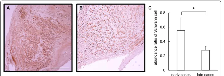
To explore the factors associated with the longer time to improve taste function in later cases than early cases, we analyzed 30 samples of traumatic neuromas fixed by formalin in the immunohistochem-istry using anti-S100β antibody to recognize Schwann cells. Twenty-two of the 30 samples were from early
cases, and eight were from later cases (Fig. 1). The
abundance ratios of Schwann cells in traumatic neur-oma were 55.3 ± 17.6% in the early cases and 27.9 ±
5.5% in the later cases (Fig. 2). The abundance ratio
was significantly lower in later cases compared with early cases (P = 0.002).
Discussion
In previous studies, lingual nerve recovery was asso-ciated with the duration from lingual nerve injury to microneurosurgery, but the methods of evaluation and the criteria of duration from injury to
micro-neurosurgery varied between them (Table 5) [13–
16]. Functional sensory recovery is often evaluated
by 2PD and/or Semmes-Weinstein (SW) tests, and
FSR achievement is defined as proper patient
sensory perception, generally evaluated by 2PD and
allodynia appearance. FSR may therefore better
reflect patient’s sensory function than the results of a functional sensory test only. The patients undergo-ing microneurosurgery within 3 or 6 months after lingual nerve injury was defined as early case in
previous studies (Table 5). Vincent et al. and Renton
et al. indicate that some patients can spontaneously recover the sensory and taste function within 3
months [26, 27]. Thus, we perform
microneurosur-gery after the 3-month follow-up of the lingual nerve injury to exclude the patients who can spon-taneously recover the sensory and taste function. In the current study, the patients undergoing micro-neurosurgery within 6 months after lingual nerve in-jury was defined as early case.
In this study, there were no significant differences in the ratio of achievement of FSR between early and later cases, although the ratio of allodynia ap-pearance before microneurosurgery tended to be lower in early cases than in later cases. Susarla et al. reported that the ratio of FSR achievement was sig-nificantly high in patients who underwent surgery within 3 months of lingual nerve injury. Bagheri et al. also reported that the odds ratio of FSR achievement was significantly high in patients who underwent surgery within 6 months of lingual nerve
injury (Table 6). However, reanalysis of data used by
Bagheri et al. using the chi-square test resulted in a P value of 0.056, and the 95% confidence interval
for the odds ratio was between − 0.01 and 0.18
(Table 6). The sample size in the study of Bagheri
et al. was, however, larger than that of other studies. We calculated the effect sizes of the odds ratio for FSR achievement on the duration from injury to surgery on order to adjust the sample size among the studies, and we compared the difference of odds ratio for FSR among the studies. The effect size re-ported by Susarla et al. was as much as double that
Table 4 Treatment period to improve sensory and taste function in early and later cases
Early cases Later cases
12 months 24 months 12 months 24 months P value FSR achievement 36 5 18 1 0.654 Recovery of taste 25 2 9 6 0.016
Numbers/percentages represent the number and ratio of cases. P values were calculated using Fisher’s exact test
Table 3 Functional sensory and taste recovery in early and later cases at 24 months after surgery
Early cases Later Cases
Improvement No improvement Improvement No improvement P value
FSR achievement 41 (93.2%) 3 (6.8%) 19 (90.5%) 2 (9.5%) 0.655
Recovery of taste 27 (61.4%) 17 (38.6%) 15 (71.4%) 6 (28.6%) 0.586
Numbers/percentages represent the number or ratio of cases. P values were calculated using the Fisher’s exact test
Table 2 Comparison of early and later treatment after lingual nerve injury at 12 months after surgery
Early cases Later cases P value
2PD test (mm) 12.6 ± 4.2 11.1 ± 3.7 0.173
Allodynia appearance 6 (13.0%) 4 (16.7%) 0.857
FSR achievement 38 (82.6%) 18 (75.0%) 0.517
Recovery of taste 25 (54.4%) 9 (37.5%) 0.127
Plus–minus values are means ± SD. P values were calculated using Student’s t test or Fisher’s exact test. In the two-point discrimination test, four patients could not distinguish between two points 20 mm apart. The result of the two-point discrimination test in the four patients was 20 mm
reported by both Bagheri et al. and the current study. The surgery within 6 months after lingual nerve injury may not therefore be fully associated with FSR achievement, although surgery within 3 months of the injury may have some association.
The current study did not show significant improve-ment of sensory or taste functions in early cases at 12 and 24 months after the microneurosurgery (Tables
2 and 3). Focusing on the patients with improving
sensory and taste functions, taste function was recov-ered within 12 months after the microneurosurgery in almost all of the early cases. In contrast, over a year was needed for improvement of taste function in 40% of later cases whose taste function recovered (Table
4). Thus, patients who undergo microneurosurgery
more than 6 months after lingual nerve injury may re-quire more time to improved taste function compared with patients who undergo microneurosurgery within 6 months after lingual nerve injury.
Traumatic neuroma occurs at the proximal end of an injured site because of differentiation and prolifer-ation of Schwann cells [28]. Schwann cells are critical components in nerve regeneration, including promo-tion of axon growth and myelinapromo-tion in damaged
per-ipheral nerves [29]. Hall et al. reported that the no
myelination for long time in absent axon induced apoptosis in Schwann cells, and Schwann cells were
disappeared [30]. In the present study, the number of
Schwann cells in late cases was significantly reduced compared with early cases based on histopathological analysis. The past reports also indicated that the number of Schwann cells was therefore decreased in the traumatic neuromas owing to the delay of the
microneurosurgery [31]. Thus, the reduction of
Schwann cells could suppress the nerve regeneration in sensory or taste function. The differences in the duration from injury to surgery did not affect the ra-tio of improvement of sensory or taste disorder but affect a period to recover taste function in this study. Therefore, the delay of the microneurosurgery after the injury may need more time to recover taste func-tion followed by decreasing the number of Schwann cells. This histological analysis to measure the num-ber of Schwann cells in removed traumatic neuroma may predict the period to recover taste function, and further studies should validate this hypothesis.
Conclusion
Microneurosurgery within 6 months after lingual nerve injury was not associated with improvement of sensory and taste functions. However, a longer period was re-quired to recover taste function in patients undergoing
Table 5 Criteria of surgery and the evaluation methods in previous studies
Author Criteria of period to undergo
surgery after lingual nerve injury
Evaluation methods
Pogrel et al. [2] < 3 Pogrel’s score
Susarla et al. [13] < 3 FSR
Ziccardi et al. [15] < 6 2PD, SW
Mozsary et al. [32] < 6 FSR
Bagheri et al. [14] < 6 MRCS
Robinson et al. [17] None 2PD
Fig. 2 Effect of duration from lingual nerve injury to the microneurosurgery on Schwann cells in traumatic neuroma.
Immunohistochemistry using anti-S100β antibody was performed on traumatic neuroma samples from early cases (a) and later cases (b). The abundance ratio of Schwann cells was significantly lower in the traumatic neuroma samples from later cases (c). Scale bar indicates 500μm. Asterisk indicates significant difference between early and later cases (P < 0.05)
microneurosurgery more than 6 months after lingual nerve injury. This may be partially associated with the decrease in Schwann cells in traumatic neuromas. Clini-cians should inform patients on the possible necessity of 2 years until improvement of taste function if they undergo microneurosurgery more than 6 months after lingual nerve injury.
Abbreviations
2PD:Two-point discrimination; FSR: Functional sensory recovery; SW: Semmes-Weinstein
Acknowledgements
I would like to thank the members in the Department of Anatomy and Cell Biology and Oral and Maxillofacial Surgery at Wakayama Medical University for the technical assistance with the experiments. The study was supported by a grant from the Grant-in-Aid for Scientific Research from Japan Society for the Promotion of Science (No.15 K11318) and scholarship donation from NIPRO Medical Corporation.
We acknowledge the proofreading and editing by Benjamin Phillis at the Clinical Study Support Center at Wakayama Medical University.
Authors’ contributions
TN read and wrote the manuscript. TN, SY, and IT prepared the retrospective data. KT analyzed and interpreted this data. YY revised and corrected the manuscript. YY and SF designed and wrote the entire article. All authors read and approved the final manuscript.
Funding
There is no funding related to this article.
Availability of data and materials Please contact the author for data requests.
Ethics approval and consent to participate
This study was performed in accordance with the Declaration of Helsinki for medical protocols and was approved by the Wakayama Medical University Institutional Review Board (Nos. 1689 and 1698). General consent was given by the patients.
Competing interests
The authors declare that they have no competing interests. Author details
1Department of Oral and Maxillofacial Surgery, Wakayama Medical University,
811-1 Kimiidera, Wakayama, Wakayama 641-8509, Japan.2Department of
Anatomy and Cell Biology, Wakayama Medical University, 811-1 Kimiidera, Wakayama, Wakayama 641-8509, Japan.3Clinical Study Support Center,
Wakayama Medical University, 811-1 Kimiidera, Wakayama, Wakayama 641-8509, Japan.
Received: 23 September 2019 Accepted: 10 December 2019
References
1. Martos-Fernandez M, de-Pablo-Garcia-Cuenca A, Bescós-Atín MS (2014) Lingual nerve injury after third molar removal: unilateral atrophy of fungiform papillae. J Clin Exp Dent; 6(2): e193-e196.
2. Pogrel MA, Renaut A, Schmidt B, Ammar A (1995) The relationship of the lingual nerve to the mandibular third molar region: an anatomic study. J Oral Maxillofac Surg 53:1178–1181
3. Behnia H, Kheradvar A, Shahrokhi M (2000) An anatomic study of the lingual nerve in the third molar region. J Oral Maxillofac Surg 58:649–651 4. Andreasen JO, Laskin DM, Petersen JK (1997) Textbook and color atlas of
tooth impactions: diagnosis, treatment, prevention. Munksgaard, Copenhagen, Denmark, pp 87–126
5. Bataineh AB (2001) Sensory nerve impairment following mandibular third molar surgery. J Oral Maxillofac Surg 59:1012–1017
6. Kim SY, Hu KS, Chung IH, Kim HJ (2004) Topographic anatomy of the lingual nerve and variations in communication pattern of the mandibular nerve branches. Surg Radiol Anat 26:128–135
7. Guerrero ME, Nackaerts O, Beinsberger J, Hoer K, Schoenaers J, Jacobs R (2012) Inferior alveolar nerve sensory disturbance after impacted mandibular third molar evaluation using cone beam computed tomography and panoramic radiography: a pilot study. J Oral Maxillofac Surg 70:2264–2270 8. Kipp DP, Goldstein BH, Weiss WW (1980) Dysesthesia after mandibular third
molar surgery: a retrospective study and analysis of 1,377 surgical procedures. J Am Dent Assoc 100:185–192
9. Mason DA (1988) Lingual nerve damage following lower third molar surgery. Int J Oral Maxillofac Surg 17:290–294
10. Gregg JM (1990) Studies of traumatic neuralgias in the maxillofacial region: surgical pathology and neural mechanisms. J Oral Maxillofac Surg 48:228–237
11. Fujita S, Tojyo I, Yamada M, Go Y, Matsumoto T, Kiga N (2014) Outcome following lingual nerve repair with vein graft cuff: a preliminary report. J Oral Maxillofac Surg; 72: 1433.e1-7.
12. Mackinnon SE (1989) Surgical management of the peripheral nerve gap. Clin Plast Surg 16:587–603
13. Susarla SM, Kaban LB, Donoff RB, Dodson TB (2007) Does early repair of lingual nerve injuries improve functional sensory recovery? J Oral Maxillofac Surg 65:1070–1076
14. Bagheri SC, Meyer RA, Khan HA, Kuhmichel A, Steed MB (2010)
Retrospective measures of microsurgical repair of 222 lingual nerve injuries. J Oral Maxillofac Surg 68:715–723
15. Ziccardi VB, Rivera L, Gomes J (2009) Comparison of lingual and inferior alveolar nerve microsurgery outcomes. Quintessence Int 40:295–301 16. Cornelius CP, Roser M, Ehrenfeld M (1997) Microneural reconstruction after
iatrogenic lesions of the lingual nerve and the inferior alveolar nerve. Critical evaluation. Mund Kiefer Gesichtschir 1:213–223
17. Robinson PP, Loescher AR, Smith KG (2000) A prospective, quantitative study on the clinical outcome of lingual nerve repair. Br J Oral Maxillofac Surg 38:255–263
18. Rasmussen OC (1980) Painful traumatic neuromas in the oral cavity. Oral Surg Oral Med Oral Pathol Oral Radiol Endod 49:191–195
19. Swanson HH (1961) Traumatic neuromas: a review of the literature. Oral Surg Oral Med Oral Pathol Oral Radiol Endod 14:317–326
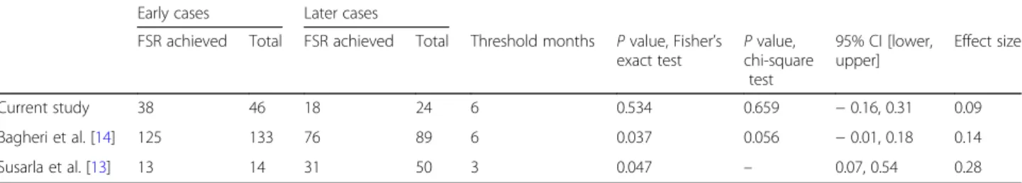
Table 6 Relationship between FSR achievement and the duration from injury to the microneurosurgery in our study and in past studies
Early cases Later cases
FSR achieved Total FSR achieved Total Threshold months P value, Fisher’s exact test P value, chi-square test 95% CI [lower, upper] Effect size Current study 38 46 18 24 6 0.534 0.659 − 0.16, 0.31 0.09 Bagheri et al. [14] 125 133 76 89 6 0.037 0.056 − 0.01, 0.18 0.14 Susarla et al. [13] 13 14 31 50 3 0.047 – 0.07, 0.54 0.28
Effect sizes were calculated with Fisher’s exact test. P value was not calculated with chi-square test in the study by Susarla et al. because the number of patients that did not achieve FSR among the early cases was less than five
20. Seddon HJ (1965) Nerve injuries. J Univ Mich Med Center 31:4–10 21. Swaim SF (1972) Peripheral nerve surgery in the dog. J Am Vet Med Assoc
161:905–911
22. Raffe MR (1979) Compendium on Continuing Education for the Small Animal Practitioner; 1: 269-276.
23. Vora AR, Bodell SM, Loescher AR, Smith KG, Robinson PP, Boissonade FM (2007) Inflammatory cell accumulation in traumatic neuromas of the human lingual nerve. Arch Oral Biol 52:74–82
24. Vora AR, Loescher AR, Craig GT, Boissonade FM, Robinson PP (2005) A light microscopical study on the structure of traumatic neuromas of the human lingual nerve. Oral Surg Oral Med Oral Pathol Oral Radiol Endod 99:395–403 25. Bird EV, Christmas CR, Loescher AR, Smith KG, Robinson PP, Black JA,
Waxman SG, Boissonade FM (2013) Correlation of Nav1. 8 and Nav1. 9 sodium channel expression with neuropathic pain in human subjects with lingual nerve neuromas. Molecular Pain 9:52–62
26. Vincent IN, Miloro M (2013) Trigeminal nerve injuries. Springer-Verlag, Berlin Heidelberg, Chicago, Illinois, USA, pp 149–166
27. Renton T, Yilmaz Z (2012) Managing iatrogenic trigeminal nerve injury: a case series and review of the literature. Int J Oral Maxillofac Surg 41:629–637 28. Kline DG, Nulsen FE (1972) The neuroma in continuity: its preoperative and
operative management. Surg Clin North Am 52:1189–1209 29. Steed MB (2013) Nerve injury and regeneration. IN: Miloro M.
Trigeminal Nerve Injuries. Springer-Verlag Berlin Heidelberg, Chicago, Illinois, USA; 149-166.
30. Hall S (2005) The response to injury in the peripheral nervous system. J Bone Joint Surg 87-B:1309–1319
31. Terenghi G, Calder JS, Birch R, Hall SM (1998) A morphological study of Schwann cells and axonal regeneration in chronically transected human peripheral nerves. J Hand Surg [Br] 23:583–587
32. Mozsary PG (1987) Inferior alveolar and lingual nerve injuries: regeneration or operation? J Oral Maxillofac Surg 45(3):204–286
Publisher’s Note
Springer Nature remains neutral with regard to jurisdictional claims in published maps and institutional affiliations.