†† 주 저자 (e-mail: [email protected]) call: 054-770-2651 †† 교신저자 (e-mail: [email protected]) call: 054-770-2651 †† 교신저자 (e-mail: [email protected]) call: 054-770-2366 †† 교신저자 (e-mail: [email protected]) call: 054-770-2665
Vol. 47, No. 1, March 2021, 23-30 http://dx.doi.org/10.15230/SCSK.2021.47.1.23
자생식물 Essential Oil 5 종의 항 아토피피부염 활성 연구
정 정 화*,†ㆍThao Kim Nu Nguyen**ㆍ최 민 진1ㆍ응 웬 리1ㆍ신 흥 묵2,††ㆍ이 병 욱3,††ㆍ양 인 준2,††
*㈜동제메디칼 **하노이 베트남 국립대학교 미생물공학연구소 1동국대학교 한의학과 생리학교실, 대학원생 2동국대학교 한의학과 생리학교실, 교수 3동국대학교 한의학과 원전의사학교실, 교수 (2020년 11월 13일 접수, 2021년 02월 25일 수정, 2021년 03월 03일 채택)
A Study on the Activities of Five Natural Plant Essential Oils on Atopic Dermatitis
Jeong-Hwa Jeong*,†, Thao Kim Nu Nguyen**, Min-Jin Choi1, Ly Thi Huong Nguyen1,
Heung-Mook Shin2,††, Byung-Wook Lee3,††, and In-Jun Yang2,††
*Dongje Medical, 2nd floor, 46 Jibeom-ro 23-gil, Suseong-gu, Daegu 42196, Korea
1,2Department of Physiology, College of Korean Medicine, Dongguk University, Gyeongju 38066, Korea **Vietnam Type Culture Collection, Institute of Microbiology and Biotechnology, Vietnam National University
3Department of Medical Classics and History, College of Korean Medicine, Dongguk University
(Received November 13, 2020; Revised February 25, 2021; Accepted March 03, 2021)
1)
요 약: 본 연구는 개똥쑥(Artemisia annua L., AA), 유자(Citrus junos Sieb. ex TANAKA, CJ), 산국화(Chrysanthemum boreale Makino, CB), 잣나무(Pinus koraiensis, PK), 금강송(Pinus densiflora for. erecta, PD) 총 5 종의 자생식물 essential oil의 항 아토피 효능을 확인하기 위한 실험이다. 항균 효과를 확인하기 위해 자생식물 essential oil 5 종을 Staphylococcus aureus, Escherichia coli, Pseudomonas aeruginosa, Candida albicans 총 4 종류의 균에 처리했다. 항염증 효과를 확인하기 위해 TNF-α와 IFN-γ (TI)를 처리한 HaCaT 세포에 5 종의 자생식물 essential oil을 처리했다. AA, CJ, CB, PK, PD은 10 mg/mL 농도에서 Candida albicans에 대한 항균 효과를 나타냈다. AA (1 μg/mL), CB (1 μg/mL), PK (0.1, 1 μg/mL)에서 thymus and activation-regulated chemokine (TARC) 생성량이 억제되었고, AA 및 PK (1 μg/mL)에서 macrophage-derived chemokine (MDC) 생성량이 억제되었으며, AA (0.1, 1 μg/mL), PK (0.1, 1 μg/mL)에서 IL-6 생성량이 억제되었다. AA, CB, PK essential oil의 항균 및 항염증 효과가 확인되었으며, 이에 아토피 피부염 완화에 기여할 수 있음을 기대한다.
Abstract: This study is an experiment to evaluate the anti-atopy efficacy of five kinds of natural plant essential oils; Artemisia annua L. (AA), Citrus junos Sieb. ex TANAKA (CJ), Chrysanthemum boreale Makino (CB), Pinus koraiensis (PK), and Pinus densiflora for. erecta (PD). Through Agar diffusion test, five species of native plant essential oils were treated in a total of four strains, Staphylococcus aureus, Escherichia coli, Pseudomonas aeruginosa, and Candida albicans. In order to invest the anti-inflammatory effect, five kinds of natural plant essential oils were treated in HaCaT cells-induced
1. 서 론
아토피피부염(atopic dermatitis, AD)은 피부의 습진과 소 양감을 주요 증상으로 하는 만성 염증성 피부질환이다. 아 토피피부염은 영유아기의 16.3%에서 발병할 정도로 흔한 질병이며, 지난 30 년간 유병률이 지속적인 증가 추세에 있는 것으로 보고되었다[1,2]. 아토피피부염은 피부에 존재 하는 항원 제시 세포(antigen presenting cells, APC)인 Langerhans cell이 집먼지진드기, Staphylococcus aureus (S. aureus), Candida albicans (C. albicans) 등의 항원을 인지하 여 Th2 cell 및 B cell의 면역반응을 유도하여 발생하는 것 으로 알려져 있다. 특히 S. aureus, C. albicans와 같은 균 감염이 발생했을 시 각질 형성 세포에서 pro-inflammatory cytokines (TNF-α, IL-6 등)의 분비가 촉진된다[3-5]. TNF-α 는 IL-6와 함께 B cell의 분화를 촉진하고 IgE의 분비를 증 가시키며 소양증을 유발한다[6,7]. 결과적으로 가려움증, 피부장벽의 손상, 균감염의 증가로 이어지는 악순환의 고 리가 생성되며 병증이 더욱 심해지게 된다[8]. 피부를 구성하는 세포 중 가장 많은 수로 존재하는 각 질형성세포는 외부 자극에 대해 pro-inflammatory cytokines 및 chemokines 분비를 통해 피부 면역반응에 참여한다. 일 반적으로 각질 형성 세포를 이용하여 아토피피부염 in vitro 모델을 구축하기 위해 TNF-α와 IFN-γ를 10 ng/mL씩 처리한다[9,10]. 각질 형성 세포에 TNF-α와 IFN-γ를 동시에 처리함으로써 thymus and activation-regulated chemokine (TARC)와 macrophage-derived chemokine (MDC)와 같은 Th2 attractive chemokine을 생성하여 T cell 표면에 발현된 C-C chemokine receptor type 4 (CCR4)와 작용하여 표적 세 포로 T cell을 이동시킨 후 염증반응을 강화시킨다[10,11]. 아토피피부염 환자의 피부 병변 부위에서 TARC, MDC의 발현이 증가되어 있음을 확인한 연구 결과를 보면, 이들이 아토피피부염의 발생 및 악화에 기여한다고 볼 수 있다 [10]. 따라서 아토피피부염을 효과적으로 치료하기 위해
pro-inflammatory cytokines 생성을 억제하거나, Th2 cell이 피부 병변 부위로 이동하는 것을 억제하는 것이 중요하다. 아토피피부염 치료제로써 다용되는 스테로이드 외용제 의 경우 피부 위축증, 혈관 확장증, 이차 감염 등의 부작용 을 가진다. 이러한 스테로이드제의 사용을 줄이기 위해 보 완 대체 요법을 택한 환자 중 essential oil을 사용한다는 경 우가 22%에 다다랐다[10]. 이에 아토피피부염에 essential oil을 적용하는 것이 효과가 있는지에 대한 과학적 근거를 제시하려는 연구가 활발히 이어져 오고 있으나 자생식물 에 관한 연구는 드물다[12]. Essential oil은 식물에서 추출 한 고농축 된 방향성 오일로 구성 분자들의 크기가 작아 모낭에 흡수되기 용이한 특성을 가진다[13]. Lavandula angustifolia, Melaleuca alternifolia 등의 essential oil은 피부 도포 시 보습, 항염증 및 항균 활성 등을 하는 것으로 보 고되었다[14,15]. 아토피피부염 치료를 목적으로 Cupressus sempervirens L., Chamaecyparis obtusa 등의 essential oil을 활용한 연구 결과가 보고되었으나, 우리나라 자생식물 essential oil의 항 아토피 활성 및 기전에 대한 연구보고는 많지 않다[16,17]. 또한 앞서 언급된 essential oil들은 주로 수입에 의존되어왔고, 이를 극복하고자 자생식물로부터 추 출된 essential oil의 효능 연구를 진행하였다[18]. 이에 저자 는 개똥쑥(Artemisia annua L., AA), 유자(Citrus junos Sieb ex TANAKA, CJ), 산국화(Chrysanthemum boreale Makino, CB), 잣나무(Pinus koraiensis, PK), 금강송(Pinus densiflora for. erecta, PD) 총 5 종의 자생식물 essential oil을 이용하여 각 질 형성 세포 및 C. albicans 균에서 항 아토피 효능과 관 련한 유의한 결과를 얻었기에 보고하는 바이다.
2. 재료 및 방법
2.1. 세포 배양
HaCaT cell을 10% fetal bovine serum (FBS, Merck, Germany)와 1% penicillin/streptomycin (P/S, Invitrogen, USA)
by TNF-α and IFN-γ (TI). AA, CJ, CB, PK and PD showed antibacterial effects on Candida albicans at a concentration of 10 mg/mL. We also found that the thymus and activation-regulated chemokine (TARC) expression was suppressed in 0.1 μg/mL of PK, 1 μg/mL of AA, CB, and PK. macrophage-derived chemokine (MDC) expression was suppressed in 1 μg/mL of AA and PK. IL-6 expression was suppressed in 0.1, 1 μg/mL of AA, PK in HaCaT cells. Hence it suggests that AA, CB, and PK have the anti-inflammatory effects, and it could contribute to atopic dermatitis relief by reducing the infiltration of immune cells to inflamed area.
이 함유된 Dulbecco's Modified Eagle's Medium (DMEM, Welgene, Korea)을 사용하여 37 ℃, 5% CO2 조건에서 배양
하였다. 80 ~ 90% confluence로 자랐을 때 계대 배양하였으 며 각 실험에 알맞은 농도로 분주하여 진행하였다. 2.2. 세포 독성평가
96 well plate (Hyundai micro, Korea) 5 x 10⁴ cells/mL 농
도의 HaCaT cell을 100 μL씩 분주하고 37 ℃, 5% CO2 조
건에서 12 h 배양해 주었다. 배양된 cell에 1, 10, 50, 100 μg/mL의 농도로 essential oil (㈜벤자롱, Korea) 샘플을 처 리한 후 24 h 배양한 뒤 2, 3-bis (2-methoxy-4-nitro-5- sulfophenyl)-2H-tetrazolium-5-carboxanilide (XTT, Roche, Swiss) 시약을 50 μL씩 각 well에 넣고 4 h 동안 CO2 incubator에
서 반응시켰다. 이후 microplate reader (Sunrise-basic, Tecan, Switzerland)를 사용하여 450 - 650 nm에서 흡광도를 측정 하였다. Essential oil 샘플은 oil을 dimethyl sulfoxide (DMSO, Junsei, Japan)에 10 mg/mL 농도로 선 희석한 후 FBS를 첨가하지 않은 DMEM에 10 μg/mL 농도로 재 희석 하여 실험에 사용하였다.
2.3. 염증성 케모카인과 사이토카인 생성 억제능 평가 HaCaT cell을 1 x 10⁶ cells/mL의 농도로 6 well plate
(SPL, Korea)에 분주하고 12 h 배양 후 FBS를 첨가하지 않 은 DMEM으로 교체하여 추가 배양하였다. 60% confluence 로 배양된 cell에 essential oil 샘플을 0.1, 1 μg/mL 농도로 1 h 전처리하고, TNF-α와 IFN-γ (Koma biotech Inc., Korea)를 10 ng/mL 농도로 각각 처리하여 24 h 배양하였다. 24 h 배 양 후 얻은 배지를 100 μL씩 사용하였고, TARC, MDC ELISA kit (R&D Systems, USA), IL-6 ELISA kit (Koma biotech Inc., Korea)를 사용하여 실험을 진행하였다. 이후 microplate reader를 사용하여 TARC, MDC의 경우 450~540 nm, IL-6의 경우 450 nm에서 흡광도를 측정하였다.
2.4. 항균 효과
S. aureus, E. coli, P. aeruginosa (Vietnam Type Culture Collection (VTCC), Vietnam)는 Luria-Bertani (LB) 배지에서 C. albicans (Vietnam Type Culture Collection (VTCC), Vietnam)는 Yeast Malt (YM) 배지에서 배양되었다. LB 배 지의 성분은 yeast extract (Himedia, India) 5 g/L, peptone (Himedia, India) 10 g/L, NaCl (JHD Chemical, China) 10 g/L 으로 구성되었으며, YM 배지는 glucose (JHD Chemical,
China) 10 g/L, peptone 5 g/L, yeast extract 3 g/L, malt (Himedia, India) 3 g/L으로 구성되었다. 각 미생물들은 액 체배지에서 16 h 동안 배양된 후 40 ℃의 agar 배지에 더 해졌고 petri dish에 25 mL 담겨 실온에서 굳혀졌다. 굳혀 진 배지에 5 mm 직경의 홈을 내고, 각 essential oil 샘플들 과 DMSO를 홈에 40 μL씩 넣어 37 ℃에서 24 h 동안 반응 시켰다. 반응 후 홈 주변에 생긴 저해환의 직경을 자로 측 정하여 항균 효과를 확인하였다. 2.5. 통계 결과는 중복하여 수행된 최소 3 개의 독립적 실험의 mean ± standard deviation (SD)로 나타내었다. 그룹 간 비교 는 student’s t-test를 사용하여 수행되었으며, p 값이 0.05 미만인 경우 통계적 유의성이 인정되었다.
3. 결과 및 고찰
3.1. 자생식물 Essential Oil이 HaCaT cell 생존율에 미 치는 영향
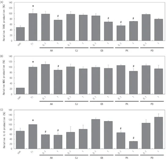
HaCaT cell에서 자생식물 essential oil의 독성을 확인하 기 위해서 XTT assay를 통해 세포생존율을 측정하였다. AA, C J, C B, PK, PD에서 10 μg/mL 이상의 농도 처리 시 80% 이하의 생존율을 보이며, 독성이 확인됐다. 이에 우리 는 세포 독성이 없는 0.1, 1 μg/mL의 농도에서 항염증 실 험을 진행하였다(Figure 1). 3.2. 자생식물 Essential Oil의 항염증 효과 TARC과 MDC는 Th2 cell을 염증 부위로 유도하는 것으 로 알려져 있다. 염증 부위로 유도된 Th2 cell은 IL-6를 분 비하여 B cell 및 mast cell을 활성화시켜 염증을 유도하는 역할을 한다[19]. 따라서 본 연구에서는 TNF-α와 IFN-γ (TI) 처리를 통해 염증 반응을 유도한 HaCaT cell에서 AA, CJ, CB, PK, PD essential oil 처리에 따른 TARC, MDC, IL-6 발현 억제능에 대해 실험하였다. 그 결과 TI 처리 그 룹(100%)에 비해 AA 1 μg/mL, CB 1 μg/mL, PK 0.1, 1 μg/mL 처리 시 TARC 생성량이 각각 24.0, 31.0, 44.6, 29.0%씩 감소하였다(Figure 2A). AA 1 μg/mL에서 TI 처리 그룹에 비해 MDC 생성량이 11.1% 감소했고, PK 1 μg/mL 에서도 15.2% 감소했다(Figure 2B). 또한 AA와 PK 0.1, 1 μg/mL 에서 TI 처리 그룹에 비해 IL-6 생성량이 각각 41.5, 43.4, 33.3, 69.5%의 감소를 보였다(Figure 2C). AA essential
Figure 1. The effect of native plant essential oil on the cell viability in HaCaT cells. Cells were treated with essential oils (concentration is μg/mL) for 24 h, 37 ℃. Viability was measured by XTT assay. Con means control. The result is presented as the mean ± SD of the experiment (N = 3). AA (Artemisia annua L.), CJ (Citrus junos Sieb. ex TAN AKA), CB (Chrysanthemum boreale
Makino), PK (Pinus koraiensis), and PD (Pinus densiflora for. erecta).
Figure 2. The anti-inflammation effect of native plant essential oil. HaCaT cells were pre-treated with essential oils (concentration is μg/mL) for 1 h, 37 ℃, and then treated with TI (concentration is 10 ng/mL) for 24 h, 37 ℃. (A) TARC, (B) MDC, (C) IL-6. Con means control. The result is presented as the mean ± SD of the experiment (N = 3). *p < 0.05 (vs. control), #p < 0.05 (vs. TI). AA (Artemisia annua L.), CJ
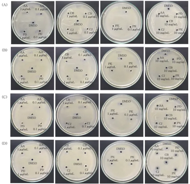
Concentration Strains AA CJ CB PK PD 0.1 1 10 0.1 1 10 0.1 1 10 0.1 1 10 0.1 1 10 S. aureus - - - -E. coli - - - -P. aeruginosa - - - -C. albicans - - 9* - - 9* - - 9* - - 9* - - 9*
-: No inhibition, *: Inhibition zone (Diameter of clear zone, unit: mm)
Table 1. The Anti-Microbial Effect of Native Plant Essential Oil (Oil concentration: 0.1, 1 μg/mL, 10 mg/mL) Staphylococcus aureus
(S. aureus), Escherichia coli (E. coli), Pseudomonas aeruginosa (P. aeruginosa), Candida albicans (C. albicans)
Figure 3. The anti-microbial effect of native plant essential oil. AA (Artemisia annua L.), CJ (Citrus junos Sieb. ex TAN AKA), CB (Chrysanthemum boreale Makino), PK (Pinus koraiensis), and PD (Pinus densiflora for. erecta) (A) S. aureus VTCC 12275, (B) E. coli VTCC 12272, (C) P. aeruginosa VTCC 12273, (D) C. albicansVTCC 40674.
oil의 구성성분은 camphor, germacrene D, trans- pinocarveol, β-selinene, β-caryophyllene 등이 있다[20]. 이 중 β -caryophyllene은 balb/c mice를 이용한 이전 연구에서 피부 조직 내 IL-6 mRNA, serum에서 IL-6 protein 발현량이 감소 하는 결과를 보였다[21]. CB의 구성 성분으로는 α-pinene, β-caryophyllene, caryophyllene oxide, camphor, α-thujone, cis-chrysanthenol 등이 있으며, PK의 구성성분은 α-pinene, β-pinene, β-myrcene, Ɩ-limonene, α-thujene, bornylacetate, epi-bicyclosesquiphellandrene 등이 있다[22,23]. 이 중 공통 성분인 α-pinene은 MAPKs, NF-κB pathway의 억제를 통해 항염증 효과를 나타낸다는 결과가 보고되었다[24]. CB, PK essential oil에서 확인된 TARC 생성 억제는 앞서 보고된 α -pinene의 항염증 기전인 MAPKs, NF-κB pathway에 기인하 는 것으로 예상된다. 따라서 AA, CB, PK가 TARC, MDC, IL-6 생성을 억제하는 것으로 보아 아토피피부염 완화에 기여할 수 있을 것으로 사료된다. 3.3. 자생식물 Essential Oil의 항균 효과 아토피피부염 환자의 병변 부위에서 정상 대조군에서는 분리되지 않았던 S. aureus와 P. aeruginosa가 검출되었으며 아토피피부염 환자의 구강에서는 정상 대조군에서는 분리 되지 않은 S. aureus, P. aeruginosa, E. coli, C. albicans가 검출되었다[25]. 따라서 S. aureus, E. coli, P. aeruginosa, C. albicans 총 4 종류의 균주에서 항균 효능을 확인한 결과를 Table 1에 나타내었다. S. aureus, E. coli, P. aeruginosa에서 는 AA, CJ, CB, PK, PD 모두 저해환이 관측되지 않았다. C. albicans에서는 10 mg/mL의 농도에서만 AA, CJ, CB, PK, PD 모두 지름 9 mm의 저해환을 형성하였고 0.1, 1 μg/mL 농도에서는 저해환이 나타나지 않았다(Figure 3). 저해환 을 형성한 10 mg/mL의 농도에 대해 기존의 논문을 찾아본 결과, C. albicans에 대해 minimum inhibitory concentration (MIC) 가 10 mg/mL로 측정 되었으며 이를 화장품의 천연 보존제 로서 사용 가능함을 제시하는 연구 결과를 확인하였다[26]. 이전의 연구에 따르면 아토피피부염의 악화가 C. albicans 특이적인 IgE 항체의 생산과 연관이 있고 이는 피부뿐 아 니라 구강, 장내에서도 존재하여 끊임없이 면역계를 자극할 수 있다는 연구 결과가 보고되었다[27]. 따라서 AA, CJ, CB, PK, PD가 C. albicans에 대한 항균 효과를 통해 아토피피부 염 완화에 기여할 수 있을 것으로 사료된다.
4. 결 론
아토피피부염 치료제로 주로 사용되는 스테로이드제는 장기간 사용 시 피부 위축증, 혈관 확장증 등 부작용이 나 타날 수 있다. 최근 들어 nanoparticle을 활용한 아토피피부 염 치료제가 주목받고 있으며, essential oil은 구성 분자들 의 크기가 작아 흡수되기 쉽고, 보습·항염증·항균 효과 등 의 기능을 보유하고 있어 이를 통해 아토피피부염 완화 효 과를 보기 위한 연구들이 활발해지고 있다[28,29]. 본 실험 에서 5 종의 자생식물 essential oil (AA, CJ, CB, PK, PD)을 이용하여 항균 효과를 확인한 결과 5종 모두 C. albicans에 서 항균 효과를 보였다. C. albicans는 피부와 점막을 손상 시키기도 하지만 혈액 감염까지 일으킬 수 있는 매우 치명 적인 곰팡이균이다. Lanosterol 14α-demethylase enzyme inhibitor 는 현재까지 흔히 항균제로 사용되는 화합물이지 만, 남용하게 되면 저항성이 생길 뿐 아니라 심각한 감염 의 위험을 증가시킬 수 있기 때문에 새로운 고활성, 저독 성 항진균제를 개발해야 한다[30]. 이전연구에 따르면 4ESW는 Thi5 H66G mutant를 가지고 있는 C. albicans 단백 질이며, 진균의 대사증식에 관여한다고 보고되었다[31,32]. 최근의 Piper betle L., Cleistocalyx operculatus L., Ageratum conyzoides L. essential oil을 이용한 연구에 따르면 chavicol acetate, eugenol acetate, eugenol, caryophyllene oxide, cis-Lanceol, precocene II, precocene I 가 4ESW억제를 통해 C. albicans의 증식을 억제시킬 수 있는 성분으로 확인되었 으며 이 중 caryophyllene oxide는 CB의 구성 성분이기도 하다[32]. HaCaT cell에서 Th2 관련 염증 인자 발현 정도를 확인한 결과 본 실험에서는 AA, CB, PK는 TARC 생성을 억제하였으며, AA, PK는 TARC, MDC, IL-6의 생성을 모 두 억제하는 것을 확인하였다. 따라서 본 연구는 AA, CB, PK의 아토피피부염 완화 소재 물질로 쓰일 수 있음을 제 시하는 바이다.Acknowledgement
본 연구는 2020년도 과학기술정보통신부의 재원으로 한 국연구재단 (NO. NRF-2019R1F1A1059856)과 동국대학교의 지원(2020)을 받아 수행된 연구결과입니다.References
1. S. J. Park, J. S. Lee, K. M. Ahn, and S. J. C hung, The comparison of growth and nutrient intakes in children with and without atopic dermatitis, Korean J. Community Nutr., 17(3), 271 (2012).
2. C. Y. Han, J. G. Park, D. W. Kang, S. Y. Park, B. H. Kim, Y. B. Kim, and K. S. Kim, Analysis of Case Studies of Treating Atopic Dermatitis, J. Korean Med. Ophthalmol. Otolaryngol. Dermatol., 32(3), 151 (2019). 3. U. Wollina, W. Künkel, L. Bulling, C. Fünfstück, B.
Knöll, I. Vennewald, and U. C. Hipler, Candida albicans‐ induced inflammatory response in human keratinocytes, Mycoses, 47(5‐6), 193 (2004).
4. J. B. Travers, A. Kozman, N. Mousdicas, C. Saha, M. Landis, M. Al-Hassani, W. Yao, Y. Yao, A.-M. Hyatt, and M. P. Sheehan, Infected atopic dermatitis lesions contain pharmacologic amounts of lipoteichoic acid, J/ Allergy Clin. Immunol., 125(1), 146 (2010).
5. A. K. Syed, T. J. Reed, K. L. C lark, B. R. Boles, and J. M. Kahlenberg, Staphlyococcus aureus phenol-soluble modulins stimulate the release of proinflammatory cytokines from keratinocytes and are required for induction of skin inflammation, Infect. Immun., 83(9), 3428 (2015).
6. N. Dargahi, J. Johnson, O. Donkor, T. Vasiljevic, and V. Apostolopoulos, Immunomodulatory effects of probiotics: C an they be used to treat allergies and autoimmune diseases?, Maturitas, 119, 25 (2019).
7. A. Vijayaraghava and V. Doreswamy, Exercise and the cytokines-interleukin-6 (IL-6) and tumor necrosis factor-α (TNF-factor-α): A review, Ann. Med. Physiol., DOI, (2017). 8. K. S. Li, Itch in atopic dermatitis: from pathogenesis to
treatment, Allergy Asthma Respir. Dis., 2(1), 8 (2014). 9. Y. Ha, W. H. Lee, J. W. Jeong, M. R. Park, J. Y. Ko,
O. W. Kwon, J. S. Lee, and Y. J. Kim, Pyropia yezoensis Extract suppresses IFN-Gamma-and TNF-alpha- induced proinflammatory chemokine production in HaCaT cells via the down-regulation of NF-κB, Nutrients, 12(5), 1238 (2020). 10. S. Y. Eun, J. J. Yoon, H. Y. Kim, Y. M. Ahn, B. H.
Han, M. H. Hong, C. O. Son, S. W. Na, Y. J. Lee, D. G. Kang, and H. S. Lee, Protective effects of Chijabaegpi-
tang on atopic dermatitis in TNF-α/IFNγ-induced HaCaT cells., J. Physiol. & Pathol. Korean Med., 32(4), 226 (2018). 11. T. W. Song, B. C. Kwon, S. Y. Choi, Y. H. Shin, K. E.
Lee, H. S. Yang, K. W. Kim, E. S. Kim, M. H. Sohn, and K. E. Kim, Increased serum thymus and activation- regulated chemokine (TARC) levels in children with atopic dermatitis, Pediatr. Allergy Respir. Dis., 15(3), 250 (2005).
12. E. J. Jeong and J. D. Kim, A study on the utilization and effect of essential oil, J. Korean Soc. Cosmet. Cosmetol., 1, 93 (2011).
13. R. S. Young and K. K. Ha, Effects of aroma massage on pruritus, skin pH, skin hydration and sleep in elders in long-term care hospitals, J. Korean Acad. Nurs., 43(6), 726 (2013).
14. Y. S. Yoo and M. S. Na, Inhibitory effect on acne using anti-bacteria of lavender essential oil in adolescents, Kor. J. Aesthet. Cosmetol., 8(4), (2010).
15. S. D. Cox, C. M. Mann, J. L. Markham, H. C. Bell, J. E. Gustafson, J. R. Warmington, and S. G. Wyllie, The mode of antimicrobial action of the essential oil of Melaleuca alternifolia (tea tree oil), J. Appl. Microbiol., 88(1), 170 (2000).
16. G. S. Lim, R. Kim, H. Cho, Y. S. Moon, and C. N. Choi, Comparison of volatile compounds of Chamaecyparis obtusa essential oil and its application on the improvement of atopic dermatitis, KSBB J., 28(2), 115 (2013).
17. C. I. Park, A study on the skin protection effects of Cypress essential oil on the DNCB-induced atopic dermatitis in NC/Nga mice, Korea J. Herbol., 32(3), 37 (2017). 18. E. C. Choi, Ph. D. Dissertation, Hoseo Univ., Chungnam,
Korea (2018).
19. R. Y. Kang, B. K. Park, S. B. Gim, H. J. Choi, and D. H. Kim, The effects of HYT on various immunological factors related to pathogenesis of allergic dermatitis in NC/Nga mice induced by Biostir AD, J. Haehwa Med., 18(2), 47 (2009).
20. F. Juteau, V. Masotti, J. M. Bessière, M. Dherbomez, and J. Viano, Antibacterial and antioxidant activities of Artemisia annua essential oil, Fitoterapia, 73(6), 532 (2002). 21. A. F. Bento, R. Marcon, R. C. Dutra, R. F. Claudino, M.
C ola, D. F. P. Leite, and J. B. C alixto, β-Caryophyllene inhibits dextran sulfate sodium-induced colitis in mice through CB2 receptor activation and PPARγ pathway, Am. J. Pathol., 178(3), 1153 (2011).
22. J. M. Lee, S. Han, S. L. Lee, J. Y. Park, H. M. Kim, and S. H. P. Lee, GC/MS analysis of volatile constituents from Zizyphus jujuba var. inermis, Zanthoxylum piperitum, Gardenia jasminoides for. grandiflora, and Pinus koraiensis, Korean J. Hortic. Sci. Technol., 26(3), 338 (2008).
23. K. J. Kim, Y. H. Kim, H. H. Yu, S. I. Jeong, J. D. Cha, B. S. Kil, and Y. O. You, Antibacterial activity and chemical composition of essential oil of Chrysanthemum boreale, Planta Med., 69(03), 274 (2003).
24. D. S. Kim, H. J. Lee, Y. D. Jeon, Y. H. Han, J. Y. Kee, H. J. Kim, H. J. Shin, J. W. Kang, B. S. Lee, and S. H. Kim, Alpha-pinene exhibits anti-inflammatory activity through the suppression of MAPKs and the NF-κB pathway in mouse peritoneal macrophages, Am. J. Chin. Med., 43(04), 731 (2015).
25. M. K. Lee, K. Y. Park, T. W. Jin, W. J. Oh, and S. J. Seo, Distribution of skin and oral microorganisms in atopic dermatitis, Korean J. Dermatol., 54(1), 1 (2016). 26. D. Y. Jang and J. C. Yang, A Study on the evaluation of
antimicrobial activity of extracts from Rhus javanica L fruit, J. Korean Appl. Sci. Technol., 37(1), 145 (2020). 27. E. Morita, M. Hide, Y. Yoneya, M. Kannbe, A. Tanaka,
and S. Yamamoto, An assessment of the role of Candida
albicans antigen in atopic dermatitis, J. Dermatol., 26(5), 282 (1999).
28. A. R. Bilia, C. Guccione, B. Isacchi, C. Righeschi, F. Firenzuoli, and M. C. Bergonzi, Essential oils loaded in nanosystems: a developing strategy for a successful therapeutic approach, Evid. Based Complement. Alternat. Med., 2014, (2014).
29. E. V. Ramos Campos, P. L. F. Proenca, L. Doretto-Silva, V. Andrade-Oliveira, L. F. Fraceto, and D. R. de Araujo, Trends in nanoformulations for atopic dermatitis treatment, Expert Opin. Drug Deliv., 17(11), 1615 (2020).
30. N. Rani, P. Kumar, R. Singh, and A. Sharma, Molecular docking evaluation of Imidazole analogues as potent Candida albicans 14α-Demethylase inhibitors, Curr. Comput. Aided Drug Des., 11(1), 8 (2015).
31. R.-Y. Lai, S. Huang, M. K. Fenwick, A. Hazra, Y. Zhang, K. Rajashankar, B. Philmus, C . Kinsland, J. M. Sanders, and S. E. Ealick, Thiamin pyrimidine biosynthesis in Candida albicans: a remarkable reaction between histidine and pyridoxal phosphate, J. Am. Chem. Soc., 134(22), 9157 (2012).
32. B. T. P. Thuy, T. T. A. My, N. T. T. Hai, H. T. P. Loan, N. T. T. Thuy, N. T. Triet, T. T. Van Anh, N. T. X. Dieu, P. T. Quy, and N. Van Trung, Screening for Streptococcus pyogenes antibacterial and Candida albicans antifungal bioactivities of organic compounds in natural essential oils of Piper betle L., Cleistocalyx operculatus L. and Ageratum conyzoides L., Chem. Pap., 75, 1507 (2020).