박봉욱∙변준호∙이성균∙하영술*∙김덕룡*∙조영철**∙성일용**∙김종렬***
경상대학교 의과대학/의학전문대학원 구강악안면외과학교실, 경상대학교 건강과학연구원, 경상대학교 생명과학연구원,
*경상대학교 의과대학/의학전문대학원 생화학교실, 경상대학교 건강과학연구원, 경상대학교 생명과학연구원
**울산대학교 의과대학 구강악안면외과학교실, ***부산대학교 치과대학 구강악안면외과학교실
배양된 인간 골막기원세포의 조골활성 및 골기질 형성의 평가
EVALUATION OF OSTEOGENIC ACTIVITY AND MINERALIZATION OF CULTURED HUMAN PERIOSTEAL-DERIVED CELLS
Bong-Wook Park, June-Ho Byun, Sung-Gyoon Lee, Young-Sool Hah
*
, Deok-Ryong Kim*
, Yeong-Cheol Cho**
, Iel-Yong Sung**
, Jong-Ryoul Kim***
Department of Oral and Maxillofacial Surgery, College of Medicine and Institute of Health Sciences, Research Institute of Life Science, Gyeongsang National University School of Medicine
*Department of Biochemistry, College of Medicine and Institute of Health Sciences, Research Institute of Life Science, Gyeongsang National University School of Medicine
**Department of Oral and Maxillofacial Surgery, College of Medicine, Ulsan University
***Department of Oral and Maxillofacial Surgery, School of Dentistry, Pusan National University
Autogenous bone grafts have been considered the gold standard for maxillofacial bony defects. However, this procedure could entail a complicated surgical procedure as well as potential donor site morbidity.
Possibly the best solution for bone-defect regeneration is a tissue engineering approach, i.e. the use of a combination of a suitable scaffold with osteogenic cells. A major source of osteogenic cells is the bone mar- row. Bone marrow-derived mesenchymal stem cells are multipotent and have the ability to differentiate into osteoblastic, chondrocytic, and adipocytic lineage cells. However, the isolation of cells from bone mar- row has someproblems when used in clinical setting. Bone marrow aspiration is sometimes potentially more invasive and painful procedure and carries of a risk of morbidity and infection. A minimally invasive, easily accessible alternative would be cells derived from periosteum. The periosteum also contains multipotent cells that have the potential to differentiate into osteoblasts and chondrocytes.
In the present study, we evaluated the osteogenic activity and mineralization of cultured human periosteal-derived cells. Periosteal explants were harvested from mandibule during surgical extraction of lower impacted third molar. The periosteal cells were cultured in the osteogenic inductive medium consist- ing of DMEM supplemented with 10% fetal calf serum, 50g/ml L-ascorbic acid 2-phosphate, 10 nmol dex- amethasone and 10 mM -glycerophosphate for 42 days. Periosteal-derived cells showed positive alkaline phosphatase (ALP) staining during 42 days of culture period. The formation of ALP stain showed its maxi- mal manifestation at day 14 of culture period, then decreased in intensity during the culture period. ALP mRNA expression increased up to day 14 with a decrease thereafter. Osteocalcin mRNA expression appeared at day 7 in culture, after that its expression continuously increased in a time-dependent manner
Abstract
※ 이 논문은 2006년도 경상대학교 학술진흥지원사업 지원 연구비에 의하여 수행되었음 (This work was supported by
the fund of Research Promotion Program (RPP-2006-061), Gyeongsang National University).
Ⅰ. 서 론
기술과 장비의 지속적인 개발로 악안면영역의 골결손에 대하여서는 일반적으로는 자가골이식이 선호되어 왔다. 하 지만 이러한 자가골 이식에는 술후 합병증과 공여부 손상 가능성이라는 피할 수 없는 단점이 나타날 수 있다. 이러한 단점에 따라 최근에는 골신장술이 많이 선호되고 있다. 이 러한 골신장술은 골절단술 후 점진적인 신장을 통한 골조직 의 늘림에 의하여 나타나는 긴장-응력 (tension-stress) 효 과를 통하여 살아있는 골조직에서 세포학적, 그리고 저세포 학적 수준의 반응으로 대사적 활성, 혈관형성, 그리고 본질 적으로 새로운 골이 절단된 골의 가장자리에서 분리된 골편 의 중앙으로 구심성으로 형성되게 하는 것이다. 성공적인 골신장술을 위해서는 골막의 보존이 중요하며 시술시 골막 이 손상될 경우 새로운 골형성이 저하될 수 있다
1-3). 최근 골 조직공학을 통한 골결손의 회복이 상당히 주목받고 있다.
조직공학이란 생명 과학과 공학의 기본 개념과 기술을 통합 응용하여 생체 조직의 구조와 기능 사이의 상관 관계를 이 해하고 응용하여 생체 조직의 대체물을 만들어 이식함으로 써 신체의 기능을 유지, 복원 또는 향상시키는 것을 목적으 로 하는 학문이다. 이러한 면을 고려할 때 상대적으로 골결 손부위와 관련이 적은 부분에서 국소마취하에 적은 양의 골 관련 절편을 채취하여 골관련 세포를 추출하고 이를 증식, 분화시켜 실질 조직을 형성하는 골조직공학을 이용하는 것 이 가장 이상적인 골결손 수복 방법일 것이다
4-8). 골조직공 학을 통한 성공적인 골형성을 위해서 가장 중요한 것은 자 가 골형성 세포를 추출하는 것이다. 현재 이의 원천으로 가 장 잘 알려진 것은 골수이다. 주로 흡입방법 (aspiration) 을 통해 획득되는 골수는 중간엽줄기세포 (mesenchymal stem cells)의 풍부한 근원을 제공한다. 이러한 골수기원줄
cells)는 다능성 (multipotent) 세포로 조골세포, 연골세포 및 지방세포등으로 분화할 수 있는 능력을 가지고 있다. 이 미 여러 연구에서 골수기원줄기세포를 통한 골생성은 만족 할 만한 결과를 산출하고 있다. 그러나 이러한 흡입방법을 통한 골수세포의 흡입은 다소 침습적으로 여겨지고 동통 및 감염가능성과 같은 단점이 존재할 뿐 아니라 임상적으로 이 를 획득하는 것이 불리한 경우가 많다. 그리고 골조직공학 을 통한 골형성이 골수기원줄기세포를 통해서만 이루어지 는 것은 아니다. 외래 환자에서 골결손 부위와 상대적으로 덜 관계된 부위에서 국소마취하에 적은양의 골관련 절편을 쉽게 채취하고 이를 통하여 관련 세포를 추출하고 증식시켜 며 분화시키는 것이 골조직공학의 본연의 장점임을 고려할 때, 흡입방법을 통한 골수기원줄기세포의 채취는 임상적인 면에서 다소 불리한 것이 사실이다. 그리하여 골기원세포를 가지며 치과적으로 국소마취하에 매복치 발치등을 포함한 일반적인 시술을 통하여 쉽게 채취할 수 있는 골막을 이용 하는 것이 골조직공학의 임상적인 면에서 상당히 유리할 수 있다
9-14).
골막 또한 골수와 마찬가지로 조골세포와 연골세포등으로 분화할 수 있는 다능성 세포를 함유하고 있으며 채취된 골 막이 세포배양과정을 위하여 콜라겐분해효소 (collage- nase)에 처리되어도 골막세포의 증식능, 분화능은 유지되 는 것으로 알려져 있으므로 골막기원세포 (periosteal- derived cells)를 통한 조골세포로의 분화는 큰 의미를 가진 다고 할 수 있다
9). 최근 골조직공학에 대한 관심의 증가로 골막기원세포를 통한 조골세포의 형성이 일부 보고되고 있 으나 골막기원세포의 조골세포 분화과정에서 시기별로 나 타나는 조골활성 정도 및 골기질 형성정도는 아직 완전히 알려져 있지 않다. 이에 본 연구의 목적은 골막기원세포의 조골세포로의 분화과정에서 시기별로 나타나는 조골활성 정도와 골기질 형성정도를 평가하고자 한다.
In conclusion, our study showed that cultured human periosteal-derived cells differentiated into active osteoblastic cells that were involved in synthesis of bone matrix and the subsequent mineralization of the matrix. As the periosteal-derived cells, easily harvested from intraoral procedure such as surgical extrac- tion of impacted third molar, has the excellent potential of osteogenic capacity, tissue-engineered bone using periosteal-derived cells could be the best choice in reconstruction of maxillofacial bony defects.
Key words: Periosteal-derived cell, Osteoblastic differentiation, Mineralization
Ⅱ. 연구재료 및 방법
1. 골막기원세포의 추출 및 증식
본 병원의 윤리위원회를 따르고 환자 동의하에 매복된 하 악 제3대구치의 발치과정에서 약 5 × 20 mm의 골막을 채 취하여 몇조각으로 다시 자른다. 이를 100-mm culture dish에 넣은 후 넣은 후 10% fetal bovine serum, 100 IU/mL penicillin, 그리고 100 μg/mL streptomycin이 함유된 Dulbecco’s modified Eagle’s medium (DMEM) 배지에서 37℃, 5% CO
2배양기 (Model 3546, Forma Scientific Inc, OH, USA)를 통하여 배양한다. 약 90%의 세포군집 (confluence)을 나타내면 증식된 세포들을 0.02% 트립신과 0.02% EDTA로 5분간 트립신 처리시키 고 1,500 rpm에서 원심분리하여 계대배양을 실시한다.
2. 골막기원세포의 조골세포로의 분화
Passage 3을 거친 후, 골막기원세포들은 3 × 10
4cells/well의 밀도로 6-well plate에 주입하고 10% fetal calf serum, 50μg/ml
L-ascorbic acid 2-phosphate, 100 nmol dexamethasone, 그리고 10 mM β-glycerophos- phate이 포함된 DMEM로 구성된 골형성 유도 배지에서 6 주동안 배양한다. 50μg/ml
L-ascorbic acid 2-phosphate, 100 nmol dexamethasone, 그리고 10 mM β-glyc- erophosphate이 포함된 골형성 유도 배지를 매 일주일마 다 교체해주며 6주동안 배양하였다.
3. 골막기원세포 표면 표지자 분석 (Cell surface markers analysis)
일차배양 후, 골막기원세포들을 트립신 처리하고 5 × 10
5개 정도의 세포들을 30분간 얼음에 일차항체와 배양시 켰다. CD44, CD90, 그리고 CD166에 대한 Fluorescein isothiocyanate (FITC)-conjugated 항체를 사용하였으며 mouse IgG-FITC를 음성 대조군으로 이용하였으며 그 결 과는 fluorescence-activated cell sorter (FACS) calibur flow cytometer를 통하여 히스토그램 점도 (histogram plot)로 평가하였다.
4. 배양된 골막기원세포의 조골세포관련 유전인자의 발현
골형성 유도 배지에서 7, 14, 21, 28, 35, 그리고 42일의 배양기간동안 골막기원세포의 조골활성 정도를 평가하 였다.
(1) 알칼리성 인산분해효소에 대한 조직화학적 검사 (Histochemical detection of Alkaline phos- phatase, ALP)
인산염 식염수로 세포층을 세척한 후, 3.7% 포름알데히 드와 90%에탄올로 2분간 고정하고 10분간 TBS (Tris Buffer saline)에 세척하였다. 이후 5-bromo-4-chloro-3- indolyl phosphate와 nitroblue tetrazolium (BCIP/
NBT, Amresco, Ohio, USA) 알칼리성 인산분해효소 기 질로 실온에서 10분간 염색하였다.
(2) 알칼리성 인산분해효소와 osteocalcin에 대한 Reverse Transcription-Polymerase Chain Rea- ction (RT-PCR) 분석
총 RNA를 각 주의 세포층에서 TRIzol reagent를 처리하 여 추출하였고 oligo (dT) 시발체 (primer)와 Superscript First-Strand Synthesis System (Invitrogen Life Technologies, CA, USA)을 이용한 역전사반응으로 cDNA를 합성하였다. 적절한 시발체를 이용하여 합성된 cDNA로부터 알칼리성 인산분해효소, osteocalcin 및 GAPDH에 대한 PCR 증폭을 실시하였다. PCR을 위하여 사용체 시발체는 다음과 같다 (sense / antisense) : 5’- CCCTCACACTCCTCGCCCTAT-3’, 5’-TCAGC- CAACTCGTCACAGTCC-3’, 246bp, osteocalcin, 5‘- CCTCCTCGGAAGACACTCTG-3’, 5’-AGACTGCGC- CTGGTAGTTGT-3’, 238bp, 알칼리성 인산분해효소.
RT-PCR 산물은 1.5% 아가로스겔을 사용하여 전기영동으 로 확인하였다. 그리고 이러한 유전인자들의 mRNA 발현 은 음영계측 (Densitometry, Bio-Rad Laboratories, CA, USA)을 통하여 상대적으로 평가하였다.
5. 배양된 골막기원세포의 골기질 형성정도의 평가
골형성 유도 배지에서 7, 14, 21, 28, 35 그리고 42일의 배양기간 동안 골막기원세포의 골기질 형성정도를 평가하 였다.
(1) Von Kossa 염색
무기질이 침착된 기질을 갈색으로 표현되게 하는 Von
Kossa 염색을 통하여 골기질 형성정도를 평가하였다. 배양
된 세포를 인산염 식염수로 세척하고 4% 포름알데히드로
10분간 고정하였다. 증류수로 세척한 후, 5% 질산은 용액
으로 처리하고 30분간 암실에서 보관하였다. 과잉의 질산
은 용액을 증류수로 여러번 세척하고 발색을 위하여 중탄산
나트륨/포름알데히드 용액을 7분간 적용하였다. 5% 티오
황산염나트륨으로 여분의 질산은을 중성화시켰다.
다. 골막기원세포는 중배엽 줄기세포 (mesenchymal stem cells) 표지자로 알려져 있는 CD44, CD90, 그리고 CD166에 양성을 나타내어 배양된 인간 골막기원세포의 표 현형이 중배엽 줄기세포와 유사함을 관찰하였다 (Fig. 1).
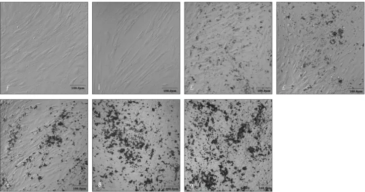
amethasone, 그리고 β-glycerophosphate이 포함된 골형 성 유도 배지에서는 다층구조로 입방형의 세포형태를 나타 냈으며 배양 2주에서 미성숙하나 무기질 결절을 형성하는 것으로 보였다 (Fig. 2).
Fig. 2.Confocal laser microscopy photomicrographs of cultured human periosteal-derived cells. Periosteal-derived cells presented a stretched fibroblastic morphology in culture medium for early passages (A). The morphology of periosteal-derived cellschanged to a cuboidal shaped form for culture times of 7 (B), 14 (C), 21 (D), 28 (E), 35 (F) and 42 days (G) in osteogenic inductive culture medium. At day 14 of culture period, the periosteal-derived cells first seemed to form immature mineralized matrix.
E F G
D
Fig. 1.FACS analysis of cultured human periosteal-derived cells. The gray histograms represent the fluorescence intensity of the cells with negative control mouse IgG-FITC. The green histogramsshow the fluorecence intensity of the cells with each anti- body. These results indicate that the cultured human periosteal-derived cells own the phenotypic characterization of mesenchy- mal stem cells.
A B C
CD44 CD90 CD166
3. 배양된 골막기원세포의 조골세포관련 유전인자의 발현
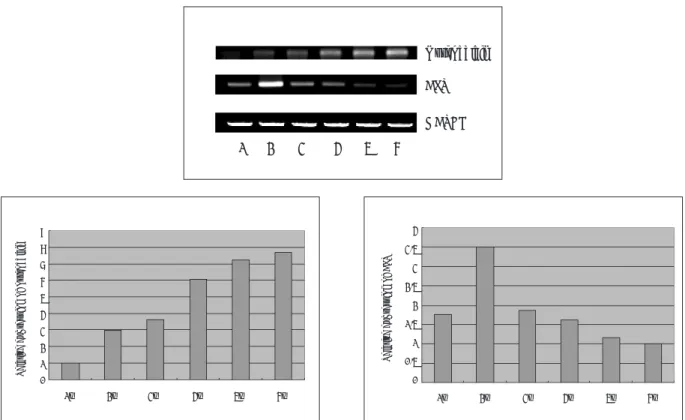
(1) 알칼리성 인산분해효소에 대한 조직화학적 검사 알칼리성 인산분해효소의 발현은 배양 14일까지 증가하 는 양상을 나타내었으며 이후 배양기간동안 이의 발현은 약 간 감소하는 경향을 나타내었다 (Fig. 3).
(2) 알칼리성 인산분해효소와 osteocalcin에 대한 RT- PCR 분석
ALP mRNA 발현은 배양 7일째부터 나타났고 배양 14일
에 가장 강하였으며 이후 이의 발현은 계속 감소하는 경향 을 나타내었다. Osteocalcin mRNA 발현은 배양 7일째에 처음으로 매우 약하게 나타났으며 이후 배양기간동안 지속 적으로 증가하는 경향을 나타내었다 (Fig. 4).
4. 배양된 골막기원세포의 골기질 형성정도
(1) Von Kossa 염색
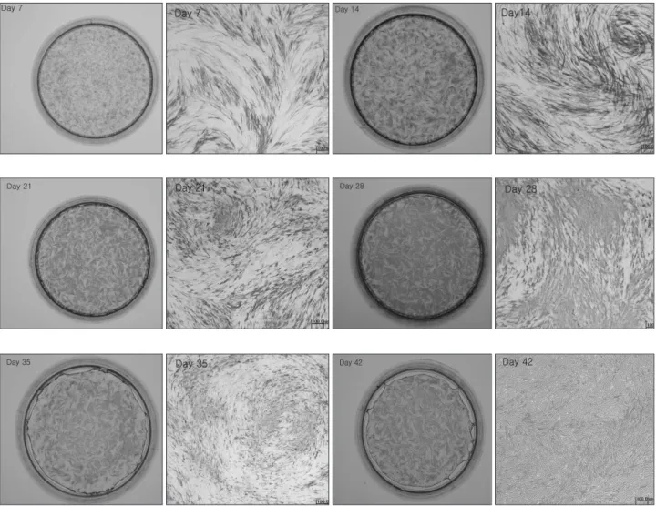
무기질이 침착되어 갈색으로 인기되는 의미있는 골기질 형성은 배양 14일에 나타나 이후 배양기간동안 이의 양상 은 계속적으로 증가하였다 (Fig. 5).
Fig. 3. Histochemical expression of ALP. Periosteal-derived cells stained blue represent positive ALP activity. Periosteal-derived cells showed positive ALP staining during 42 days of culture period. ALP showed intense staining in day 14 of culture period, followed by slightly decreased expression during the culture period in periosteal-derived cells.
Fig. 5. Von Kossa staining for mineralized nodule formation in periosteal-derived cells cultures at sequential time points. The dish covered with brown staining represents mineralized nodule. The first appearance of von Kossa-positive mineralization nodules (arrows) first as indicated day 14 in culture. Subsequently mineralization gradually increased during the entire duration of the culture period.
Fig. 4. Gene expressions in periosteal-derived cells. RNA was isolated from the cultures and after standardization for GAPDH expression, equal amounts of cDNA were subjected to PCR to amplify osteocalcin expression and ALP expres- sion. Lane 1, day 7; lane 2, day 14; lane 3, day 21; lane 4, day 28; lane 5, day 35, lane 6, day 42 in culture.
Quantifications of these genes are expressed as relative mRNA levels by densitometry. Bands with the lowest intensity were regarded as 1.
GAPDH
1 2 3 4 5 6
1w 2w 3w 4w 5w 6w
1w 2w 3w 4w 5w 6w
1w
3w 4w 5w 6w
2w
9 8 7 6 5 4 3 2 1 Relative expression of osteocalcin 0
4 3.5 3 2.5 2 1.5 1 0.5 0
Relative expression of ALP
Ⅳ. 총괄 및 고찰
악안면영역의 골결손에 대하여 거의 최소한의 공여부 손 상을 가지는 조직공학적 해결 방법은 자가골 이식과 비교할 때 분명히 많은 장점을 제공한다. 조직공학적 골형성을 위 하여 현재 골수기원줄기세포, 골막기원세포, 그리고 지방기 원세포 (adipose-derived cells)등과 같은 다양한 세포들이 이용되고 있다
15-18). 일반적으로 조직공학적 골형성에서 가 장 주요한 골세포의 원천은 골수이다. 골수는 골, 연골, 그 리고 지방과 같은 간엽조직을 형성할 수 있는 간엽줄기세포 (mesenchymal stem cells)를 함유하고 있으며 골수기원 줄기세포는 자가 골형성 세포의 우수한 원천을 제공한다.
골수기원줄기세포의 조골세포로의 분화에 대해서는 이미 많은 연구들이 보고되고 있다
10,12-14). 주로 흡입 과정을 통하 여 채취되는 골수기원줄기세포가 그 분화능력으로 인하여 조직공학적 골형성에 있어서 무기질을 침착시킬 수 있는 훌 륭한 원천이 될 수 있지만 이러한 골수기원줄기세포를 얻는 과정이 구강악안면외과의 임상적인 면에서 불리한 점이 있 는 것이 사실이다. 외래 환자에서 골결손 부위와 상대적으 로 덜 관계된 부위에서 국소마취하에 적은양의 골관련 절편 을 쉽게 채취하고 이를 통하여 관련 세포를 추출하고 증식 시켜며 분화시키는 것이 골조직공학의 본연의 장점임을 고 려할 때, 흡입방법을 통한 골수기원줄기세포의 채취는 그 준비과정의 복잡함과 패혈증 및 시술후 감염과 같은 합병증 의 존재가능성으로 임상적인 면에서 유리하지 못한 여건을 제공할 수 있다. 이에 조직공학적 골형성을 위하여 좀 더 쉽 게 접근할 수 있는 원천으로 고려할 수 있는 것이 골막이다.
매복치 발치 등을 포함한 일반적인 시술을 통하여 쉽게 채 취할 수 있는 골막 또한 골수와 마찬가지로 골을 포함하여 여러 가지 간엽조직 세포들로 분화할 수 있는 다양한 전구 세포 (progenitor cells)를 함유하고 있다. 임상적인 면에서 골수기원줄기세포보다 골막기원세포를 사용하는 잇점중의 하나는 그 채취의 쉬움에 있다. 조직공학적 골형성을 통하 여 손상된 골부위를 대치함에 있어서 그 원천이 되는 세포 를 쉽게 채취할 수 있다는 점은 골조직공학의 기본적인 장 점이 될 수 있다. 골막기원세포를 통한 조직공학적 골형성 은 이미 보고되고 있으며 Zhu 등
19)은 성견을 이용한 동물실 험에서 조직공학적 방법을 통한 골형성에 있어서 골막세포 가 가장 훌륭한 원천이 될 수 있다고 하였다. 그리고 인간 골막기원세포를 이용함에 있어서 또 다른 장점으로는 공여 자의 연령이 조골세포로의 기능적 분화에 크게 영향을 미치 지 않으므로 골결손을 나타내는 고연령의 환자에서도 골막 을 이용하여 충분히 자가 조직공학적 골형성이 가능할 수 있다.
본 연구에서 저자들은 배양된 인간 골막기원세포의 조골 세포로의 분화과정동안 골형성 관련인자의 표현형 (pheno-
types)을 관찰하였다. 일반적으로 미분화 상태에서 기능을 나타내는 활동성 조골세포로의 분화과정은 알칼리성 인산 분해효소와 osteocalcin의 발현 및 무기질 침착과 같이 각 각의 단계에서 특정 유전인자들이 관여하는 일련의 과정을 통하여 이루어지며 알칼리성 인산분해효소의 발현은 분화 초기에 나타나며 osteocalcin의 분비 및 무기질 침착은 조 골세포로의 분화 마지막 단계에서 이루어진다. 알칼리성 인 산분해효소는 조골세포에서 형성되는 외효소 (ectoen- zyme)로, 무기성 피로인산염 (inorganic pyrophosphate) 의 분해에 관여하여 무기질 침착을 위한 인산염 혹은 무기 성 피로인산염의 국소적 증가를 이루게 하는 역할을 한다.
일반적으로 알칼리성 인산분해효소는 조골세포의 분화 초 기에 나타나 성숙한 조골세포에서 그 발현의 최고치를 나타 내며 만기 조골세포 혹은 골세포에서는 그 발현이 감소하게
된다
20,21). 성숙한 조골세포의 특이 표지자로 알려져 있는
osteocalcin 또한 골형성에 있어서 중요한 요소로 무기질이
침착되는 단계에서 합성되는 인자로 알려져 있다. 이는 골
에서 발견되는 가장 흔한 비교원질성 단백질로 분자내에 칼
슘과 결합할 수 있는 감마-카르복실글루탐산 (γ-carboxyl-
glutamate)인 Gla잔기를 2~3부위 함유하며 아미노산 49
잔기로 구성되어 있는 비타민 K 의존성 칼슘 결합 단백질이
다. 조골세포에 의해 합성된 후 세포내에서 비타민 K 의존
성 카르복실라아제 (carboxylase)에 의하여 Gla화 되는데
이 Gla화 된 osteocalcin은 골속의 수산화인회석과 결합하
여 골기질 중에 축적되어 국소적으로 석회화를 조절하거나
골과 체액간 칼슘이온의 움직임을 제어하는 등 등 골 대사
와 관련된 중요한 생리적 기능을 통하여 골의 광화 등에 관
여하며 Gla화 되지 않은 osteocalcin은 골기질과의 친화성
이 약하여 혈액으로 방출되게 된다. 그리하여 osteocalcin
의 발현은 골이 석회화되어 결절을 형성하는 시기와 일치하
는 것으로 알려져 있으며 골기질의 석회화를 촉진시키는 요
인으로 작용하여 골 광화 및 재생과 밀접한 관계가 있는 것
으로 알려져 있다
22-25). 성숙한 조골세포에 대한 최종 표지자
는 석회화된 세포외 기질이므로 석회화된 결절 형성정도를
통하여 골막기원세포의 조골세포로의 분화 정도를 평가할
수 있다. 일반적으로 석회화된 골기질을 평가하는데 가장
흔히 사용되는 방법은 Von Kossa 염색 혹은 alizarin red
S 염색으로 알려져 있다. 본 연구에서 알칼리성 인산분해효
소는 6주까지의 배양기간동안 그 발현을 나타내었으며 배
양 14일까지 증가하다가 감소하는 경향을 나타내었고
osteocalcin은 배양 7일째에 처음으로 나타나 이후 지속적
으로 증가하는 경향을 보였다. 배양 14일째에 Von Kossa
양성의 의미있는 석회화된 골결절이 나타나 이후 계속적으
로 침착되는 양상을 나타냈다. 이는 앞서 언급한 알칼리성
인산분해효소와 osteocalcin의 발현에 대한 일반적인 사실
과도 어느 정도 일치하는 경향을 나타내었다.
가 우수한 조골활성을 가지고 있다고 할 수 있을 것이다. 향 후 골조직공학을 이용한 악안면영역의 골결손 치료에 이러 한 골막기원세포는 임상적으로 상당히 유용한 가치를 가질 것으로 생각되며 적절한 담체 (scaffold)와의 적용시 훌륭 한 자가골 이식재의 한 축이 될 수 있을 것으로 사료된다.
Ⅴ. 결 론
본 병원의 윤리위원회를 따르고 환자 동의하에 매복된 하 악 제3대구치의 발치과정에서 5 × 20 mm의 골막을 채취 하여 일차배양 및 계대배양을 실시하고 passage 3을 거친 골막기원세포를 50μg/ml L-ascorbic acid 2-phosphate, 100 nmol dexamethasone, 그리고 10 mM β-glyc- erophosphate이 포함된 DMEM 배지에서 6주동안 배양하 여 다음과 같은 결론을 얻었다.
1. 골막기원세포는 일차배양 및 계대배양을 거치는 동안 섬 유아세포 유사형태를 나타내었으며 이후 L-ascorbic acid 2-phosphate, 100 nmol dexamethasone, 그리 고 10 mM β-glycerophosphate이 포함된 골형성 유도 배지에서는 다층 구조로 입방형의 세포형태를 나타내 었다.
2. 골막기원세포에서 배양 14일까지 알칼리성 인산분해효 소 발현의 증가를 나타났으며 이후 배양기간동안 이의 발현은 감소하는 경향을 나타내었다.
3. 골막기원세포에서 배양 7일에 처음으로 osteocalcin의 발현이 나타났으며, 이후 배양기간동안 이의 발현은 지 속적으로 증가하는 경향을 나타내었다.
4. 골막기원세포에서 무기질이 침착되어 갈색으로 나타나 는 Von Kossa 염색에서 침착된 골기질은 배양 14일에 나타나 이후 배양기간동안 계속적으로 증가하였다.
상기 결론을 통하여 골막으로부터 기원한 골막기원세포는 우수한 골기질 형성정도를 나타냄을 확인하여 향후 악안면 영역의 골결손 치료에 있어서 적절한 담체 (scaffold)와의 적용시 훌륭한 자가골 이식재의 한 축이 될 수 있을 것으로 사료된다.
참고문헌
1. Cillo JE Jr, Gassner R, Koepsel RR et al : Growth factor and cytokine gene expression in mechanically strained human osteoblast-like cells: implications for distraction osteogenesis. Oral Surg Oral Med Oral Pathol Oral Radiol
4. Laurencin CT, Ambrocio AM, Borden MD et al : Tissue engineering: orthopedic applications. Annu Rev Biomed Eng 1 : 19, 1999.
5. Bianoco P, Robey PG : Stem cells in tissue engineering.
Nature 414 : 118, 2001.
6. Spitzer RS, Perka C, Lindenhayn K et al : Matrix engi- neering for osteogenic differentiation of rabbit periosteal cells using alpha-tricalcium phosphate particles in a three- dimensional fibrin culture. J Biomed Mater Res 59 : 690, 2002.
7. Dragoo JL, Samimi B, Zhu M et al : Tissue-engineered cartilage and bone using stem cells from human infrapatel- lar fat pads. J Bone Joint Surg Br 85 : 740, 2003.
8. Leach JK, Mooney DJ : Bone engineering by controlled delivery of osteoinductive molecules and cells. Expert Opin Biol Ther 4 : 1015, 2004.
9. Takushima A, Kitano Y, Harii K : Osteogenic potential of cultured periosteal cells in a distracted bone gap in rab- bits. J Surg Res 78 : 68, 1998.
10. Meirelles Lda S, Nardi NB : Murine marrow-derived mes- enchymal stem cell: isolation, in vitro expansion, and characterization. Br J Haematol 123 : 702, 2003.
11. Hutmacher DW, Sittinger M : Periosteal cells in bone tis- sue engineering. Tissue Eng 9 Suppl : S45, 2003.
12. Kiramura S, Ohgushi H, Hirose M et al : Osteogenic dif- ferentiation of human bone marrow-derived mesenchymal stem cells cultured on alumina ceramics. Artif Organs 28 : 72, 2004.
13. Lee JH, kim IS, Cho TH et al : Problems in osteogenic dif- ferentiation of rat bone marrow stromal cells. J Kor Maxillofac Plast Reconstr Surg 27 : 1, 2005.
14. Lee JH, kim IS, Cho TH et al : The effect of rhBMP-2 in human bone marrow-derived stem cells as osteogenic inducers. J Kor Maxillofac Plast Reconstr Surg 27 : 16, 2005.
15. Meinel L, Karageorgiou V, Fajardo R et al : Bone tissue engineering using human mesenchymal stem cells: effects of scaffold material and medium flow. Ann Biomed Eng 32 : 112, 2004.
16. Sun JS, Wu SY, Lin FH : The role of muscle-derived stem cells in bone tissue engineering. Biomaterials 26 : 3953, 2005.
17. Laino G, Graziano A, d’Aquino R et al : An approachable human adult stem cell source for hard-tissue engineering.
J Cell Physiol 206 : 693, 2006.
18. Dudas JR, Marra KG, Cooper GM et al : The osteogenic potential of adipose-derived stem cells for the repair of rabbit calvarial defects. Ann Plast Surg 56 : 543, 2006.
19. Zhu SJ, Choi BH, Huh JY et al : A comparative qualita- tive histological analysis of tissue-engineered bone using bone marrow mesenchymal stem cells, alveolar bone cells, and periosteal cells. Oral Surg Oral Med Oral Pathol Oral Radiol Endod 101 : 164, 2006.
20. Safadi FF, Xu J, Smock SL et al : Expression of connec- tive tissue growth factor in bone: its role in osteoblast pro- liferation and differentiation in vitro and bone formation in vivo. J Cell Physiol 196 : 51, 2003.
21. Mcduffee LA, Anderson GI : In vitro comparison of equine
cancellous bone graft donor sites and tibial periosteum as sources of viable osteoprogenitors. Vet Surg 32 : 455, 2003.
22. Ducy P, Desbois C, Boyce B et al : Increased bone forma- tion in osteocalcin-deficient mice. Nature 382 : 448, 1996.
23. Lawton DM, Andrew JG, Marsh DR et al : Expression of the gene encoding the matrix gla protein by mature osteoblasts in human fracture non-unions. Mol Pathol 52 : 92, 1999.
24. Mora S, Pitukcheewanont P, Kaufman FR et al : Biochemical markers of bone turnover and the volume and the density of bone in children at different stages of sexual development. J Bone Miner Res 14 : 1664, 1999.
25. Shirakawa Y, Shirahata A, Fukuda M : Differences in reactivity to vitamin K administration of the vitamin K- dependent procoagulant factors, protein C and S, and osteocalcin. Semin Thromb Hemost 26 : 119, 2000.
저자 연락처
우편번호 660-702
경상남도 진주시 칠암동 90번지 경상대학교 의과대학 치과학교실 변 준 호
원고 접수일 2006년 9월 27일 게재 확정일 2006년 11월 1일
Reprint Requests June-Ho Byun
Dept. of OMFS, College of Medicine, Gyeongsang National University.
90 Chilam-dong, Jinju-city, Gyeongsangnam-do, 660-702, South Korea Tel: 82-55-750-8264 Fax: 82-55-761-7024
E-mail: [email protected] Paper received 27 September 2006 Paper accepted 1 November 2006