• 검색 결과가 없습니다.

Surgical Treatment of Mediastinal Schwannoma Showing Malignant Potentiality on Chest Magnetic Resonance Imaging Study : A Case Report

N/A
N/A
Protected

Academic year: 2021

Share "Surgical Treatment of Mediastinal Schwannoma Showing Malignant Potentiality on Chest Magnetic Resonance Imaging Study : A Case Report"

Copied!
3
0
0

로드 중.... (전체 텍스트 보기)

전체 글

(1)

- 70 -

大 韓 氣 管 食 道 科 學 會 誌

□증 례□

Vol.14. No. 2, Dec. 2008

흉부 자기 공명 영상에서 악성 소견을 보인 종격동 신경집종의 수술적 치료 : 1예 보고

인제대학교 의과대학 일산백병원 흉부외과학 교실

1

, 인제대학교 의과대학 일산백병원 해부병리학 교실

2

김 연 수

1

․박 경 택

1

․류 지 윤

1

․김 창 영

1

․장 우 익

1

․장 선 희

2

=Abstract=

Surgical Treatment of Mediastinal Schwannoma Showing Malignant Potentiality on Chest Magnetic Resonance Imaging Study : A Case Report

Yeon Soo Kim, M.D.

1

, Kyoung Taek Park, M.D.

1

, Ji Yoon Ryoo, M.D.

1

, Chang Young Kim, M.D.

1

, Woo Ik Chang, M.D.

1

, Sun Hee Chang, M.D.

2

1

Department of Thoracic and Cardiovascular Surgery, Ilsan Paik Hospital, College of Medicine, Inje University, Ilsan, Republic of Korea

2

Department of Anatomical Pathology, Ilsan Paik Hospital, College of Medicine, Inje University, Ilsan, Republic of Korea

Nerogenic tumor of various histologic types may arise in the posterior mediastinum.

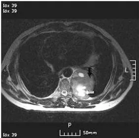
Mediastinal schwannoma is a frequent paraspinal neurogenic tumor, but malignant mediastinal schwannoma is rare tumor which is derived from Schwann cells. Although there are some reports dealing with approach for screening patients with symptoms suggesting malignancy and the imaging criteria for distinguishing malignant from benign schwannoma but the results are not clearly defined. We present a case of hugh mediastinal schwannoma which was taken for malignancy in imaging studies because of its invasiveness.

Key Words : Schwannoma

교신저자 : Yeon Soo Kim , M.D.

Department of Thoracic & Cardiovascular surgery, ilsan paik hospital, 2240, Daehwadong, Ilsanseo- gu, Goyang-si, Gyeonggi-do 411-706, Korea Tel :+82-31-910-7367 Fax :+82-31-910-7614 E-mail : [email protected]

Ⅰ. Case Report

A 56-year-old male patient was referred to our department because of a left hugh mediastinal mass on a chest roentgenogram. Both chest computed tom- ographic scan and magnetic resonance imaging showed a solid, inhomogeneous left posterior mediastinal mass that was 8 cm in diameter, accompanying by irregular 7,8,9th ribs and 8ththoracic vertebrae body erosion.

The mass showed inhomogeneous intermediate signal

intensity on Ti -weighted images(Fig. 1), high inten-

sity on T2-weighted images, and necrotic foci within

it(Fig. 2). Despite the fact that a histologic examin-

ation using percutaneous needle biopsy did not reveal

malignat cells, we suspected the tumor to be malignant

according to its definitely invasive characteristics

revealed on radiologic examinations. Because there

was no evidence of metastasis, we performed surgery

to confirm the diagnosis.At endoscopic first look, the

tumor was well encapsulated and firmly adherant to

the chest wall. The tumor was excised en bloc with

left 7,8,9th ribs. The intercostal nerve was ligated and

resected in the intervertebral foramen between 8th and

(2)

- 71 - Fig. 1. T1-weighted axial MR image shows a mass

lesion with intermediate signal intensity in paraspinal region. Central portion(white arrow) of mass has a somewhat lower intensity than does peripheral portion (black arrow). The mass erodes vertebral body and rib(black ar- row heads).

Fig. 2. T2-weighted axial MR image shows a signal intensity of central portion(white arrow) of mass lesion is higher than that of peripheral portion(black arrow). Per- ipheral portion includes a high signal intensity lesion.

Fig. 3. Specimen is three ribs with attached round mass. The 9x8x5cm sized mass is located in the internal surface of ribs. It is encapsulated and glistening. Cut surface of the mass shows myxoid appearance, necrosis and cystic change.

9ththoracic vertebrae. Chest wall was reconstructed using 2mm GORE-TEX® soft tissue patch securely.

The cut surface of the resected specimen were yellow- brown myxoid appearance with necrotic foci and had the consistency of rubber with a thick capsule(Fig. 3).

Histologic examination and Immunocytochemistry re- vealed benign schwannoma. There was no tumor extension to the margin of resected ribs. The posto- perative course was uneventful and the patient was discharged 17days after surgery

Ⅱ. Review

Intrathoracic neurogenic tumors were reported to

represent 75% to 95% of tumors in the posterior

mediastinum.

1, 2)

Schwannoma is a frequent paraspinal

neurogenic tumor but malignant schwannoma is an

aggressive rare neoplasm and its incidence is about

10% of soft tissue sarcoma. Malignant schwannoma

may manifest as a solitary tumor or may be one

manifestation of neurofibromatosis. The reported

incidences about the latter ranges from 2% to 29%.

3-6)

(3)

- 72 - The prognosisof malignant schwannoma is stern. The 5 year survival of malignant schwannoma combined with neurofibromatosis is as low as 23%, 47% in malignant schwannoma alone by contrast.

7)

At present, surgical indications may be carefully determined in cases with no evidence of metastsis and with imaging that indicate the possibility of malignancy. However, the problem is how to make an accurate preoperative diagnosis of malignant schwannoma. MRI findings have been reported to enable histological comparison of intrathoracic neurogenic tumors. Sakai et al.

reported that typical MRI findings among frequent intrathoracic neurogenic tumors

8)

but it is still que- stionable that MRI has been useful to distinguish benign intrathoracic neurogenic tumor from malignant variant histologically. In our case, en bloc excision seemed feasible as a curative resection because the tumor revealed aggressive behavior with typical MRI findings of schwannoma.

References

1. Davidson KG, Walbaum PR, McCormack RJ. Intr-

athoracic neural tumours. Thorax 1978;33:359–67.

2. Ribet ME, Cardot GR. Neurogenic tumors of the thorax. Ann Thorac Surg 1994;58:1091–5.

3. Crowe FW, Schull WJ, Nee1 JV. A Clinical, Pat- hological, and Genetic Study of Multiple Neurofibro- matosis. Springfield, IL: Charles C. Thomas, 1956.

4. Preston FW, Walsh WS, Clarke TH. Cutaneous neurofibromatosis(von Recklinghausen’s disease):

Clinical manifestations and incidence of sarcoma in 61 male patients. Arch Surg 1952;64:813-827.

5. Brasfield RD, Das Gupta TK. von Reckling- hausen’s disease: A clinicopathological study. Ann Surg 1972;175, 236-104.

6. Maseritz IH. Neurogenic sarcoma. J Bone Joint Surg 1942;24:586-594.

7. Sordillo PP, Helson L, Hajdu SI, et al. Malignant schwammoma-clinical characteristics, survival and response to therapy. Cancer 1981;47:2503-9 8. Sakai F, Sone S, Kiyono K, et al. Intrathoracic

neurogenic tumors: MR-pathologic correlation. AJR

Am J Roentgenol 1992;159:279–83.

수치

Fig. 3. Specimen is three ribs with attached round  mass. The 9x8x5cm sized mass is located in the internal surface of ribs

참조

관련 문서

95 Effects of the heat treatment of the substrate on residual stress distributions for the case of the deposition of multiple lines and layers

Consequently, Zr-Cu binary alloys have the potential to be used as biomaterials with nullifying magnetic properties for magnetic resonance imaging diagnosis and

Purpose: Giant cell tumor of the tendon sheath are the most common tumors after ganglionic cysts in benign soft tissue tumors which could be recurred after surgical

Results : The expression of p21 was increased in boderline serous tumor and serous cystadenocarcinoma in contrast to benign serous tumors. The expression of

근래에 연구되는 격자형 모델은 각 경계범위에서 각기 다른 변수의 영향을 정확 하게 산출하지 못하고 있으나 , 수용모델링을 병행하는 경우 높은 정확도를 추정할

For an electron, the magnetic moment is antiparallel to the orbital angular momentum, but proportional to it. For spin angular momentum, there is a factor 2, which

출처 : IAEA 발표 자료(Comprehensive inspection exercise at bulk handling facilities, “U-235 Enrichment measurements by gamma-ray spectroscopy”) 13.  Uranium

This study aimed to evaluate the site and extent of injury, injury mechanism, player position, and the reinjury incidence in the hamstring by using magnetic