48
한 국 균 학 회 지
The Korean Journal of Mycology
First Report of Metacordyceps chlamydosporia (Cordyceps chlamydosporia) Isolated from Soil in Korea
Hyun Seung Kim
1, Mahesh Adhikari
1, Dil Raj Yadav
1, Sang Woo Kim
1, Yong Hyun Um
1, Hyang Burm Lee
2and Youn Su Lee
1*
1Division of Biological Resource Sciences, Department of Applied Plant Sciences, Kangwon National University, Chuncheon 24341, Korea
2Division of Applied Bioscience and Biotechnology, College of Agriculture and Life Sciences, Chonnam National University, Gwangju 61186, Korea
ABSTRACT : A previously unrecorded species, Metacordyceps chlamydosporia KNU14-22, was isolated from soil in Korea.
Identification of the fungal species was based on morphological and molecular characteristics. This species has not been previously reported in Korea and herein we present data with detailed descriptions and figures.
KEYWORDS : Metacordyceps chlamydosporia, Molecular identification, Morphology
Metacordyceps chlamydosporia is an entomophagous fun- gus belonging to the Clavicipitaceae family of the phylum Ascomycota and the order Hypocreales. Metacordyceps chlamydosporia was previously named Cordyceps chlamy- dosporia [1]. The genus Metacordyceps, which was erected from the genus Cordyceps along with other genera, Ophi- ocordyceps and Elaphocordyceps, contains more than 400 species attacking a range of insects and fungal hosts [2, 3]. Metacordyceps includes species with solitary or several simple and branched stromata, fleshy or tough stipe, par- tially or completely immersed perithecia in stromata, and cylindrical ascospores [3].
Metacordyceps species are particularly abundant and diverse in humid temperate and tropical forests of Asia.
However, a wide range of entomopathogenic fungi are still hidden in the island and mountain regions of Korea.
Ulleung-do, one of Korea’s many islands, is located 150 km
east of the Korean Peninsula. The island is volcanic in origin and has high variation in geographical and ecolo- gical indices, including temperature, altitude, biodiversity, soil physicochemical properties, etc. [4]. These factors contribute to a rich fungal diversity. Thus, examination of the fungal diversity of this solitary area so far away from the main land of Korea is important. The aim of this study was to isolate soil fungi from Ulleung-do, Korea, and confirm the first record of fungal species in Korea.
The current study compares a previously unknown fun- gal species, M. chlamydosporia KNU14-22, with previ- ously described Metacordyceps species with respect to morphological and phylogenetic characteristics.
Soil sample collection and isolation of fungi. Soil samples were collected in 2014 from the forest of Nari Basin, Ulleungdo Island, Korea (37o 31'06.67'' N, 120o 51' 54.27'' E). The samples were taken from a depth of 0~15 cm, air dried, and stored in plastic bags at 4oC until used.
The fungus was isolated using a conventional dilution me- thod [5] and grown on potato dextrose agar (PDA) for 5~7 days at 25oC. The isolate was preserved at 20oC on PDA slants for further studies.
Morphological characterization. The isolate KNU14-22 was cultured on PDA at 25oC for 14 days and colony cha- racteristics including color, shape, and size were recorded.
Photomicrographs were taken with an HK 3.1 CMOS dig- ital camera (KOPTIC, Seoul, Korea) attached to an Olym-
Research Note
*Corresponding author E-mail: [email protected] Received December 3, 2015 Revised December 16, 2015 Accepted December 20, 2015
This is an Open Access article distributed under the terms of the Creative Commons Attribution Non-Commercial License (http://
creativecommons.org/licenses/by-nc/3.0/) which permits unrestricted non-commercial use, distribution, and reproduction in any medium, provided the original work is properly cited.
Kor. J. Mycol. 2016 March, 44(1): 48-50 http://dx.doi.org/10.4489/KJM.2016.44.1.48 pISSN 0253-651X • eISSN 2383-5249
© The Korean Society of Mycology
New Record of Metacordyceps chlamydosporia in Korea
49
pus BX50F-3 microscope (Olympus, Tokyo, Japan) and a scanning electron microscope (LEO Model 1450VP; Carl Zeiss, Oberkochen, Germany). The morphological charac- teristics of the identified species are summarized in Table 1. The colony attained a diameter of 80~90 mm. The col- ony was white to pale white in color, floccose mycelium, margin entire, and lacking exudate (Fig. 1A, 1B). Phiali- des were usually scanty and produced on prostrate aerial hyphae, solitary, sometimes in whorls of 2~3 numbers, tapered towards the tip and 15~25 μm in diameter. Coni- dia were ellipsoidal, smooth walled, one-celled, and 2.5~
3.5 μm in size. Dichlomydospores were abundant, produ- ced in the aerial mycelium, and stalked (Fig. 1C~1F).
Genomic DNA extraction, sequencing, and data ana- lysis. The genomic DNA of the isolate was extracted using the DNeasy Plant Mini Kit (QIAGEN, Germantown, MD, USA) following the manufacturer's instructions. The int- ernal transcribed spacer (ITS) region of rDNA was ampli- fied using the ITS1 and ITS4 primers [6]. The purified DNA was sequenced directly using the Macrogen Sequen- cing Service (Macrogen, Seoul, Korea). The obtained 579- Table 1. Morphological characteristics of Metacordyceps chlamydosporia isolated in this study
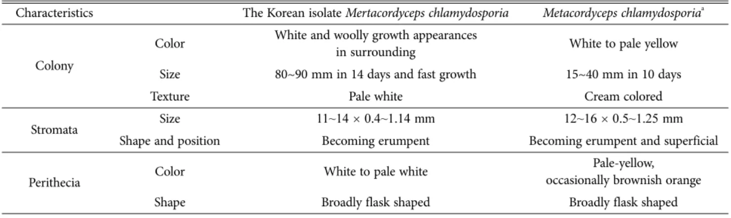
Characteristics The Korean isolate Mertacordyceps chlamydosporia Metacordyceps chlamydosporiaa
Colony
Color White and woolly growth appearances
in surrounding White to pale yellow
Size 80~90 mm in 14 days and fast growth 15~40 mm in 10 days
Texture Pale white Cream colored
Stromata Size 11~14 × 0.4~1.14 mm 12~16 × 0.5~1.25 mm
Shape and position Becoming erumpent Becoming erumpent and superficial
Perithecia Color White to pale white Pale-yellow,
occasionally brownish orange
Shape Broadly flask shaped Broadly flask shaped
aSource of description [1].
Fig. 1. Morphological characterization of Metacordyceps chlamydosporia KNU14-22 using a compound microscope and scanning electron microscope (SEM). A, Colony in front; B, Colony in reverse; C, D, Microscopic pictures of dichlamydospores; E, F, SEM of dichlamydospores.
50
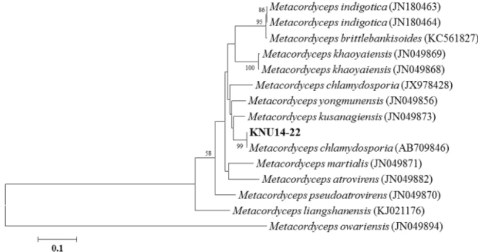
Hyun Seung Kim, Mahesh Adhikari, Dil Raj Yadav, Sang Woo Kim, Yong Hyun Um, Hyang Burm Lee and Youn Su Leebp sequence was deposited in GenBank at the National Center for Biotechnology Information under accession no. KP055595. In a GenBank BLAST search the ITS seq- uence showed 99% similarity with M. chlamydosporia (AB 709846). For phylogenetic analyses, all available ITS rDNA sequences of reference species belonging to the Metacor- dyceps were retrieved from GenBank. M. owariensis (JN 049894) was used as an outgroup taxon. All retrieved se- quences were aligned using the Multalin program. The phylogenetic tree was constructed using neighbor-joining method with Kimura 2-parameter model in MEGA6 soft- ware [7]. Bootstrap analysis was performed with 1,000 replications to determine the support for each clade. The ITS sequence of KNU14-22 matched with M. chlamydo- sporia (AB709846) with 99% similarity (576/576 bp) and clustered together in a clade with a 99% bootstrap value (Fig. 2).
This fungal species is a new record for Korea and pre- vious research results have proved that M. chlamydosporia is pathogenic to nematodes and insects. Thus, further inv- estigations on the biocontrol aspect of the current isolate would be worthwhile.
Acknowledgements
This work was supported by a grant (NIBR2014-01205)
from the National Institute of Biological Resources (NIBR), funded by the Ministry of Environment (MOE) of the Re- public of Korea for projects on the survey and discovery of indigenous Korean fungal species.
REFERENCES
1. Zare R, Gams W, Evans HC. A revision of Verticillium section Prostrata. V. The genus Pochonia, with notes on Rotiferophth- ora. Nova Hedwigia 2001;73:51-86.
2. Kepler RM, Sung GH, Ban S, Nakagiri A, Chen MJ, Huang B, Li Z, Spatafora JW. New teleomorph combinations in the entomopathogenic genus Metacordyceps. Mycologia 2012;104:
182-97.
3. Sung GH, Hywel-Jones NL, Sung JM, Luangsa-Ard JJ, Shres- tha B, Spatafora JW. Phylogenetic classification of Cordyceps and the clavicipitaceous fungi. Stud Mycol 2007;57:5-59.
4. Ryu SH, Jang KH, Choi EH, Kim SK, Song SJ, Cho HJ, Ryu JS, Kim YM, Sagong J, Lee JH, et al. Biodiversity of marine invertebrates on rocky shores of Dokdo, Korea. Zool Stud 2012;51:710-26.
5. Davet P, Rouxel F. Detection and isolation of soil fungi. En- field: Science Publishers; 2000.
6. White TJ, Bruns TD, Lee SB, Taylor JW. Amplification and direct sequencing of fungal ribosomal RNA genes for phylo- genetics. In: Innis MA, Gelfand DH, Sninsky JJ, editors. PCR protocols: a guide to methods and applications. San Diego:
Academic Press; 1990. p. 315-22.
7. Tamura K, Stecher G, Peterson D, Filipski A, Kumar S.
MEGA6: Molecular Evolutionary Genetics Analysis version 6.0. Mol Biol Evol 2013;30:2725-9.
Fig. 2. Neighbor-joining tree of the partial 18S, ITS1-5.8S-ITS2 and partial 28S rDNA sequence of Metacordyceps chlamydo- sporia KNU14-22. The sequence was compared with the reference sequences in NCBI GenBank. The sequences obtained in the present study are shown in boldface. Numerical values (>50) above/below branches are the percentage of 1,000 bootstrap repli- cates that support the branch. Metacordyceps owariensis was used as an outgroup. The scale bar represents the number of sub- stitutions per site.