Korean J Intern Med 2016;31:971-976 http://dx.doi.org/10.3904/kjim.2015.125
INTRODUCTION
Hypouricemia is generally defined as a serum uric acid (SUA) concentration of less than 2.0 mg/dL [1]. It has no recognizable symptoms that require treatment. It is characterized by increased uric acid clearance or de- creased uric acid production [1,2]. While hyperuricemia and gout are significant health concerns and have been the subject of a large number of studies [3,4], less atten- tion has been paid to hypouricemia, even though it is clinically important. Nonetheless, interest in hypourice- mia has begun to increase, due to a better understanding of the role of uric acid transporters. The human urate
transporter 1 (URAT1) and human glucose transporter- like protein 9 (GLUT9) are two representative renal urate transporters [2,5-11]. A genetic defect in these two urate transporters is responsible for idiopathic hypouricemia.
The reported prevalence of hypouricemia has varied in previous studies, depending on the type of study subject and the definition of hypouricemia [5,12-20].
Some studies have reported that it was between 0.15%
and 3.30% in the general population and in outpatients and between 1.24% and 2.54% in hospitalized patients.
A higher prevalence of hypouricemia has been found among inpatients than among outpatients. In addi- tion to a genetic defect of the renal urate transporter,
1Department of Internal Medicine, Keimyung University Dongsan Medical Center, Daegu; 2Department of Rheumatology, Hospital for Rheumatic Diseases, Hanyang University College of Medicine, Seoul, Korea
Received : April 30, 2015 Revised : June 4, 2015 Accepted : June 25, 2015 Correspondence to Jae-Bum Jun, M.D.
Department of Rheumatology, Hospital for Rheumatic Diseases, Hanyang University College of Medicine, 222-1 Wangsimni-ro, Seongdong-gu, Seoul 04763, Korea Tel: +82-2-2290-9216
Fax: +82-2-2298-8231
E-mail: [email protected]
Background/Aims: We aimed to investigate the prevalence and possible causes of hypouricemia in the Korean population and to compare our findings with pub- lished results of other populations.
Methods: We examined the serum uric acid levels of 30,757 subjects who had their uric acid values measured at least once during a 1-year period. All individuals with hypouricemia (serum uric acid < 2.0 mg/dL, n = 424) were reviewed with re- spect to medical drug history and concomitant diseases previously identified as being associated with hypouricemia.
Results: The prevalence of hypouricemia was 4.14% (299/7,223) among inpa- tients and 0.53% (125/23,534) among outpatients, for an overall prevalence of 1.39%
(424/30,757). Possible causes associated with hypouricemia were found to be solid or hematologic malignancies (n = 86), diabetes mellitus (n = 56), and therapeutic drugs (n = 29). The medications were allopurinol (n = 11), angiotensin II receptor blockers (n = 10), salicylates (n = 6), febuxostat (n = 1), and warfarin (n = 1). In the remaining 226 individuals, the cause of hypouricemia was not identified.
Conclusions: Hypouricemia is relatively common in the Korean population com- pared to those of other countries. The possible causes associated with hypourice- mia are related to underlying diseases and medications.
Keywords: Hypouricemia; Uric acid
Prevalence and possible causes of hypouricemia at a tertiary care hospital
Chang-Nam Son1, Ji-Min Kim1, Sang-Hyon Kim1, Soo-Kyung Cho2, Chan-Bum Choi2, Yoon-Kyoung Sung2, Tae-Hwan Kim2, Sang-Cheol Bae2, Dae-Hyun Yoo2, and Jae-Bum Jun2
enhanced uric acid excretion due to medical diseases or medications can also result in hypouricemia [1].
The prevalence of hypouricemia has not been investi- gated previously in Korea and few studies have focused on the factors affecting hypouricemia in the Korean population. Therefore, we aimed to investigate the prev- alence and possible causes of hypouricemia in the Ko- rean population and to compare our findings with those reported for other populations.
METHODS
Population sampleA total of 48,613 SUA values were obtained from patients who visited a tertiary care hospital for medical treat- ment or a checkup during the period January to Decem- ber 2012. In these 12 months 30,757 patients received at least one SUA test, and 7,223 patients had the test while they were hospitalized. Although 24,970 subjects visit- ed the outpatient clinic, the number of outpatients was calculated as 23,534 because those who had already had an SUA test while hospitalized during that period were excluded if they had the test again as outpatients. If a patient had multiple SUA values during the test period, if at least one of those values was less than 2.0 mg/dL, he/she was classified in the hypouricemia patient group.
The total number of hypouricemia patients was 424.
SUA levels of the study subjects
The medical information team at the hospital used a computer program to record the patients’ SUA levels.
Hypouricemia was identified as cases in which the SUA level was less than 2.0 mg/dL [1,19]. An average of each patient’s SUA values was taken as a representative SUA value for each patient. However, in the hypouricemia group, if a patient only had one SUA value that was less than 2.0 mg/dL, that measurement was taken as his/her representative value. Moreover, in cases where there were more than two values of SUA less than 2.0 mg/dL, the values exceeding 2.0 mg/dL were excluded, and only those less than 2.0 mg/dL were used for calculating the average to obtain the representative value.
Review of possible causes of hypouricemia
The hospitalization records, outpatient medication re-
cords, and underlying diseases of the subjects with hy- pouricemia were assessed for the 3 months before the occurrence of hypouricemia. If there were recorded medications from another hospital, they were included as etiologies, and the most probable etiologies were se- lected.
Statistical analysis
Continuous variables, such as the representative SUA levels, are expressed as the mean ± standard deviation.
Categorical variables, such as the cause of hypouricemia, are shown as frequencies. Student t test was used to ex- amine differences in the SUA levels of the subjects with hypouricemia. A p value of less than 0.05 was considered statistically significant. All statistical analyses were per- formed using SPSS version 20.0 (IBM Co., Armonk, NY, USA).
Ethics statement
This study was approved by the Institutional Review Board of Hanyang University Hospital. Informed con- sent from the patients was not required because we ex- amined the data retrospectively from medical records and de-identified it after collection to ensure patient confidentiality.
RESULTS
Characteristics of the study subjects
Among the 30,757 subjects, 57.6% were female (n = 17,725) and 42.4% were male (n = 13,032). The total mean age was 50.8 ± 15.7 years; the mean age of the female subjects was 51.5 ± 15.5 years and the mean age of the male subjects was 49.9 ± 16.0 years. Mean SUA level was 5.16 ± 1.62 mg/
dL; the mean SUA level of the male subjects (6.05 ± 1.60 mg/dL) was higher than that of the female subjects (4.50
± 1.27 mg/dL, p < 0.05).
Prevalence of hypouricemia
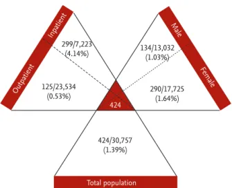
The prevalence of hypouricemia among all of the sub- jects was 1.39% (424/30,757) (Fig. 1). Among the 424 sub- jects with hypouricemia, 68.4% (n = 290) were female and 31.6% (n = 134) were male. The prevalence of hypourice- mia was 4.14% (299/7,223) among inpatients and 0.53%
(125/23,534) among outpatients, and 1.03% (134/13,032) in
male patients and 1.64% (290/17,725) in female patients.
Possible causes associated with hypouricemia
Table 1 presents the possible causes of hypouricemia and their rates among the patients enrolled in the study.
The possible causes were identified as solid or hema- tologic malignancies (n = 86), diabetes mellitus (n = 56), and therapeutic drugs (n = 29). The medications were allopurinol (n = 11), angiotensin II receptor blockers (n = 10), salicylates (n = 6), febuxostat (n = 1), and warfarin (n = 1). In the remaining 226 individuals, the cause of hypou- ricemia was not identified.
DISCUSSION
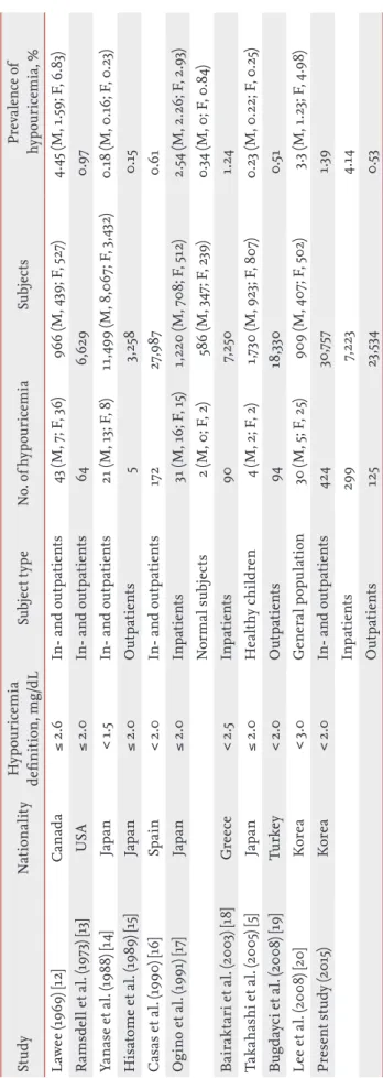
In the present study, the prevalence of hypouricemia among Koreans was relatively high, and it was detected in inpatients as well as outpatients and healthy groups [12-19]. As in other studies, the prevalence of hypouri- cemia was higher in inpatients than in outpatients or healthy people [15,17-19]. According to data collected in studies that used a similar definition of hypouricemia, the total prevalence rate of hypouricemia among west- erners varied from 0.61% to 0.97% [13,16]. In Japan, the prevalence rate for outpatients and healthy individuals ranged between 0.15% and 0.34%, whereas that of inpa- tients was 2.54% [15,17]. In our study, the total prevalence
rate was 1.39%, which is higher than that of westerners;
the prevalence rate of outpatients and checkup patients was 0.53%, and that of inpatients was 4.14%. The prev- alence rates were higher compared to those reported for Japanese patients. The prevalence rates are sum- marized in Table 2. In a previous study, the prevalence of hypouricemia among 909 healthy Koreans was 3.3%
[20]. However, the criterion for hypouricemia in that study was less than 3.0 mg/dL, so the results cannot be compared with those of our study. Applying the defini- tion of an SUA value of less than 3.0 mg/dL in our data analysis, the prevalence rate was 7.66% (2,355/30,757), and even when comparing only the outpatients and check-up patients, it was as high as 5.23% (1,306/24,971).
The prevalence was higher among women than among men [12,14,17,20]. This finding is natural, because the number of medications or diseases that might trigger hypouricemia in inpatients is greater [18], and it is well known that the SUAs of premenopausal women are low- er than men’s [21]. The cause of the higher prevalence of hypouricemia among Koreans than among other ethnicities is not clear. Although the exact reason is un- known, it should be associated with genetic differences, body weight, and dietary habits. Nevertheless, given the prevalence rate, more research and attention must be Table 1. Possible causes associated with hypouricemia in Korean population
Possible causes No. (%)
Neoplasia 86 (43.4)
Solid 79 (39.9)
Hematologic 7 (3.5)
Diabetes mellitus 56 (28.3)
Therapeutic drugs 29 (14.6)
Allopurinol 11 (5.6)
Angiotensin II receptor blockers 10 (5.1)
Salicylates 6 (3.0)
Febuxostat 1 (0.5)
Warfarin 1 (0.5)
Intracranial disease 18 (9.1)
Obstructive jaundice 7 (3.5)
Renal tubular acidosis 1 (0.5)
Ulcerative colitis 1 (0.5)
Total 198
Figure 1. The prevalence of hypouricemia in total popula- tion, male, female, inpatient, and outpatient.
299/7,223
(4.14%) 134/13,032
(1.03%)
290/17,725 (1.64%)
424/30,757 (1.39%) 125/23,534
(0.53%)
424
Total population
Female Male
Outpatient Inpatient
applied to hypouricemia, considering the great deal of interest in hyperuricemia.
Malignancy ranked first as a possible etiology of hy- pouricemia [22,23], followed by diabetes mellitus and concomitant medication [24,25]. The rate of obstructive jaundice or liver disease as etiologies was relatively low [19]. These results are consistent with previous research outcomes. In order to differentiate hypouricemia, a frac- tional excretion of uric acid (FEUA) is necessary to evalu- ate renal tubular defects [1,2]. When FEUA is over 10%, it is considered that the subject has pathologic uricosuria.
When FEUA decreases, it means that uric acid produc- tion has declined, while the reason FEUA increases is a decrease in reabsorption or an increase in the renal excretion of uric acid. When hypouricemia is detected in a hospital, efforts should be made to find the cause.
Whether there is another disease causing it should also be checked; hypouricemia can be a tool for finding un- derlying disease.
Hypouricemia patients are known to have no symp- toms, so any symptoms that appear are those of a caus- ative disease of hypouricemia [1]. According to reports on idiopathic renal hypouricemia (iRHUC), patients exhibits exercise-induced acute renal failure, urolithia- sis, and hematuria along with fatigue, nausea, vomiting, and diffuse abdominal discomfort as causative diseases of hypouricemia [1,2,10,26]. Uric acid is a strong anti- oxidant, and anaerobic exercise increases oxygen-free radicals, which then trigger kidney vasoconstriction and reduce the glomerular filtration rate [27]. In Japan, it is argued that iRHUC can be found by testing SUA before the initiation of a physical education class [26]. In or- der to diagnose iRHUC, hypouricemia and increased FEUA should first be checked [2]. There are two differ- ent types of genetic defects of the renal urate transporter [10,28]. The first type is a defect of the SLC22A12 gene encoding in URAT1 [29], and the second type is a defect of the SLC2A9 gene encoding GLUT9 [30]. Among the 424 subjects with hypouricemia in our study, 226 cases were of an unknown cause. Unfortunately, only a little FEUA was measured during the measurement of SUA, and therefore, the possibility of iRHUC was not verified.
However, it was possible in a considerable number of cases. Therefore, when the cause of hypouricemia is un- clear, efforts to find iRHUC through FEUA and genetic testing should be made.
Table 2. The prevalence of hypouricemia, data from previous published studies StudyNationalityHypouricemia definition, mg/dLSubject typeNo. of hypouricemiaSubjectsPrevalence of hypouricemia, % Lawee (1969) [12]Canada≤ 2.6In- and outpatients43 (M, 7; F, 36)966 (M, 439; F, 527)4.45 (M, 1.59; F, 6.83) Ramsdell et al. (1973) [13] USA≤ 2.0In- and outpatients646,6290.97 Yanase et al. (1988) [14]Japan< 1.5In- and outpatients21 (M, 13; F, 8)11,499 (M, 8,067; F, 3,432)0.18 (M, 0.16; F, 0.23) Hisatome et al. (1989) [15]Japan≤ 2.0Outpatients53,2580.15 Casas et al. (1990) [16]Spain< 2.0In- and outpatients17227,9870.61 Ogino et al. (1991) [17]Japan≤ 2.0Inpatients31 (M, 16; F, 15)1,220 (M, 708; F, 512)2.54 (M, 2.26; F, 2.93) Normal subjects2 (M, 0; F, 2)586 (M, 347; F, 239)0.34 (M, 0; F, 0.84) Bairaktari et al. (2003) [18]Greece< 2.5Inpatients 907,2501.24 Takahashi et al. (2005) [5]Japan≤ 2.0Healthy children4 (M, 2; F, 2)1,730 (M, 923; F, 807)0.23 (M, 0.22; F, 0.25) Bugdayci et al. (2008) [19]Turkey< 2.0Outpatients9418,3300.51 Lee et al. (2008) [20]Korea< 3.0General population30 (M, 5; F, 25)909 (M, 407; F, 502)3.3 (M, 1.23; F, 4.98) Present study (2015)Korea< 2.0In- and outpatients42430,7571.39 Inpatients2997,2234.14 Outpatients12523,5340.53
The present study had some limitations. As men- tioned earlier, an investigation of test results in a single medical institution was conducted. While a previous study of Koreans involved 30 hypouricemia patients, this study is meaningful because we analyzed the prevalence and causes of this disease with a large number of sub- jects. Second, there might be information missing, such as height, weight, and renal function, because this study only investigated medical records of medication and co- morbidities to determine the causes of hypouricemia.
Interviews with patients, FEUA, and genetic testing will all lead to a more precise analysis. Lastly, comparisons with other ethnicities with respect to the prevalence of hypouricemia and its causes require caution in inter- pretation, because the results might differ according to the prevalence of underlying diseases and the frequency of using medication.
In conclusion, hypouricemia is relatively common in the Korean population. The possible causes associated with hypouricemia are related to underlying diseases and medications. In addition, idiopathic hypouricemia seems not to be rare.
Conflict of interest
No potential conflict of interest relevant to this article was reported.
Acknowledgments
This work was supported by the research fund of Han- yang University (HY-2012-MC).
REFERENCES
1. Esparza Martin N, Garcia Nieto V. Hypouricemia and tu- bular transport of uric acid. Nefrologia 2011;31:44-50.
2. Sebesta I, Stiburkova B, Bartl J, et al. Diagnostic tests for primary renal hypouricemia. Nucleosides Nucleotides Nucleic Acids 2011;30:1112-1116.
3. Kim HS. Carpal tunnel syndrome caused by tophaceous gout. Korean J Intern Med 2014;29:544-545.
4. Park JW, Ko DJ, Yoo JJ, et al. Clinical factors and treat- ment outcomes associated with failure in the detection of urate crystal in patients with acute gouty arthritis. Korean J Intern Med 2014;29:361-369.
5. Takahashi T, Tsuchida S, Oyamada T, et al. Recurrent URAT1 gene mutations and prevalence of renal hypouri- cemia in Japanese. Pediatr Nephrol 2005;20:576-578.
6. Cheong HI, Kang JH, Lee JH, et al. Mutational analysis of idiopathic renal hypouricemia in Korea. Pediatr Nephrol 2005;20:886-890.
7. Ichida K, Hosoyamada M, Hisatome I, et al. Clinical and molecular analysis of patients with renal hypouricemia in Japan-influence of URAT1 gene on urinary urate ex- cretion. J Am Soc Nephrol 2004;15:164-173.
8. Iwai N, Mino Y, Hosoyamada M, Tago N, Kokubo Y, En- dou H. A high prevalence of renal hypouricemia caused by inactive SLC22A12 in Japanese. Kidney Int 2004;66:935- 944.
9. Hamajima N, Naito M, Hishida A, Okada R, Asai Y, Wakai K. Serum uric acid distribution according to SLC22A12 W258X genotype in a cross-sectional study of a general Japanese population. BMC Med Genet 2011;12:33.
10. Shen H, Feng C, Jin X, et al. Recurrent exercise-induced acute kidney injury by idiopathic renal hypouricemia with a novel mutation in the SLC2A9 gene and literature review. BMC Pediatr 2014;14:73.
11. Jeannin G, Chiarelli N, Gaggiotti M, et al. Recurrent exer- cise-induced acute renal failure in a young Pakistani man with severe renal hypouricemia and SLC2A9 compound heterozygosity. BMC Med Genet 2014;15:3.
12. Lawee D. Uric acid: the clinical application of 1000 unso- licited determinations. Can Med Assoc J 1969;100:838-841.
13. Ramsdell CM, Kelley WN. The clinical significance of hy- pouricemia. Ann Intern Med 1973;78:239-242.
14. Yanase M, Nakahama H, Mikami H, Fukuhara Y, Orita Y, Yoshikawa H. Prevalence of hypouricemia in apparently normal population. Nephron 1988;48:80.
15. Hisatome I, Ogino K, Kotake H, et al. Cause of persistent hypouricemia in outpatients. Nephron 1989;51:13-16.
16. Casas E, Serrano C, Daimiel E, Michan A, Mateos F, Gar- cia Puig J. Prevalence, physiopathology and processes as-
KEY MESSAGE
1. Hypouricemia is relatively common in the Ko- rean population compared to those of other countries.
2. The possible causes associated with hypourice- mia are related to underlying diseases and med- ications.
sociated with hypouricemia in a hospitalized population:
analysis of 27,987 analytic determinations. Rev Clin Esp 1990;186:211-215.
17. Ogino K, Hisatome I, Saitoh M, et al. Clinical signifi- cance of hypouricemia in hospitalized patients. J Med 1991;22:76-82.
18. Bairaktari ET, Kakafika AI, Pritsivelis N, et al. Hypouri- cemia in individuals admitted to an inpatient hospi- tal-based facility. Am J Kidney Dis 2003;41:1225-1232.
19. Bugdayci G, Balaban Y, Sahin O. Causes of hypouricemia among outpatients. Lab Med 2008;39:550-552.
20. Lee JH, Choi HJ, Lee BH, et al. Prevalence of hypouricae- mia and SLC22A12 mutations in healthy Korean subjects.
Nephrology (Carlton) 2008;13:661-666.
21. Johnson RJ, Rideout BA. Uric acid and diet: insights into the epidemic of cardiovascular disease. N Engl J Med 2004;350:1071-1073.
22. Lesmes A, Diaz-Curiel M, Castrillo JM. Tumoural hy- pouricemia. Adv Exp Med Biol 1980;122A:145-148.
23. Kelly WN. Hypouricemia. Arthritis Rheum 1975;18(6 Sup- pl):731-737.
24. Shichiri M, Iwamoto H, Shiigai T. Diabetic renal hypouri-
cemia. Arch Intern Med 1987;147:225-228.
25. Bo S, Cavallo-Perin P, Gentile L, Repetti E, Pagano G.
Hypouricemia and hyperuricemia in type 2 diabetes: two different phenotypes. Eur J Clin Invest 2001;31:318-321.
26. Nakamura A, Niimi R, Yanagawa Y. Renal hypouricemia in school-aged children: screening of serum uric acid level before physical training. Pediatr Nephrol 2006;21:1898- 1900.
27. Ames BN, Cathcart R, Schwiers E, Hochstein P. Uric acid provides an antioxidant defense in humans against oxi- dant- and radical-caused aging and cancer: a hypothesis.
Proc Natl Acad Sci U S A 1981;78:6858-6862.
28. Sebesta I, Stiburkova B. Purine disorders with hypourice- mia. Prilozi 2014;35:87-92.
29. Stiburkova B, Sebesta I, Ichida K, et al. Novel allelic vari- ants and evidence for a prevalent mutation in URAT1 causing renal hypouricemia: biochemical, genetics and functional analysis. Eur J Hum Genet 2013;21:1067-1073.
30. Vitart V, Rudan I, Hayward C, et al. SLC2A9 is a newly identified urate transporter influencing serum urate concentration, urate excretion and gout. Nat Genet 2008;40:437-442.