약학회 지 제 45 권 제 1 호 71-77(2001) Yakhak Hoeji Vol. 45, No. 1
口
f
우스에서VEGF
발현Naked DNA
벡터인pCK-VEGF
의약동력학 및 조직내 분포
도현미 ■ 고준일 • 이종진 • 손미원 • 조홍찬* • 김종묵* • 김병문 • 김선영#
동아제약 (주) 연구소, *(주) 바이로메드, #서울대학교 생명과학부 (Received October 31,2000; Revised December 8, 2000)
Pharmacokinetics and Biodistribution in Mice of pCK-VEGF Expressing Human Vascular Endothelial Growth Factor
Hyounmie Doh, Jun-IL Ko, Jong-Jin Lee, Miwon Son, Hongchan Cho*, Jong-Mook Kim *, Byong-Moon Kim and Sunyoung Kim #
Research Laboratories of Dong-A Pharmaceutical Co., Ltd,
*ViroMed Limited
#School of Biological Sciences, Seoul National University
Abstract —— We recently developed a high efficiency expression vector, pCK, which drives a high level of gene expression in the skeletal muscles of mice. In this study, we investigated the pharmacokinetics and biodistribution of pCK-VEGF expressing human VEGF165 after intravenous or intramuscular admin
istration. The quantity of pCK-VEGF in the tissues of mice was measured by the PCR method which has a detection limit of approximately 1 pg of the exogenously added plasmid. In the case of intravenous admin
istration, the half life of the pCK-VEGF plasmid in the bloodstream was 1.68 min. After intra-muscular administration, the half life of pCK-VEGF plasmid in the bloodstream was 6.78 min. At 90 min post-admin- istration, 30% of the injected pCK-VEGF was found at the site of injection, where it persisted for up to 8 hours. Less than 1.6% of the injected pCK-VEGF plasmid DNA was detected in highly vascularized tissues such as the lung, kidney, and liver at 90 min post-administration, but the plasmid was undetectable at later time points. These results suggested that intramuscularly administrated pCK-VEGF persisted for longer periods of time in muscles than in other tissues and that direct intra-muscular injection of pCK-VEGF might be useful for local therapeutic angiogenesis.
Keywords □ pCK-VEGF; pharmacokinetics, biodistribution
말초 동맥 질환(peripheral artery disease) 즉, 허 혈성 지체질환(limb ischemia)은하지 동맥의 협착또 는폐색으로 인하여 다리에 허혈증상을일으키는질 환으로 혈관 협착 정도에 따라, 걸으면 다리에 쥐가 나는듯한통증이 발생하는 파행 (claudication) 증상부 터휴식기의 통증, 심하면족부에 궤양(ulceration), 괴
저 (gangrene)를동반하여 다리를 절단해야하는경우
까지 다양한 임상양상을보인다.D 현재 허혈성 지체
# 본 논문에 관한 문의는 이 저자에게로 (전화) 82-2-880-7529 (팩스) 82-2-875-0907
질환 환자에게 여러 가지 내과적인 치료 방법들이 시 도되고 있으나 아직 효과적인 치료법이 없는 상태로,
증상이 심한 만성적 허혈증상을 갖는환자들에게 경 피적 혈관확장술 (percutaneous transluminal angiopla
sty; PTA) 이나 혈관 우회(bypass) 수술이 시행되고 있으나, 여러 차례 PTA나우회술을 시행하였거나, 혈 관이 너무 작거나, 다른동반 질환으로 인하여 더 이 상의 시술이 불가능한 경우가 많다.2} 따라서, 허혈성 지체 질환에 대한 새로운 치료전략이 요구되고 있는 실정이다.
최근들어, 혈관생성의 세포분자학적 기전이 밝혀짐
72 도현미 ■ 고준일 • 이종진 • 손미원 • 조흥찬 • 김종묵 • 김병문 • 김선영
에따라혈관생성인자들을단백질이나유전자형태로 서허혈부위에 투여하^ 새로운 혈관즉, 측부혈관을 생성시켜 허혈성 지체질환을 치료하고자하는 신혈관 조성 치료법이 제시되고 있다.3) 특히, 혈관 내피세포 에 특이적으로 작용하는 내피세포 성장인자(vascular endothelial growth factor; VEGF)를 이용한 신혈관 조성 치료법이 안전성 및 유효성면에서 가장 우수한 것으로나타나임상연구가활발히 진행되고 있다.4> 지 금까지의 연구결과에서, VEGF의 4개의 이형체 (isofrom)중활성이 가장우수한것은 VEGF165로알 려져 있고,5> 유전자 형태의 치료제가 단백질 형태의 치료제에 비해 안전성과생물학적 활성이우수하며,토 끼를이용한동물실험과 허혈성 지체질환환자 9명을 대상으로한 1상 임상시험에서 안전성과탁월한치료 효과가보고되고 있다.6’7)
본 연구팀과 (주) 바이로메드는사람의 혈관평활근 세포(vascular smooth muscle cells)로부터 VEGF165
cDNA를 합성하고 고발현 운반체인 pCK에 클로닝하
여 pCK-VEGF plasmid DNA 유전자치료제를 개발
하고,8> 동물모델에서 높은유전자발현효율을 나타내
는것을확인하였다.9)
본연구에서는 pCK-VEGF에대한미우스의 약동력
학 연구를 수행함에 있어, 생체시료 중 pCK-VEGF
정량법을개발하H자하였으며, 이를통하여, 정맥주사 및근육주사세 의한약동력학적 반감기와조직내 분포 를평가하고자하였다.
(Ultrapure 100; Qiagen, Germany) 를 이용하여 plasmid DNA를최종 정제하였다.10
pCK-VEGF 의 정맥투여 및 혈액분리
6주령의 웅성 ICR 마우스(30-35 인의 꼬리 정맥 에, pCK-VEGF 10(Hig/head의 용량으로 멸균생리 식염수에 용해시켜 정맥주사하였다. 투여 후 1분, 5 분, 15분, 30분, 45분,60분,90분, 2시간 및 8시간 에 안와정맥총으로부터 혈액을 채취하여 적당량의 표이브를 첨가하여 잘 혼합한 후,즉시 액체 질소에
담가 동결시킨 후 분석 전까지 -750C냉동고에 보관
하였다.
pCK-VEGF 의 근육투여와 혈액 및 조직의 분리 6주령의 웅성 ICR 口!우스(30~35g)의왼쪽대퇴부 에 pCK-VEGF 100 ng^iead 용량(3 mg/kg 임상용량 의 90배)으로멸균 생리식염수에용해시켜 근육주사하 였다. 투여 후 5분,15분, 30분, 90분, 2시간, 8시간, 3일, 30일에 혈액과각조직들을분리하였다. 투여 부 위인 근육을 비롯하여 뇌, 심장,위, 간, 폐, 비장, 소 장, 대장,신장,고환등의 장기는 채취하는즉시, 액
체질소에 담가동결시킨 후분석 전까지 -75°C 냉동
고에 보관하였고, 혈액은 안와정맥총으로부터 일정량
을 채취하여 이 중 약 500야를 취하여 적정량의
EDTA를첨가하여 잘혼합한후, 분석 전까지 조직과
같은방법으로저장하였다.
심험 재 료 및 방법
pCK-VEGF 의 제조
VEGF 유전자를 함유하고 있는 plasmid DNA인 pCK-VEGF(4261 bp)는앞서 보고된 바와 같이 (주)바 이로메드에서 제조하였다.8’9)
동<가제약(주) 연구소생물의약팀은 (주 ) 로 메 드 로 부터 pCK-VEGF를 제공받아 CaCl2 방법을 이용하여 E. coli DH5a 균주에 형질도입시킨 후, pCK-VEGF
plasmid를 보유하고있는 균주를선별하여 생산 세포
주은행을 만들었다.10) pCK-VEGF는 생산세포주은행
에서균주 한비이알을선택하여 카나마이신 함유 LB
배지에서 종균 배양한 후 15 리터 대량발효를 하여
제조하였다. 배양액에서 세포를원심분리로분리한후 염기성 분해와 음이온 교환수지 크로마토그래피
생체시료로부터의 pCK-VEGF 의 분리
동물조직으로부터 plasmid DNA를 분리하기위해 DNA extraction kit(Promega, USA) 를 이용하였다- 각 조직에 현탁용액(50 mM Tris-HCl, pH 7.5, 10 mM EDTA, 100 g/m/ RNase A )i:첨가한후 Dounce homogenizer(Glas-Col Co., USA)를 이용하여 균질화 하고 염기성 용액(0.2 M NaOH, 1% SDS)을 첨가하 여세포막을분해하였다. 중화용액 (1.32 M potassium acetate, pH 4.8)을 첨가한 후 10,000Xg에서 5~10 분간원심분리하며 상등액을취하고, 적정량의 bead와
혼합하고 감압을 걸어 kit에포함된 컬럼에 충진하였
다. 세척용액(80 mM potassium acetate, 8.3 mM Tris- HCl, pH 7.5, 40 M EDTA in 55% ethanol)으로 컬 럼을 세척한 후 컬럼을 1.5m/ microtube로 옮기고 미리 준비한 40oC TE 완충액(10mM Tris-HCl, 1
J. Pharm. Soc. Korea
VEGF 발현 Naked DNA 벡터의 약동력학 및 조직내 분포 73
실험결고 F
pCK-VEGF 정량법의 validation
시료로부터 분리 정제된 pCK-VEGF의양을 분석하
는 데에 사용된 PCR 방법은 약 lp g의 정량한계를
가지며, 0.03—L0 ng 구간에서 정량성을 나타내었다
(Fig. 1). 조직으로부터의 회수율을 측정하기 위하여,
조직 중에 0.001 〜 10 ng 일정량 가하고 재료 및 방
법에 제시된 바와 같이 추출한 후 PCR을 통하여 중
폭시키고, agarose gel 상에서 전기영동시켜 densito-
o
0.01 0.1 1 10 100 1000
pCK-VEGF(ng/100^1)
Fig. 1 - Standard curve for quantification using PCR analysis. PCR was performed with various concentrations of template. Samples were separated on 1% agarose gel, and the band intensity was analyzed by densitometry.
1 ng VEGF 0.1 ng VEGF control
Fig. 2 - Agarose gel electrophoresis of pCK-VEGF from tissue spiked with the DNA. Purified DNA from muscle tissue injected with a known con
centration of pCK-VEGF was amplified by PCR and separated in 1% agarose gel. Control lanes indicate 10,1,0.1 ng of template, respectively.
The pCK-VEGF recovery rate from tissue or blood was obtained using this process. 1 (ig of 1 kb plus DNA ladder (GIBCO BRL) was used as a DNA size marker.
mM EDTA, pH 8.0) 100 |i/를 가하여 plasmid DNA 를용출, 분리하였다.
Polymerase chain reaction(PCR) 을이용한 pCK- VEGF 의증폭
생체 시료로부터 정제된 DNA 시료에 pCK-VEGF plasmid DNA의 VEGF165 유전자 직전과 직후 부위 의 염기서열로부터 합성된 2개의 primers(5'-GG- CTTGTCACATCTGCAAGT-3', 5'-TCTGCTGTCTT- GGGTGCATT-3’)를 첨가한 후 RTC-100 thermo- cycler(MJ Research Co., USA) PCR 기기를 이용하 여 PCR 빈응I: 수행하였다. 반응을 위한 혼합물에는 250 mM의 dNTR 50 pmoles 의 primers, 2.5 units 의 Taq polymerase 등이 100 의 Taq polymerase buffer(10 mM Tris-HCl, pH 8.3, 50 mM KC1, 1.5 mM MgCl2)에 포함되게 하였다. PCR 반응은 35 cycle 반복하였으며, 각 cycle마다 denaturation반응은 94°C에서 1분, annealing반■§•은 56°C에서 1분, exten- siorffrg은 740C에서 1분씩하였다. 반응후 PCR 생성 물(557 bp)은 1% agarose gel상에서의 전기영동 분 석법과 Ethidium bromide(EtBr) 염색법으로 VEGF165 유전자임를 확인하였다.
밀도정량을이용한분석
밀도를 이용한 정량 분석 프로그램(BioiD, version 96, Vilberlourmat Co. France) 을 이용하여 직선성 성립 구간내에서 조직내의 pCK-VEGF DNA의 양을 산출하였다.
약동력학적 분석
시간 0에서 무한대까지의 혈중농도곡선하 면적 (AUC) 은 trapezoidal rule extrapolation method 에 의하여 구하였다. 이방법은 혈중농도가감소하는동 안에는 logarithmic trapezoidal rule을,증가히는 동 안에는 trapezoidal rule을시용한다. 마지막 data로부 터무한대까지의 면적은마지막농도를 속도상수로나 누어 계산하였다. 반감기와분포용적의 평균은조화평 균을이용하며 구하였다.
통계 처리
Unpaired data의평균간에 t-test를사용■하여통계처 리하였으며, 유의 수준 p < 0.05으로판단하였다.
9
6
-3
' OS
.^suea
74 도현미 • 고준일 • 이종진 . 손미원 • 조흥■찬 • 김종묵 . 김병문 . 김선영
-
0 10 20 30 40 50 60 70 80
time (min)
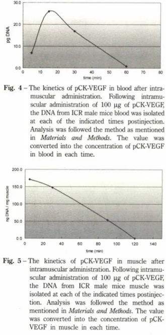
Fig. 4 - The kinetics of pCK-VEGF in blood after intra
muscular administration. Following intramu
scular administration of 100 jo.g of pCK-VEGR the DNA from ICR male mice blood was isolated at each of the indicated times postinjection.
Analysis was followed the method as mentioned in Materials and Methods. The value was converted into the concentration of pCK-VEGF in blood in each time.
5 10 15 20 25 30
time (min)
Fig. 3 - The kinetics of pCK-VEGF in blood after intra
venous administration. Following intravenous administration of 100 jig of pCK-VEGF; the DNA from ICR male mice blood was isolated at each of the indicated times postinjection. PCR, agarose gel electrophoresis and densitometry were followed the method as mentioned in Materials and Methods. The value was converted into the concentration of pCK-VEGF in blood in each time.
口(무스의 근육내 투여 후 익동력학
마f 스근육에 pCK-VEGF 100 ng/head를투여히여 얻은 혈액중의 농도•시간곡선을 Fig. 4에나타내었다. 혈액으로이행하여 15분에서 혈중최고농도를보였고,
혈중 최고 농도는 26 pg/m/이었으며, 빠른 속도로 소 실되어 60분이후에는 관찰되지 않았다. 혈중 반감기 는 6.78±2.090 min이었으며,AUC는 781.9±327pg • min/m/ 이었다. 마우스 근육에 pCK-VEGF 100 |ig/
head를투여한후, 근육내 농도•시간곡선을 Fig. 5에 나타내었다. 투여 후 90분에는투여용량의 약 30%가 투여부위인 근육에 존재하였고, &시간후에는 극미량
의 DNA만이 검출되었다. 근육에 투여된 pCK-VEGF 는빠른속도로근육으로부터 소실되는것으로관찰되 었는데, 이중 일부는혈중으로이행되어 대사되거나 2 차적으로다른조직에 재분포되었으며, 많은양은근 육조직 내에서 대사되는 것으로 나타났다.
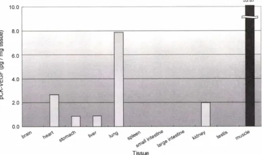
근육투여 후 pCK-VEGF의다른 장기로의 분포 정
도는 Fig. 6에 나타난 바와 같다. 투여 후 30분에 pCK-VEGF는 심장, 폐, 신장 등과 같이 혈류량이 많 은 장기에서 검출되기 시작하였으나그농도는 lpg / mg 미만이었고, 투여 후 90분에 최고농도에 도달하 여, 근육을 제외할 때,폐(7.873pg/mg tissue), 심장 meter로 정량하였다(Fig. 2). 근육 중농도가 0.001-3
ng인검체를이추출법으로 5회분리, 정량한결과희 수율은 약 85.59%이였으며, 표준편차는 약 8.66%이내 로 양호한 결과를나타내었다. 혈액에 대하여 동일한 방법으로 측정한 결과, l~10pg의 pCK-VEGF가 포 함된검체로부터는약 80.18%의회수율을보였으나,1 pg 미만의 검체에서는낮은 회수율을나타내었다.
마무스의 정맥투여 후의 약동력학
마우스 꼬리 정맥에 pCK-VEGF lOOpg/head를투 여한 후,혈액중의 농도•시간곡선을 Fig. 3에나타내 었다. 일단혈액내에존재하게되면, 빠른속도로분해 되기 시작하여, 약 1.68±0.51min의 혈중 반감기를 보이며, AUC는 149.5± 102.4 lig-min/m/ 이었다.
150.0
100.0
0 20 40 60 80 100 120 140
time (min)
Fig. 5 - The kinetics of pCK-VEGF in muscle after intramuscular administration. Following intramu
scular administration of 100 |ig of pCK-VEGF, the DNA from ICR male mice muscle was isolated at each of the indicated times postinjec
tion. Analysis was followed the method as mentioned in Materials and Methods. The value was converted into the concentration of pCK- VEGF in muscle in each time.
ᅳ .스 ) 뚫 느 . ᅳ J
J. Pharm. Soc. Korea
VEGF 발현 Naked DNA 벡터의 익동력학 및 조직내 분포 75
53.87
丁issue
Fig. 6 - The tissue distribution of pCK-VEGE Tissue distribution of pCK-VEGF was analyzed at 90 min postinjection.
Following intramuscular administration of 100 fig pCK-VEGF; the DNA from various tissues of ICR male mice was isolated. PCR, agarose gel electrophoresis and densitometry were followed the method as mentioned in Materials and Methods. The value was converted into the concentration of pCK-VEGF in tissue.
Table I - Existence ratio in organs at 90 min after intermuscular administration
Organ Existence (%)
Brain Nd*
heart 0.40
stomach 0.22
liver 0.65
lung 1.62
spleen nd*
small intestine nd*
large intestine nd*
kidney 1.21
testis nd*
muscle 28.71
The existence ratio of pck-VEGF in respective organs were calculated from the concentration of the DNA in each tissue at 90 min after administration of 100 |ig pCK-VEGE
*nd: non-detected.
(2.680 pg/mg tissue), 신장(1.980 pg/mg tissue) 등
에서 단위 조직량에 존재하는 pCK-VEGF 농도가높
았다. 또한, 각조직의 평균무게를 고려한조직 분포 율은폐,심장,간의 순으로나타났다. 그러나,'근육을 제외할 때각 장기에서의 분포율은 약 1.6% 이하로 무시할만한 적은분포율을 나타내었으며(Table I), 이 들은시간이경과하면서 빠르게소실되어 약 8시간경
과후에는 거의 모든장기에서 검출되지 않았다-
고 찰
본 연구는 국내 최초로 시도되는 허혈성 지체질환
유전자 치료제인 pCK-VEGF의 생체내 동태에 관한
연구로서 혈액 또는조직에서의 약동력학적 반감기와 타장기로의 분포 및 소실을 관찰하며,pCK-VEGF의 흡수와분포의 변화를 알아보고자 하였다.
pCK-VEGF는 정맥주사 후 빠른 속도로 분해되어,
혈중반감기가 매우舰 고 (1.68±0.507min),투여 후
30분이 경과하면 intact plasmid로는 관찰되지 않았는 데(정량한계는 약 lpg), 이러한혈액중에서의 빠른소
실은 혈중에 존재하는 핵산 분해 효소(nuclease)의 작
용에 의한 것으로 생각되며,MHC class I 유전자를
사용한암유전자치료제에서의 결과와유■사하다.121
pCK-VEGF를 근육 투여하면, 혈중으로 이행한 후
다시 각조직으로분포하여, 약 90분경과후에 조직
내 농도가 최고에 달하고, 120분을 경과하면서 모든
조직에서사라지기 시작하여 8시간경과후에는 검출되
지 않았다. 주로, 분포되어 높은 농도에 달한 조직은
폐, 신장, 심장,간등 이었으며,뇌,비장, 소장, 대장, 고환 둥에서는 관찰되지 않았다. 허혈성 지체질환 치
^
= 1 COE
16
do3A->o3
Q.
76 도현미 • 고준일 • 이종진 • 손미원 ■ 조홍찬 • 김종묵 • 김병문 . 김선영
료목적으로사용•되는 pCK-VEGF의예상임상용량은 허혈부위에 2 mg씩 2회근육주사로예측되므로, 본■시 험조건에서의 투여용량(100 ng/head)은 3 mg/kg정도이
며환자 체중을 60 kg으로환산하였을 때임상용량의
약 45배에 해당하는높은 용량이다. 또한 임상적용에 있어 허혈부위는 혈행이 억제되어, 국소 근육 주사후
전신혈을 통한 다른 장기로의 pCK-VEGF의 이행은
더욱 감소될 것으로 예측된다. pCK-VEGF는 혈액 및
조직중에서 빠르게분해되고다른장기로의 분포가낮 다는 것을 고려할 때, 임상적용에 있어 다른 장기로
이행하여 VEGF 발현을 촉진하거나다른장기에서의
약리작용에 기인한부작용발현의 위험성은 매우낮을 것으로판단된다. 투여부위인 근육에는 투여 후 90분
에투여량의 30% 정도가 잔류하였으며, 8시간이후에
도 미량의 DNA가 검출되었는데,이 결과는 DNA
luciferase expression vector를마우스근육에 투여하 여 흡수 및 분포를 관찰하며 fg이하의 정량한계로서 30일까지 plasmid의 존재를 확인하고, 60일까지
luciferase의 활성이발현되는것을확인하였던 이전의
발표 내용과유사하다.13)
결론적으로, pCK-VEGF는생체내에투여할경우원 체는혈액와장기내에서 빠르게소실되며, 투며부위인 근육에서는 고농도로 8시간까지 고농도로유지되어 유 전자의발현을유도함으로서신생혈관조성을나타내는 유전자치료법으로효용성이 클것으로예상된다.
감사의 말씀
본 연구는 보건복지부,산업자원부와과학기술부의 지원에 의해서 수행되었다.
문 헌
1) Criqui, M. H.,Fronek A., Batret-Connor, E. K., Klauber M. R., Gabriel S. and Goodman D. The prevalence of peripheral arterial disease in a defined population. Circulation 71, 510 (1985).
2) European Working Group on Critical Leg Ischemia.
Second edition consensus document on chronic critical leg ischemia, Circulation, 84,IV-1, IV-26 (1991).
3) Lee, J. S. and Feldman, A. M. Gene therapy for
therapeutic myocardial angiogenesis : a promising synthesis of two emerging technologies. Nat. Med.
4(6), 739 (1998).
4) Baumgartner, I., Pieczek, A, Manor, 0., Blair, R., Kearney, M., Walsh, K. and Isner, J. M. Constitutive expression of phVEGF165 after intramuscular gene transfer promotes collateral vessel development in the patients with critical limb ishemia, Circulation, 97, 1114 (1998).
5) Carmeliet R and Collen D. Role of vascular endothelial Growth factor and vascular endothelial growth factor receptors in vascular development.
Curr. Top. Microbiol. Immunol 237,133 (1999).
6) Takeshita, Satoshi; Pu, Li-Qun; Stein, Lawrence A.; Sniderman, Allan D.; Bunting, Stuart; Ferrara, Napoleone; Isner, Jeffi*ey M.; Symes, James E Intramuscular administration of vascular endothelial growth factor induces dose-dependent collateral artery augmentation in a rabbit model of chronic limb ischemia. Circulation, 90, 5(Pt 2),E228-II234 (1994).
7) Losordo, D. W., Vale, R R., Symes, J. E, Dunnington, C. H., Esakof, D. D.,Maysky, M.,Ashare, A. B.,
Lathi, K.,Isner, J. M. Gene therapy for myocardial angiogenesis initial clinical results with direct myocardial injection of phVEGF165 as sole therapy for myocardial ischemia. Circulation, 98,2800 (1998).
8) 이영주, 박은진,조홍찬, 서연림, 김덕경, 김선영. Naked DNA를이용한 말초동맥질환치료용 VEGF 발현벡터의 안전성시험. / Toxicol Pub. Health, 15, 373 (1999).
9) Lee, Y, Park, E. J., Yu, S. S., Kim, D. K., and Kim, S.
Improved expression of vascular endothelial growth factor by naked DNA in mouse skeletal muscles:
Implication for gene therapy of ischemic disease.
Biochem. Biophys. Res. Commun., 272, 230 (2000).
10) Sambrook J., Fritsch E. E and T. Maniatis. Molecular cloning. A laboratory manual. 2nd ed. Cold spring harbor laboratories press, New York.
11) Isner J. M.,Ann E, Robert S., Richard B., Laura H., Takayuki A., Kenneth R., Syed R., Kenneth W. and James E S. Clinical evidence of angiogenesis after arterial gene transfer of phVEGF165 in patient with ischemic limb. Lancet 348, 370 (1996).
12) Lew, D.,Parker, S. E., Latimer, T, Abai, A. M.,
/. Pharm. Soc. Korea
VEGF 발현 Naked DNA 벡터의익동력학및조직내분포 77
Kuwahara-Rundeli, A., Doh, S. G.,Yang, Z. Y., Lafece, D., Gromkowski, S. H.,Nabel, G. J., Manthorpe, M.
and Norman, Cancer gene therapy using plasmid DNA: pharmacokinetic study of DNA following injection in mice. Hum. Gene Ther. 6,553 (1995).
13) Wolff, J. A., Malone, R. W, Williams, R, Chong, W., Acsadi, G., Jani, A.and Feigner, R L. Direct gene transfer into mouse muscle in vivo. Science, 247, 1465-1468 (1990).