https://doi.org/10.22643/JRMP.2016.2.2.108
Introduction
신생혈관생성 (angiogenesis) 이란 이미 존재하는 혈관으로 부터 새로운 미세 혈관이 형성되는 생리적 과정을 일컫는 것 으로 발생기의 혈관 생성뿐만 아니라 종양, 류마티스성 관절 염, 망막증과 같은 여러가지 질병에 관여되어 있다. [1-3] 인 테그린은 혈관생성세포에 존재하는 세포 표면 수용체로서 표 피세포 이동과 성장, 생존 그리고 분화에 관련된 조절자이다.
[4, 5] 인테그린 수용체 중에서 α
vβ
3 인테그린은 신생혈관생 성 과정 중 활성화된 표피세포에는 많이 발현되지만, 휴지 상 태의 표피세포나 정상 세포에는 발현이 낮다. 특히, αvβ
3인 테그린 수용체는 종양 세포에서 그 발현이 증가되어 있으면 서 종양의 침습성 (invasiveness) 및 전이 (metastasis) 를 촉진 시킨다고 알려져 있다. 그러므로 αvβ
3인테그린의 발현 정도 를 분자 영상학적인 방법으로 평가하는 것은 종양의 진단 및 치료 전후의 효과를 모니터링하고 적절한 치료 계획을 수립
β
3인테그린의 발현 정도 를 분자 영상학적인 방법으로 평가하는 것은 종양의 진단 및 치료 전후의 효과를 모니터링하고 적절한 치료 계획을 수립
Kinetic analysis of 64 Cu-NODAGA-gluco-E[c(RGDfK)] 2 for a tumor angiogenesis PET tracer
Jae Yong Choi,
1Ji-Ae Park,
2Jung Young Kim,
2Ji Woong Lee,
2,3Minkyung Lee,
4Un Chol Shin,
2Joo Hyun Kang,
2Gwang Il An,
2Kyo Chul Lee,2 Young Hoon Ryu,
1* Kyeong Min Kim
2*
1
Department of Nuclear Medicine, Gangnam Severance Hospital, Yonsei University College of Medicine, Seoul, Korea,
2Molecular Imaging Research Center, Korea Institute of Radiological and Medical Sciences, Seoul, Korea, 3Departmet of Integrated Biomedical and Life Science, Korea University, Seoul, Korea, 4Department of Nuclear Medicine, Inha University College of Medicine, Inha University Hospital, Incheon, Korea
Molecular imaging with the radiolabeled RGD peptides for α
vβ
3integrin has been an increasing interest for tumor diagnosis and the treatment monitoring. Recently,
64Cu-NODAGA-gluco-E[c(RGDfK)]
2was developed for quantification of α
vβ
3integrin and its biological properties was elucidated. To better understand the molecular process in vivo, we performed the kinetic analysis for the
64Cu-NODAGA-gluco-E[c(RGDfK)]
2. After preparation of a radiotracer, dynamic PET images were obtained in the U87MG xenograft mice for 60 min (n
= 6). Binding potential values were estimated from the 3-tissue compartment model, reference Logan and simplified reference tissue model. In the early time frame (0-20 min), the liver, kidney, intestine, urinary bladder and tumor were visualized but these uptakes were diminished as time went by. The tumors showed a good contrast at 40 min after administration.
64Cu-NODAGA-E[c(RGDfK)]
2showed the 2-fold uptake in the tumor compared with that in the muscle. The parametric maps for binding values also provide the higher tumor-to- background contrast than the static images. A binding value obtained from the 3-tissue compartment model was comparable to other modeling methods. From these results, we conclude that
64Cu-NODAGA-gluco- E[c(RGDfK)]
2may be a promising PET radiotracer for the evaluation of angiogenesis.
J Radiopharm Mol Probes 2(2):108-112, 2016 ABSTRACT
Key Word: Integrin α
vβ
3, Angiogenesis, NODAGA,
64Cu, Bifunctional chelator, RGD
October 21, 2016 / Revised: December 01, 2016 / Accepted: December 05, 2016
Correspnding Author : Young Hoon Ryu, Kyeong Min Kim
Y. H. Ryu: Department of Nuclear Medicine, Gangnam Severance Hospital, Yonsei University College of Medicine, 211 Eonjuro, Gangnam-gu, Seoul 06273, Korea
K. M. Kim: Molecular Imaging Research Center, Korea Institute of Radiological and Medical Sciences, 75 Nowon-ro, Nowon-Gu, Seoul 01812, Korea
Tel: Y.H. Ryu: +82-2-2019-3518, Fax: +82-2-3462-5472, E-mail: [email protected]; K. M. Kim: +82-2-970-1387, Fax: +82-2-970-2436, E-mail: [email protected]
Copyright©2016 The Korean Society of Radiopharmaceuticals and Molecular Probes
하는데 있어 중요한 역할을 하므로, 많은 관심을 받고 있는 연구 분야이다. [6-9]
α
vβ
3 인테그린은 알기닌-글라이신-아스파테이트 (Arg- Gly-Asp, RGD) 그룹을 통해서 비트로넥틴, 피브리노겐, 라 미닌 그리고 콜라겐과 같은 세포외기질 단백질 (extracellular matrix protein) 과 상호 작용을 한다. 이러한 사실을 이용하 여 현재까지 다양한 RGD 펩티드가 개발되었고 그중 대표적 인 것인 것이 cyclo(Arg-Gly-Asp-D-Phe-Lys) c(RGDfK) 와 cyclo(Arg-Gly-Asp_D-Tyr-Lys) c(RGDyK) pentapeptide cyclo(-Arg-Gly-Asp) 이다. [10] . 핵의학에서 가장 잘 알 려지고 다양한 연구에 이용되고 있는 RGD 유도체로는 [18F]galacto-RGD, [
18F]AH111585, [
18F]RGD-K5, [
68Ga]
DOTA-TOC 등이 있다. 하지만 긴 합성시간 대비 낮은 표지 수율, 간에서의 높은 방사능 섭취, 인테그린에 대한 낮은 선 택성 등의 한계점을 가지고 있다. [11-13] 또한, 기존 연구에 따르면 RGD 펩티드 개수의 증가는 α
vβ
3에 대한 친화력을 증 가시키지만 동시에 비특이적 결합 증가를 유발하기 때문에 최적의 RGD 펩티드 개수는 2 개인 것이 알려져 있다. 그리 고, NODAGA conjugated 방사성화합물들이 신장에서 빠른 청소율 (renal clearance) 을 유도하여 높은 신호 대 잡음비를 제공하는 것이 알려져 있다. [20, 21]
최근 인테그린 αvβ
3에 대한 높은 선택성과 친화력을 가지면 서 간 에서의 방사능 섭취를 효과적으로 줄이기 위해 dimeric RGD 와 NODAGA 를 기본 골격으로 하고 여기에 글루코사 민을 도입한 64Cu-NODAGA-gluco-E[c(RGDfK)]2가 개발 되었지만[ 14] , 체내에서의 어떤 동력학적 특성을 나타내는지 에 대한 연구는 이루어지지 않았다. 그러므로, 본 연구에서는 종양 동물 모델에서
64Cu-NODAGA-gluco-E[c(RGDfK)]2 의 동력학 분석을 실시하여 생체기능지표를 평가하고자 한다.
가 개발 되었지만[ 14] , 체내에서의 어떤 동력학적 특성을 나타내는지 에 대한 연구는 이루어지지 않았다. 그러므로, 본 연구에서는 종양 동물 모델에서
64Cu-NODAGA-gluco-E[c(RGDfK)]2 의 동력학 분석을 실시하여 생체기능지표를 평가하고자 한다.
Materials and Methods
세포 배양 및 종양 xenograft 동물 모델 제작
Human glioma cell line, U87MG 는 American Type Culture Collection (USA) 에서 구입하여, 10% fetal bovine
serum 과 1% penicillin-streptomycin 이 들어 있는 Dulbecco’s modified Eagle’s medium (DMEM) 배지에서 37°C 5% 이 산화탄소 조건에서 배양하였다.
4~6 주령의 female BALB/c 누드마우스의 왼쪽 팔에 앞 서 배양한 5 x 10⁶ 의 U87MG 세포를 피하주사하였다 (SLC mouse, Hamamatsu, Japan, n = 6) . 그런 다음 종양의 부피가 0.7 – 0.9 cm 가 되었을 때 PET 연구에 이용하였다. 동물 실 험은 한국원자력의학원의 동물실험윤리위원회의 허가를 받 아 진행되었다.
64
Cu-NODAGA-gluco-E[c(RGDfK)]
2의 합성
Gluco-E[c(RGDfK)]
2(200mg, 0.05) 과 DIPEA (77 mg, 0.060mmol) 혼합물에 DMF (17mL) 를 넣은 후, 이 혼합액 을 NODAGA-NHS-ester (125 mg, 0.17mmol)/DMF (5ml) 용액에 천천히 추가하고 상온에서 20 시간 교반하였다. 그 후, 0.1% TFA 수용액 15 mL 를 추가하여 반응을 종결시킨 다 음 생성물을 진공에서 건조하여 흰색 파우더 형태의 crude product 를 얻을 후 고성능 크로마토그래피를 통해 정제하여 NODAGA-gluco-E[c(RGDfK)]
2를 획득하였다. ( 그림 1, A: CH3CN, B: H
2O, A:B = 10 - 45% gradient, flow rate 12 ml/min, Rt = 20.5 min.)
64
CuCl
2(37 – 370 MBq) 바이알로 추출한 다음, 100°C 에 서 질소가스를 이용하여 건조시킨 다음 1M 아세트산나트륨 를 200 μL 추가하여 pH 를 5.5 가 되도록 하였다. 그런 다음 NODAGA-gluco-E[c(RGDfK)]
2을 녹인 50% 에탄올 수용 액 (100μg/50μL) 을 첨가한 다음 50°C 에서 30 분간 반응시켰 다. 표지반응 종료 후 별도의 정제과정은 수행하지 않고 최종 생성물을 멸균 바이알에 0.22 μm 멤브레인 필터 (Millipore,
Figure 1.
64Cu-NODAGA-gluco-E[c(RGDfK)]
2의 화학 구조.
Millex-GV PVDF) 를 통과하였다. 최종생성물의 방사화학적 수율과 순도는 99% 였고, 37°C 에서 사람과 마우스 혈청에 서 24 시간 동안 순도 변화를 관찰해본 결과 방사화학적 수율 이 93% 이상이였음을 확인하였다.
PET/CT 스캔
1.5% 이소플로렌으로 마취시킨 xenograft 동물 모델을 PET/
CT 겐트리에 복와위 자세로 위치시킨 다음, 꼬리정맥에 64Cu- NODAGA-gluco-E[c(RGDfK)]
2 (7.6 ± 0.4 MB) 을 주사하면 서, 60 분 동안 동적 PET 영상을 획득하였다. 영상을 얻는 동 안 1.5% 이소플로렌으로 호흡마취를 유지하였다. 획득한 사 이노그램은 2D ordered subsets expectation maximum (2D
OSEM) 방식을 이용하여 영상을 재구성하였다. 이 때 사용
한 시간 프레임은 1 분 x10 프레임, 5 분 x10 프레임이다. 재구 성된 영상에서의 픽셀 값은 개체간 비교를 위해 표준섭취계 수 (standard uptake value, SUV) 로 변환하여 사용하였고, 관심영역 (volume of interest) 는 종양과 근육으로 하였다.
결합능은 3 구획 모델 (3 tissue compartment model, 3-TCM) , reference tissue 모델인 simplified reference tissue model (SRTM) 과 reference Logan 도표 분석법을 이용하여 구하 고 이를 서로 비교하였다. [15-19] 3-TCM 에서는 심장 좌심 실을 관심영역으로하여 영상기반 입력함수를 도출하고 이를 기반으로 결합능을 구하였고, reference tissue 모델에서는 근 육을 reference 영역으로 사용하여 결합능을 구하였다. 여기 서 좌심실은 동맥 입력함수를 추정하기 위해 사용되었고, 재 조합된 PET/CT 이미지의 초기 영상 (동적 P ET 이미지에서 두번째 프레임)에서 좌심실을 선택하였다. Guo N . 등이 제한 한 것처럼 본 연구에서 사용한 Inveon PET 스케너의 고성능 PET 기기이기 때문에 부분용적효과 (partial volume effect) 와 흘러넘침 (spill over) 에 대한 영향은 미미하다고 간주하였
다 [19] . 상기 PET 영상분석은 PMOD (ver.3.501) 을 이용하 였다. 모델링 방법 간의 상관계수는 Prism5 (GraphPad ver.
5.04) 에 포함된 Pearson 상관계수로 분석하였다.
Result and Discussion
우선 인테그린 αvβ
3에 많이 발현된 U87MG 종양과 인테 그린 αvβ
3가 거의 발현되지 않는 근육에서
64Cu-NODAGA- gluco-E[c(RGDfK)]
2의 체내 거동을 파악하기 위해 시간 경 과에 따른 관심영역에서의 평균 섭취 값부터 파악해보았다.
β
3가 거의 발현되지 않는 근육에서
64Cu-NODAGA- gluco-E[c(RGDfK)]
2의 체내 거동을 파악하기 위해 시간 경 과에 따른 관심영역에서의 평균 섭취 값부터 파악해보았다.
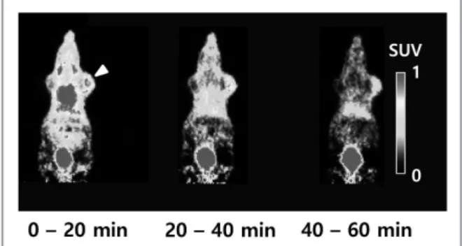
여기서 각각 시간 프레임에서의 방사능은 관심영역에서 픽셀 값의 평균을 의미한다. 주사 후 20 분 간격의 평균 PET 영상 에서 알 수 있듯이
64Cu-NODAGA-gluco-E[c(RGDfK)]
2는 초기에는 종양뿐 아니라 간, 콩팥, 내장, 방광 등에 흡수되지 만 시간이 경과함에 따라 종양을 제외한 나머지 장기에서는 방사능 섭취가 낮아졌다. 그리고 주사 40 분 이후에 종양은 높 은 대조도 영상을 나타내었다. (그림 2 . 참조).
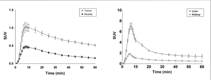
그림 3A 는 U87MG xenograft 종양과 근육에서의 시간 대 방사능 곡선을 나타낸 것으로 종양에서는 8 분 정도에 1.1 SUVmax 를 나타내면서 60 분 경과 후에도 0.8 SUV 를 유지 하지만, 근육에서는 8 분에 0.5 SUVmax 값을 보이다가 60 분 Figure 2. 6주사 후 0-20 분, 20-40분, 40-60분의 평균 micro-PET 영 상. 화살표는 U87MG xenograft 종양을 표시함. 시간이 경과함에 따라 신호 대 잡음비가 증가함.
Modeling method) 3-TCM Reference Logan SRTM
Binding potential 1.78 ± 0.2 1.78 ± 0.3 1.85 ± 0.3
Table 1. 모델링 방법에 따른 결합능 비교
*데이터는 평균값 ± 표준편차로 나타냄.
경과후에는 0.2 SUV 를 나타내었다. 이는
64Cu-NODAGA- gluco-E[c(RGDfK)]
2가 종양에 특이적으로 섭취되고 일정 시간 동안 유지됨을 의미한다. 간과 콩팥에서는 주사 6분 후 각각 1.8 SUV, 7.00 SUV 로 최대값을 나타내였고, 시간이 지 남에 따라 감소되어서 60 분 후에는 0.48 SUV, 1.39 SUV 를 나타내었다 (그림 3B ).
3 구획 모델에서 도출한 결합능을 reference Logan 도표 분석법과 SRTM 의 결과를 비교해본 결과, 결합능은 1.78 ~ 1.85 로 모두 유사한 값을 나타내었다. 이는 개체간의 결합 해리상수 (dissociation constant, Kd) 값이 비슷하다고 가정하 면, 종양에서 수용체의 분포밀도가 증가했음을 시사한다. 또 한 3 구획 모델에서의 결합능과 다른 모델링 방법에서 도출 한 결합능을 비교해보면, 모델링 방법간의 상관계수 r
2값도 0.81 또는 0.86 으로 서로 강한 상관관계가 성립하였다.
화소단위에서의 변화를 파악하기 위해 결합능에 대하여 파라 메터 영상을 구성한 결과,
64Cu-NODAGA-gluco-E[c(RGDfK)]
2는 U87MG xenograft 종양에 대하여 높은 결합능을 나타내었 고, 이는 이전의 평균 PET 영상보다 높은 신호 대 잡음비를 나타내었다. (그림 4 ). 또한 콩팥에서도 높은 결합능을 나타내 는 것으로 봐서,
64Cu-NODAGA-gluco-E[c(RGDfK)] 는 대사 되어 gastrourinary tract 을 통해 소변으로 배설 되는 것을 유 추할 수 있다. 그리고, 64Cu-NODAGA-c(RDGfK) 나 64Cu- NODAGA-E[c(RGDfK)]
2의 경우 주사 후 18 시간 경과 후에 U87MG 종양에서 선택적인 방사능 섭취를 보이는 것과 비교
Cu- NODAGA-E[c(RGDfK)]
2의 경우 주사 후 18 시간 경과 후에 U87MG 종양에서 선택적인 방사능 섭취를 보이는 것과 비교
해보면,
64Cu-NODAGA-gluco-E[c(RGDfK)]
2는 기존 신장 청 소율을 크게 개선한것으로 보인다. [14, 22]
Conclusion
64
Cu-NODAGA-gluco-E[c(RDGfK)]
2는 종양의 αvβ
3에 선택적으로 결합하여, 근육보다 2 배 높은 방사능 섭취를 가 지면서, 적절한 머무름 시간을 나타내었다. 이러한
64Cu- NODAGA-gluco-E[c(RDGfK)]
2의 종양 선택성은 결합능 Figure 3. U87MG에서 64Cu-NODAGA-gluco-E[c(RGDfK)]2의 시간 대 방사능 곡선. (A) 종양에서의 SUVmax가 근육에서보다 2배 가량 높음. (B) 콩팥에 서의 섭취가 간에서의 섭취보다 SUVmax 3.9배 높음. 데이터는 평균값 ± 표준편차로 나타냄
Cu-NODAGA-gluco-E[c(RGDfK)]2의 시간 대 방사능 곡선. (A) 종양에서의 SUVmax가 근육에서보다 2배 가량 높음. (B) 콩팥에 서의 섭취가 간에서의 섭취보다 SUVmax 3.9배 높음. 데이터는 평균값 ± 표준편차로 나타냄
Figure 4. 근육을 reference 영역으로 하여 구성한 결합능에 대한 파라
메터영상
에 대한 파라메터영상에서도 확인할 수 있었다. 또한 영상 기반입력함수 기반 3 구획모델에서 얻는 결합능은 reference Logan 도표법과 SRTM 에서 얻는 값과 높은 상호 연관성을 보였고, 빠른 신장 청소율을 나타내었다. 이러한 결과로부터 본 연구진은
64Cu-NODAGA-gluco-E[c(RGDfK)]
2가 신생 혈관생성을 평가하는 데 유용한 PET 방사성의약품이 될 것 으로 기대한다.
Acknowledgments
This work was supported by a faculty research grant of Yonsei University College of Medicine for 2007(6-2007-0169), the Nuclear R&D Program of the National Research Foundation of Korea government (MEST)(2012M2A2A7013480) and a grant of the Korea Institute of Radiological and Medical Sciences (KIRAMS) funded by the Ministry of Science, ICT & Future Planning (No. 1711021927/505302016), Republic of Korea.
References
1. Storgard CM, Stupack DG, Jonczyk A, Goodman SL, Fox RI, Cheresh DA: Decreased angiogenesis and arthritic disease in rabbits treated with an alphavbeta3 antagonist. J Clin Invest 1999;103:47-54.
2. Folkman J: Role of angiogenesis in tumor growth and metastasis. Semin Oncol 2002;29:15-18.
3. Chavakis E, Riecke B, Lin J, Linn T, Bretzel RG, Preissner KT, Brownlee M, Hammes HP: Kinetics of integrin expression in the mouse model of proliferative retinopathy and success of secondary intervention with cyclic RGD peptides. Diabetologia 2002;45:262-267.
4. Eliceiri BP, Cheresh DA: Role of alpha v integrins during angiogenesis. Cancer J 2000;6:S245-249.
5. Hynes RO, Bader BL, Hodivala-Dilke K: Integrins in vascular development. Braz J Med Biol Res 1999;32:501-510.
6. Guo WJ, Giancotti FG: Integrin signalling during tumour progression. Nat Rev Mol Cell Bio 2004;5:816-826.
7. Liu Z, Wang F, Chen X: Integrin alpha(v)beta(3)-Targeted Cancer Therapy. Drug Dev Res 2008;69:329-339.
8. Millard M, Odde S, Neamati N: Integrin targeted therapeutics.
Theranostics 2011;1:154-188.
9. Niu G, Chen X: Why integrin as a primary target for imaging and therapy. Theranostics 2011;1:30-47.
10. Ruoslahti E, Pierschbacher MD: New perspectives in cell
adhesion: RGD and integrins. Science 1987;238:491-497.
11. Haubner R, Weber WA, Beer AJ, Vabuliene E, Reim D, Sarbia M, Becker KF, Goebel M, Hein R, Wester HJ et al: Noninvasive visualization of the activated alphavbeta3 integrin in cancer patients by positron emission tomography and [
18F]Galacto-RGD. PLoS Med 2005;2:e70.
12. Kenny LM, Coombes RC, Oulie I, Contractor KB, Miller M, Spinks TJ, McParland B, Cohen PS, Hui AM, Palmieri C et al: Phase I trial of the positron-emitting Arg-Gly- Asp (RGD) peptide radioligand 18F-AH111585 in breast cancer patients. J Nucl Med 2008;49:879-886.
13. Mirfeizi L, Walsh J, Kolb H, Campbell-Verduyn L, Dierckx RA, Feringa BL, Elsinga PH, de Groot T, Sannen I, Bormans G et al: Synthesis of [18F]RGD-K5 by catalyzed [3 + 2] cycloaddition for imaging integrin alphavbeta3 expression in vivo. Nucl Med Biol 2013;40:710-716.
14. Lee JW, Park JA, Lee YJ, Shin UC, Kim SW, Kim BI, Lim SM, An GI, Kim JY, Lee KC: New Glucocyclic RGD Dimers for Positron Emission Tomography Imaging of Tumor Integrin Receptors. Cancer Biother Radiopharm 2016;31:209-216.
15. Phelps ME, Huang SC, Hoffman EJ, Selin C, Sokoloff L, Kuhl DE: Tomographic measurement of local cerebral glucose metabolic rate in humans with (F-18)2-fluoro- 2-deoxy-D-glucose: validation of method. Ann Neurol 1979;6:371-388.
16. Logan J, Fowler JS, Volkow ND, Wang GJ, Ding YS, Alexoff DL: Distribution volume ratios without blood sampling from graphical analysis of PET data. J Cereb Blood Flow Metab 1996;16:834-840.
17. Lammertsma AA, Hume SP: Simplified reference tissue model for PET receptor studies. Neuroimage 1996;4:153-158.
18. Guo N, Lang L, Li W, Kiesewetter DO, Gao H, Niu G, Xie Q, Chen X: Quantitative analysis and comparison study of [
18F]AlF-NOTA-PRGD2, [
18F]FPPRGD2 and [
68Ga]Ga- NOTA-PRGD2 using a reference tissue model. PLoS One 2012;7:e37506.
19. Guo N, Lang L, Gao H, Niu G, Kiesewetter DO, Xie Q, Chen X: Quantitative analysis and parametric imaging of
18