흰깨 추출물과 β-Sitosterol이 H1299 폐암세포의 성장, 이동, 부착에 미치는 효과
이중재․김서윤․주지형 충북대학교 식품영양학과
Effects of White Sesame Seed Extract and β-Sitosterol on Growth, Migration, and Adhesion of H1299 Human Lung Cancer Cells
Jungjae Lee, Seoyun Kim, and Jihyeung Ju
Department of Food and Nutrition, Chungbuk National University
ABSTRACT The current study aimed to investigate effects of ethanol extract of white sesame seed (WSE) as well as a major constituent of white sesame seed, β-sitosterol, on the growth, migration, and adhesion of H1299 human lung cancer cells. Treatment with WSE at concentrations of 150, 300, and 600 μg/mL dose-dependently inhibited cell growth (to 51.5∼82.6% of control). Treatment with β-sitosterol at concentrations of 3.125, 6.25, 12.5, and 25 μM inhibited cell growth to a greater extent (to 27.5∼49.0% of control) than that with WSE (P<0.05). Treatment with WSE (at concentration of 600 μg/mL) or β-sitosterol (at concentration of 25 μM) resulted in increased sub-G1 cell population, indicating their apoptosis-inducing activities. β-Sitosterol was effective in inhibiting both cell migration (to 80.8∼86.2% of control at a concentration range of 3.125∼25 μM) and adhesion (to 21.5∼37.4% of control at a concentration range of 6.25∼25 μM), whereas WSE at a concentration range of 150∼600 μg/mL was ineffective.
These results indicate that β-sitosterol is more active than WSE in inhibiting growth, migration, and adhesion of H1299 human lung cancer cells. Further studies are needed to determine if similar effects are reproduced in vivo.
Key words: white sesame seed extract, β-sitosterol, lung cancer, cancer cell characteristics
Received 15 June 2015; Accepted 24 July 2015
Corresponding author: Jihyeung Ju, Department of Food and Nutri- tion, Chungbuk National University, Cheongju, Chungbuk 28644, Korea
E-mail: [email protected], Phone: +82-43-261-2681
서 론
참깨(Sesamum indicum L.)는 통화 식물목 참깨과 참깨 속에 속하는 쌍떡잎식물로 열대성 기후에서 자란다(1,2). 참 깨는 지질 함량이 약 50%에 이르는 유료작물로(3) 그 종실 부위나 종실로부터 얻은 기름이 널리 식용되고 있다. 참깨 종실에는 단백질(약 20%), 탄수화물(약 20%)뿐 아니라 비 타민 B1, 비타민 B2, γ-tocopherol, 칼슘, 인, 칼륨 등이 함유되어 있는데, 특히 칼슘 함량(약 1.1 mg/g)이 높다(3).
참깨와 참기름은 한국인의 에너지, 지방, 칼슘 섭취를 위한 주요 식품급원으로 보고된 것처럼(4) 한국인의 식생활에서 매우 중요한 역할을 하고 있다. 참깨는 종피의 색에 따라 흰깨, 황깨, 검은깨 등으로 나뉘는데(1), 흰깨는 검은깨보다 지용성 리그난 성분인 sesamin과 sesamolin 함량이 높은 것으로 보고되었다(5). 참깨의 생리활성과 관련해서는 페놀 류, 리그난 등과 같은 항산화 성분의 함량(6,7)에 대한 연구 나 라디칼 소거능, 철환원력, 과산화물 생성 억제능과 같은 항산화 활성(7-11) 등에 대한 연구가 주를 이루며, 그 밖에
검정깨 메탄올 추출물의 대장암세포 자동사멸 유도 효과 (10), 검정깨 함유 식이의 인체에서의 항고혈압 및 항산화 효과(12), 흰깨 함유 식이의 인체에서의 고지혈증 완화 효과 (13) 등이 보고되었다.
β-Sitosterol(Fig. 1)은 campesterol, stigmasterol과 함께 식물성 식품에 광범위하게 함유되어 있는 대표적인 식 물성 스테롤 성분이다(14). β-Sitosterol의 대표적인 식품 급원은 식물성 유지이며, 참깨 등의 종실류나 곡류의 배아 등에 그 함량이 높은 것으로 알려져 있다(14). β-Sitosterol 을 포함한 식물성 스테롤의 생리활성과 관련된 선행연구로 는 콜레스테롤의 흡수 저해 효과 및 심혈관질환 억제 효과 (15), 항염 효과(16-18), 면역증진 효과(19) 등이 보고된 바 있다. 식물성 스테롤의 항암 효과에 대한 선행연구는 주 로 세포 수준에서 이루어졌는데, 대장암(20,21), 위암(22), 전립선암(23,24), 유방암(25), 골수종(26) 세포의 증식 억 제 효과가 보고된 바 있으며, 동물 수준으로는 대장암, 전립 선암, 유방암 형성 억제 효과에 대한 연구가 보고된 바 있다 (25,27-29). 또한 일찍이 보고된 case-control 연구(30)에 서는 폐의 암화를 방해할 수 있는 중요한 식이 인자로서 식 물성 스테롤이 제시된 바 있다. 그러나 이후 β-sitosterol 을 포함한 식물성 스테롤의 폐암에 대한 항암성과 관련 기전 을 규명하기 위한 세포 및 동물 수준의 후속 연구는 매우
Fig. 1. Structure of β-sitosterol.
미흡한 실정에 있다.
암은 세포가 비정상적으로 무제한 증식하면서 몸 전체로 퍼져 생명을 위협하는 질병으로(31), 전 세계적으로 보았을 때 암으로 인한 사망은 전체의 약 20%를 차지하며(32) 국내 에서도 인구 10만 명당 사망률이 가장 높은 요인이다(33).
그중 폐암은 국내외에서 그 사망률과 발생률이 모두 높은 암인데, 특히 발생률 대비 사망률이 높은 특징을 가진다 (32,33). 암이 발생되는 것을 막는 일차적 의미의 암 예방과 이미 발생이 되었더라도 암화가 진전되는 것을 저해하는 등 의 이차적 의미로서의 암 예방은 모두 중요하며, 이러한 암 예방 기능성을 가진 식품 및 식품 성분을 규명하려는 연구는 전 세계적으로 매우 활발히 진행되고 있다(34-36).
본 연구에서는 흰깨의 에탄올 추출물이 암세포의 성장, 이동, 부착 등에 미치는 영향을 인간 폐암 세포주인 H1299 세포를 이용하여 in vitro 수준에서 조사하고, 이러한 흰깨 추출물의 효과를 흰깨에 함유되어 있는 주요 활성 성분 중 하나라고 할 수 있는 β-sitosterol의 효과와 비교하고자 하 였다. 이러한 연구를 통하여 향후 흰깨 추출물과 β-sitos- terol의 폐암에 대한 in vivo 항암성 및 그 작용기전을 연구 하기 위한 기초자료를 마련하고자 하였다.
재료 및 방법
세포 배양 및 시료 제조
본 연구에 사용된 H1299 인간 폐암 세포주(한국세포주은 행, Seoul, Korea)는 10% fetal bovine serum(FBS;
Thermo Scientific, Logan, UT, USA) 및 항생제(100 units/mL penicillin, 0.1 mg/mL streptomycin; Welgene, Daegu, Korea)를 함유한 RPMI1640 medium(Welgene)을 이용하여 37°C, 5% CO2 incubator(MCO-15AC, Sanyo, Osaka, Japan)에서 배양하면서 실험에 이용하였다. 흰깨는 충북 보은군에 위치한 농업협동조합에서 구입하여 사용하 였다. 흰깨는 분쇄하고(MU5300, TC Angel, Seoul, Korea) 동결건조(PH1316, IlshinBioBase, Yangju, Korea) 한 후 중량 대비 10배 용량의 70% 에탄올과 혼합하여 4시간 동안 교반(SHO-1D, Daihan Scientific Co, Seoul, Korea)하였 다. 이어 3,000 rpm에서 3분간 원심분리(A320101, Gyrozen, Daejeon, Korea) 하여 얻은 상층액을 감압농축(NB-503
CIR, N-Biotek Inc., Bucheon, Korea) 한 후 -70°C deep freezer(DF8514, IlshinBioBase)에 보관하면서 세포 처리 에 이용하였다. 추출수율은 추출에 이용된 건조 깨 시료의 무게 대비 에탄올 용매를 완전히 휘발시킨 후 잔여물의 무게 를 %로 나타내었으며, 이렇게 산출된 수율은 15%였다. 흰 깨 추출물과 β-sitosterol(Sigma-Aldrich, St. Louis, MO, USA)은 dimethyl sulfoxide(DMSO; Biosesang, Seong- nam, Korea)에 녹여 고농도로 제조한 후 -20°C에서 보관 하였고, 실험 직전에 배지로 희석하여 세포에 처리하였다.
최종적으로 세포에 처리되는 DMSO 농도는 세포 생존 정도 에 영향이 없는 0.5%(v/v) 이하로 조절하였다.
세포의 생존 측정
암세포의 생존 정도는 3-(4,5-dimethylthiazol-2-yl)- 2,5-diphenyltetrazolium bromide(MTT)를 이용하는 방 법(37)으로 측정하였다. 세포 배양용 96-well plate(Corning Inc., New York, NY, USA)에 H1299 세포(5×10³ cells/
well)를 분주하고, 24시간 후 150, 300, 600 μg/mL 농도의 흰깨 추출물 또는 3.125, 6.25, 12.5, 25 μM 농도의 β -sitosterol을 함유한 serum-free 배지를 12시간(흰깨 추 출물 처리 시) 또는 24시간(흰깨 추출물 또는 β-sitosterol 처리 시) 동안 처리하였다. 이어서 0.5 mg/mL 농도의 MTT (Sigma-Aldrich)를 함유한 배지로 갈아주고 37°C에서 in- cubation 하였다. 4시간 후 MTT를 함유한 배지는 제거하고 DMSO를 첨가하여 환원된 formazan dye를 녹여낸 후 plate reader(Imark, Bio-Rad Laboratories, Hercules, CA, USA)를 이용하여 540 nm의 파장에서 흡광도를 측정하 였다. 흰깨 추출물 또는 β-sitosterol이 처리된 세포의 생존 정도는 대조구 세포의 생존 정도 대비 %로 나타내었다.
세포주기 분석 및 핵 형태 관찰
세포주기는 Irons 등(38)이 보고한 방법을 일부 변형하여 분석하였다. 먼저 세포 배양용 100 mm dish에 암세포(2×
105 cells/well)를 분주하고 24시간 동안 세포를 부착시켰 다. 세포의 주기를 동일하게 하기 위하여 FBS가 함유되어 있지 않은 배지로 교환하고 24시간 동안 배양하여 세포의 주기 진행을 막았다. 이후 150, 300, 600 μg/mL 농도의 흰 깨 추출물 또는 3.125, 6.25, 12.5, 25 μM 농도의 β-sitos- terol을 함유한 serum-complete 배지를 48시간 동안 처리 하였다. 이어 세포를 떼어내고 PBS로 1회 세척한 후 70%
에탄올을 이용해 -18°C의 냉동실에서 고정시켰다. 고정시 킨 세포는 PBS로 1회 세척 후 실온에서 50 µg/mL 농도의 propidium iodide(PI; Sigma-Aldrich)와 10 µg/mL 농도의 RNase(Sigma-Aldrich)를 첨가하여 DNA를 20분 동안 염 색시킨 후 1시간 이내에 FACSCalibur(BD Biosciences, Franklin Lakes, NJ, USA)에서 세포주기를 분석하였다. 분 석 프로그램은 CellQuest Pro.(BD Biosciences)를 이용하 였다.
세포핵의 형태는 Toton 등(39)이 보고한 방법에 따라 관 찰하였다. 먼저 8-well chamber slide(Thermo Fisher Scientific Inc., Waltham, MA, USA)에 H1299 세포(3×
104 cells/well)를 분주하고, 24시간 후 600 μg/mL 농도의 흰깨 추출물을 함유한 serum-free 배지를 처리하였다. 24 시간 후 부착된 세포를 PBS로 세척하고 4% formalde- hyde(Sigma-Aldrich)로 상온에서 5분간 고정시킨 후 4,6- diamidino-2-phenylindole(DAPI; Sigma-Aldrich) 용액 을 이용하여 염색하였다. 이어 slide에 cover glass(Knittel Glaser, Braunschweig, Germany)를 덮고 confocal mi- croscope(X200; MRC-1024, Bio-Rad Laboratories)에 서 핵의 형태를 관찰하였다. 핵이 응축되어 상대적으로 그 크기가 작으면서 비정상적으로 증가된 막 투과성에 의하여 DAPI 염색 강도가 상대적으로 클 때 apoptosis가 일어난 세포로 판단하였다.
세포의 이동성 측정
암세포의 이동 정도는 Han 등(40)이 보고한 방법을 일부 변형하여 측정하였다. 세포배양용 24-well plate에 serum complete 배지를 넣고 plate 위쪽에 polycarbonate filter (8.0 µm pore size)가 장착된 transwell chamber(Corning Inc.)를 위치하였다. 이어 150, 300, 600 μg/mL 농도의 흰 깨 추출물 또는 3.125, 6.25, 12.5, 25 μM 농도의 β-sitos- terol을 함유한 serum-free 배지에 H1299 세포(1×10⁵ cells/well)를 넣고 잘 섞어준 후 transwell chamber에 분 주하였다. 4시간 후 transwell chamber를 통과하지 못하여 chamber 안쪽에 위치하고 있는 세포는 면봉으로 닦아내었 다. Transwell chamber 바깥쪽 바닥에 위치하고 있는 세포 는 0.5%의 crystal violet을 이용하여 1시간 동안 염색한 후 0.5% sodium dodecyl sulfate를 이용하여 녹여내고 분 광광도계를 이용하여 540 nm에서 흡광도를 측정하였다. 흰 깨 추출물 또는 β-sitosterol이 처리된 세포의 이동 정도는 대조구 대비 %로 나타내었다.
세포의 부착성 측정
암세포의 부착 정도는 Matsuura 등(41)이 보고한 방법을 일부 변형하여 측정하였다. 96-well plate(Corning Inc.)에 fibronectin(1 μg/mL, Sigma-Aldrich)이 함유된 Hank's buffer(Welgene)를 넣고 37°C에서 2시간 동안 코팅하였 다. 이어 0.5% BSA로 37°C에서 1시간 동안 blocking 한 후 H1299 세포(1×10³ cells/well)를 150, 300, 600 μg/mL 농도의 흰깨 추출물 또는 3.125, 6.25, 12.5, 25 μM 농도의 β-sitosterol을 함유한 serum-complete 배지에 넣어 잘 섞어주고 세포 배양용 96-well plate(Corning Inc.)에 분주 하였다. 2시간 후 plate 바닥에 부착된 세포를 0.2% crystal violet으로 10분간 염색하고 1% sodium dodecyl sul- fate(Sigma-Aldrich)를 첨가하여 녹여내었다. 이어 plate reader(Bio-Rad Laboratories)를 이용하여 540 nm에서
흡광도를 측정하고, 시료가 처리된 세포의 부착 정도는 대조 구 세포의 부착 정도 대비 %로 나타내었다.
통계분석
통계분석은 SPSS 12.0(SPSS Inc., Chicago, IL, USA)을 이용하여 수행하였고, 모든 결과 값은 mean±SE로 나타내 었다. 흰깨 추출물 또는 β-sitosterol의 처리 농도에 따른 암세포 생존, 이동, 부착 정도의 차이는 one-way ANOVA를 이용하여 분석한 후 P<0.05일 때 사후 분석으로 Duncan's multiple range test를 실시하여 검증하였다. 암세포의 생 존, 이동, 부착 억제에 대한 흰깨 추출물 또는 β-sitosterol 의 활성이 각각의 농도에 의존적이었는지를 평가하기 위해 서는 선형 및 곡선형 회귀분석을 실시하였고, P<0.05일 때 유의적으로 농도 의존적 활성이 있다고 판단하였다. 흰깨 추출물과 β-sitosterol 처리군 간 또는 대조군과 처리군 간 의 활성 차이는 Student's t-test를 실시하여 검증하였다.
결과 및 고찰
세포 성장 억제 효과
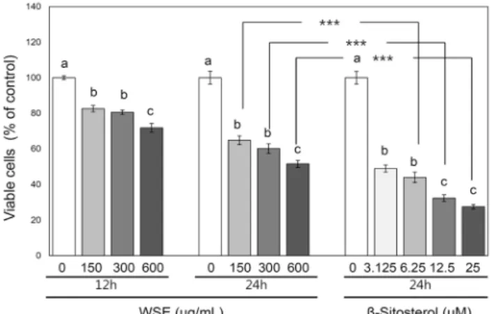
무제한적 증식은 암세포의 가장 기본적인 특징이라고 할 수 있기 때문에(42), 본 연구에서는 가장 먼저 흰깨 추출물 과 β-sitosterol의 폐암세포 성장에 대한 억제 활성을 조사 하였다. H1299 세포에 150, 300, 600 μg/mL 농도의 흰깨 추출물을 12시간 또는 24시간 동안 처리하거나, 3.125, 6.25, 12.5, 25 μM 농도의 β-sitosterol을 24시간 동안 처 리한 후 세포 생존 정도를 대조구 세포 생존 정도 대비 %로 나타내었다(Fig. 2).
흰깨 추출물이 12시간 또는 24시간 처리된 세포의 생존 정도는 흰깨 추출물이 처리되지 않은 대조구의 생존 정도에 대비하여 51.5~82.6%로, 모든 농도와 시점에서 유의적인 세포 성장 억제 활성을 보였다(P<0.05). 검정깨 메탄올 추 출물이 본 연구에서 이용된 농도보다 약 11~44배 높은 농도 범위로 24시간 처리되었을 때 인체유래 대장암세포에서 보 인 세포 성장 억제 활성(대조구 대비 약 10~80%)(10)과 비교하면 본 연구에서 관찰된 흰깨 추출물의 세포 성장 억제 활성은 처리된 농도에 비하여 높은 수준인 것으로 생각된다.
이어 흰깨 추출물의 H1299 폐암세포 성장 억제 활성이 처리 된 농도에 의존적이었는지 평가해 보기 위하여 선형 및 곡선 형 회귀분석을 실시하였다. 흰깨 추출물이 12시간과 24시 간 시점에서 보인 세포 성장 억제 활성과 흰깨 추출물의 처 리 농도는 선형, 2차 곡선, 지수형 회귀모형에서 모두 유의 한 상관관계가 있는 것으로 나타났다(R2=0.6~0.8, P<0.001).
또한 흰깨 추출물이 24시간 시점에서 보인 활성(대조구 생 존 대비 51.5~64.8%)이 12시간 시점에서 보인 활성(대조 구 생존 대비 71.8~82.6%)보다 높아, 처리 시간이 증가함 에 따라 흰깨 추출물의 세포 성장 억제 활성이 증가하는 경 향을 나타내었다.
Fig. 2. Effects of white sesame seed extract and β-sitosterol on the growth of H1299 lung cancer cells. H1299 cells were treated with either ethanol extract of white sesame seed (WSE;
at the concentrations of 150, 300, and 600 μg/mL for 12 h and 24 h) or β-sitosterol (at the concentrations of 3.125, 6.25, 12.5, and 25 μM for 24 h). Data are presented as % of control in the mean±SE of 8 determinations. Different letters (a-c) above bars mean statistical differences among different concentrations at respective time points (P<0.05). Asterisks mean statistical differences between indicated WSE- and β-sitosterol-treated groups (***P<0.001).
β-Sitosterol이 처리된 H1299 세포의 생존 정도는 24시 간 시점에서 대조구 생존 대비 27.5~49.0%로, 모든 농도에 서 유의미한 폐암세포 성장 억제 활성을 나타내었다(P<
0.05). 이는 β-sitosterol이 24시간 처리되었을 때 인체유 래 폐 선암세포에서 보인 성장 억제 활성(IC50: 약 2 μM) (43)이나 β-sitosterol이 5~20 μM 농도로 48시간 처리되 었을 때 대장암세포에서 보인 성장 억제 활성(대조구 생존 대비 약 25~50%)(21)과 비슷하였으나, β-sitosterol이 4
~64 μM 농도로 24시간 처리되었을 때 위암세포에서 보인 성장 억제 활성(대조구 생존 대비 약 8~25%)(22)보다는 다 소 낮은 활성이었다. 한편 두 가지 종류의 다발성 골수종 (26) 세포에서는 본 연구에서 이용된 농도보다 현저히 높은 농도(100 μM)의 β-sitosterol이 24시간 처리되었을 때 본 연구에서 관찰된 성장 억제 활성과 비슷한 정도의 활성(대조 구 생존 대비 약 25~50%)이 보고되었다. 연구마다 β-si- tosterol이 보인 활성에 다소 차이가 있는 것은 각 연구에서 이용된 세포의 종류나 실험 조건 등이 다르기 때문인 것으로 생각된다. 한편 β-sitosterol이 대장암세포와 정상 신장세 포의 생존에 미치는 영향을 비교한 연구(29)에서 β-sitos- terol 처리에 의하여 대장암세포의 생존 정도는 유의미하게 감소되었으나 정상 신장세포의 생존 정도는 고농도(약 100 mM) 처리 시에도 크게 변하지 않은 것으로 보고되어 암세포 에 선택적인 β-sitosterol의 성장 억제 활성이 시사되었다.
본 연구에서 나타난 β-sitosterol의 폐암세포 성장 억제 활 성이 처리 농도에 의존적이었는지 평가해 보기 위하여 선형 및 곡선형 회귀분석을 실시한 결과, 폐암세포 성장 억제 활 성과 흰깨 추출물의 처리 농도는 선형, 2차 곡선, 지수형
회귀모형에서 모두 유의한 상관관계가 있는 것으로 나타났 다(R2=0.6~0.8, P<0.001).
본 연구에서 산출된 흰깨 추출수율(15%)과 참깨의 β- sitosterol 함유량(약 2.3~3 mg/1 g)(44)으로 추정하여 보 면 600 μg/mL 농도의 흰깨 추출물과 25 μM 농도의 β- sitosterol은 동일 중량의 흰깨(약 4 mg)에서 획득되어질 수 있다. 따라서 150, 300, 600 μg/mL 농도의 흰깨 추출물 과 6.25, 12.5, 25 μM 농도의 β-sitosterol이 보인 폐암세 포 성장 억제 활성을 각각 비교하였다. 24시간 시점에서 보 인 β-sitosterol의 폐암세포 성장 억제 활성(대조구 생존 대비 27.5~44.1%)은 모든 농도에서 흰깨 추출물의 억제 활 성(대조구 생존 대비 51.5~64.8%)보다 유의적으로 높았다 (P<0.001). 이와 같은 결과로 볼 때 흰깨의 여러 함유 성분 의 혼합물인 추출물을 처리하는 것보다는 단일 함유 성분인 β-sitosterol을 처리하는 것이 H1299 폐암세포의 성장을 억제하는 데에 더 효과적인 것으로 생각된다.
세포 주기 변화
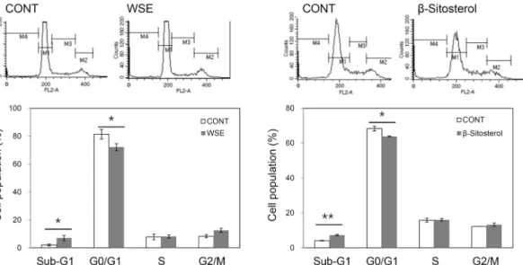
암세포는 apoptosis라고 불리는 세포 자살 프로그램이 정상적으로 일어나지 않아 잘 사멸되지 않고 성장률이 비정 상적으로 높아지는 특징을 가진다(31). 흰깨 추출물과 β -sitosterol이 세포주기를 변화시키는 효과가 있는지 여부 를 조사하기 위하여 DNA와 결합하는 붉은 빛의 형광물질인 PI와 유세포 분석기를 이용하여 H1299 폐암세포의 주기를 분석하였다. 먼저 흰깨 추출물이 600 μg/mL 농도로 처리된 세포의 sub-G1기 비율(7.1%)은 대조구 세포의 sub-G1 비 율(2.0%)보다 유의적으로 높았다(P<0.05, Fig. 3). 이어 흰 깨 추출물이 폐암세포의 apoptosis를 유도하는 지 여부를 핵산에 특이적으로 결합하는 형광물질인 DAPI를 이용하여 폐암세포의 핵을 염색한 다음 형광현미경으로 핵의 형태를 관찰하여 확인하였다. 600 μg/mL 농도의 흰깨 추출물을 24 시간 동안 처리한 후 DAPI로 염색한 결과, 대조구에서는 안정된 형태이면서 약한 DAPI 발색을 보이는 핵이 관찰되 었으나 흰깨 추출물이 처리된 세포에서는 응축된 형태를 보 이거나 DAPI 염색에 의한 강한 발색을 보이는 핵이 관찰되 었다(data not shown). Sub-G1기에 있는 세포는 곧 apop- tosis가 일어나는 것을 의미하기 때문에 이와 같은 핵 형태 관찰 결과와 종합하여 보면 흰깨 추출물은 폐암세포의 apoptosis를 유도하는 활성을 가지며 이러한 활성이 부분적 으로 세포 성장을 억제하는 결과를 초래하는 것으로 생각된 다. 한편 검정깨 메탄올 추출물은 인체 유래 대장암세포의 apoptosis를 유도하였는데, 이러한 활성은 apoptosis 저해 단백질의 유전자 발현 수준의 감소 등과 관련이 있는 것으로 보고되었다(10).
β-Sitosterol이 25 μM 농도로 처리된 세포의 sub-G1기 비율(7.3%)은 대조구 세포의 sub-G1 비율(4.1%)보다 유의 적으로 높았다(P<0.01, Fig. 3). β-Sitosterol은 대장암(21), 위암(22), 다발성 골수종(26) 세포에서 apoptosis를 유도하
Fig. 3. Effect of white sesame seed extract and β-sitosterol on cell cycle distribution of H1299 lung cancer cells. H1299 cells were treated with either ethanol extract of white sesame seed (WSE; at the concentration of 600 μg/mL) or β-sitosterol (at the concentration of 25 μM) for 48 h. Representative cell cycle distribution was shown in the upper panel. Data are presented as mean±SE of 3 determinations in the lower panel. Asterisks mean statistical differences between untreated control and treated groups (*P<0.05, **P<0.01).
Fig. 4. Effect of white sesame seed extract and β-sitosterol on the migration of H1299 lung cancer cells. H1299 cells were treated with either ethanol extract of white sesame seed (WSE;
at the concentrations of 150, 300, and 600 μg/mL) or β-sitosterol (at the concentrations of 3.125, 6.25, 12.5, and 25 μM) for 4 h. Data are presented as % of control in the mean±SE of 3 determinations. Different letters (a,b) above bars mean statistical differences among different concentrations of WSE or β-sitos- terol (P<0.05). Asterisks mean statistical differences between indicated WSE- and β-sitosterol-treated groups (*P<0.05, **P<
0.01, ***P<0.001).
는 활성이 있는 것으로 보고된 바 있으며, 이와 같은 apop- tosis 유도 활성은 β-sitosterol이 sphingomyelin cycle을 활성화시키고(24) 미토콘드리아 막 투과성을 변화시키는 (26) 등의 작용에 의한 것으로 제시된 바 있다.
세포 이동 억제 효과
암세포는 초기에 자라던 조직을 벗어나 주변 조직으로 침 윤한 후 혈액이나 림프를 통해 다른 조직으로 전이하여 새로 운 종양을 형성할 수 있는 능력을 가지게 되는데, 이러한 침윤 및 전이 과정에서 암세포가 획득하게 되는 중요한 특성 중 하나가 이동성의 증가이다(45). 따라서 본 연구에서는 β-sitosterol의 H1299 폐암세포 이동성에 대한 억제 활성 을 조사하였다(Fig. 4). 예비실험을 통하여 150~600 μg/
mL 농도의 흰깨 추출물 또는 3.125~25 μM 농도의 β-si- tosterol이 4시간 이내로 처리되었을 때 H1299 세포성장에 유의적인 변화를 일으키지 않는다는 것을 확인한 후(data not shown), 이동성 억제 활성 측정을 위한 처리시간을 4시 간으로 선정하였다. 흰깨 추출물이 150, 300, 600 μg/mL 농도로 처리된 폐암세포의 이동 정도는 대조구의 이동 정도 와 유의적으로 다르지 않았다(Fig. 4). 반면 β-sitosterol이 3.125~25 μM 범위의 농도로 처리된 세포의 이동 정도는 대조구 대비 80.8~86.2%로 약하지만 유의적인 억제 활성 이 나타났다(P<0.05, Fig. 4). 이와 같은 결과로 볼 때 H1299 폐암세포의 이동성을 억제하는 데 있어 흰깨 추출물의 형태 보다 β-sitosterol의 단독 처리가 효과적인 것으로 생각된 다. β-Sitosterol의 유방암세포 침윤에 대한 억제 효과(46) 와 전립선암세포 침윤 및 이동에 대한 억제 효과(28)에 대해 서는 보고된 바 있으나, 폐암세포의 이동에 대한 억제 효과
에 대해서는 아직까지 보고된 바 없다. 앞으로 β-sitosterol 에 의한 폐암세포의 이동성 억제와 관련된 기전 연구가 필요 할 것으로 생각된다.
세포 부착 억제 효과
암화 과정 중 침윤과 전이가 일어나기 위해서 일차적으로 일어나는 현상 중 하나가 세포 부착력의 변화이다(45). 침윤 단계에서의 암세포는 정상세포에 비하여 세포 간 부착력이
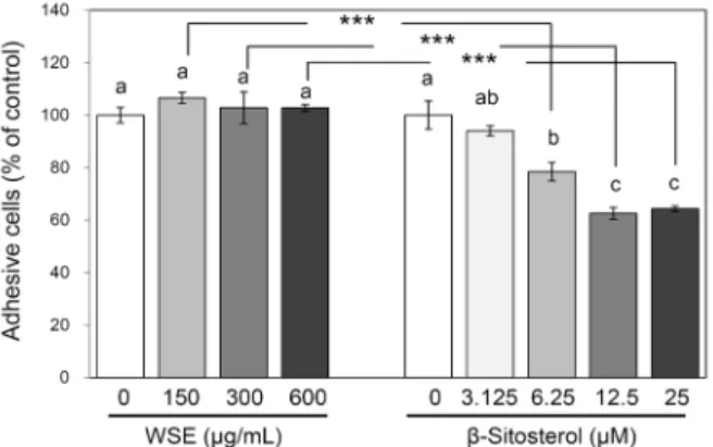
Fig. 5. Effect of white sesame seed extract and β-sitosterol on the adhesion of H1299 lung cancer cells. H1299 cells were treat- ed with either ethanol extract of white sesame seed (WSE; at the concentrations of 150, 300, and 600 μg/mL) or β-sitosterol (at the concentrations of 3.125, 6.25, 12.5, and 25 μM) for 2 h. Data are presented as % of control in the mean±SE of 6∼7 determinations. Different letters (a,b) above bars mean statistical differences among different concentrations of WSE or β-sitos- terol (P<0.05). Asterisks mean statistical differences between indicated WSE- and β-sitosterol-treated groups (***P<0.001).
감소하여 상호 결합력이 떨어지는 반면, 세포와 세포 외 기 질 간의 부착력은 증가하는 것으로 알려져 있다(45). 본 연 구에서는 세포외 기질에 존재하는 대표적인 세포 부착 물질 인 fibronectin(45)이 코팅된 세포배양용 plate를 이용하여 폐암세포가 fiboronectin에 부착되는 것을 흰깨 추출물 또 는 β-sitosterol이 억제하는 정도를 측정하였다. 흰깨 추출 물 및 β-sitosterol 처리시간은 기존에 보고된 방법(41)에 따라 2시간으로 선정하였다. 흰깨 추출물이 150, 300, 600 μg/mL 농도로 처리된 폐암세포의 부착 정도는 대조구의 부 착 정도와 유의적 차이가 없었던 반면, β-sitosterol이 6.25, 12.5, 25 μM 농도로 처리된 폐암세포의 부착 정도는 대조구 대비 21.5~37.4%(P<0.05, Fig. 5)로, 농도 의존적인 부착 억제 활성이 나타났다(곡선형 회귀모형 R2=0.6, P<0.01).
따라서 앞서 기술한 폐암세포의 성장 및 이동 등에서 나타난 결과와 마찬가지로 폐암세포의 부착을 억제하는 데에도 흰 깨 추출물보다 β-sitosterol이 더 효과적인 것으로 생각된 다. β-Sitosterol은 유방암세포에서 collagen과 laminin 같 은 기저막의 구성 성분에 부착하는 것을 억제하는 효과가 있다고 보고된 바 있다(46). 앞으로 β-sitosterol에 의한 폐 암세포의 부착 억제와 관련된 기전 연구가 필요할 것으로 생각된다.
요 약
본 연구에서는 흰깨의 에탄올 추출물이 폐암세포의 성장, 이동, 부착 등에 미치는 영향을 인체유래 폐암 세포주인 H1299 세포를 이용하여 in vitro 수준에서 조사하고, 이러 한 흰깨 추출물의 효과를 흰깨의 주요 활성 성분 중 하나인 β-sitosterol의 효과와 비교하고자 하였다. 흰깨 추출물
(150~600 μg/mL)과 β-sitosterol(3.125~25 μM)은 H1299 세포의 성장을 각각 대조구 대비 51.5~82.6%와 27.5~
49.0%로 억제하는 농도 의존적 활성을 나타내었고, 이러한 흰깨 추출물과 β-sitosterol의 세포 성장 억제 활성은 부분 적으로 apoptosis 유도 활성에서 기인되는 것으로 생각된 다. 흰깨 추출물(150~600 μg/mL)은 H1299 세포의 이동과 부착을 억제하는 활성을 나타내지 않은 반면에 β-sitosterol 은 3.125~25 μM 농도에서 세포 이동을 대조구 대비 80.8~
86.2%로, 6.25~25 μM 농도에서 세포 부착을 대조구 대비 21.5~37.4%로 각각 억제하는 활성을 나타내었다. 이상의 연구 결과를 종합하여 볼 때 H1299 폐암세포의 성장, 이동, 부착 등을 억제하는 데에는 흰깨의 추출물보다는 단일성분인 β-sitosterol이 더 효과적인 것으로 생각된다. 앞으로 이와 같은 연구 결과가 in vivo 수준에서 재현되는지 여부를 검증 하고 관련 기전을 탐색하는 것이 필요할 것으로 생각된다.
감사의 글
이 논문은 2012년도 충북대학교 학술연구지원사업의 연구 비 지원에 의하여 연구되었습니다.
REFERENCES
1. Lee CB. 2003. Coloured flora of Korea. 1st ed. Hyang Mun Sa, Seoul, Korea. p 140.
2. Kim TJ. 1996. Korean resources plants Ⅳ. Seoul National University Publisher, Seoul, Korea. p 60.
3. National Academy of Agricultural Science. 2011. Food Composition Table. 8th ed. Rural Development Administra- tion, Suwon, Korea. p 108.
4. Ministry of Health & Welfare. 2014. Korea Health Statistics 2013. Korea National Health and Nutrition Examination Survey (KNHANES) VI-1. Ministry of Health & Welfare, Sejong, Korea. p 475-489.
5. Lee SU, Kang CW, Kang DH, Yasumoto S, Kasuta M.
1999. Varietal variation of sesamin, sesamolin, and oil con- tents according to seed-coat colors in sesame. Korean J Breed 31: 286-292.
6. Lee J, Lee SR. 1994. Analysis of phenolic substances con- tent in Korean plant foods. Korean J Food Sci Technol 26:
310-316.
7. Park JL, Chae KY, Hong JS. 2007. A comparison of anti- oxidant activities in black sesame seeds according to prepa- ration and cooking conditions. J East Asian Soc Dietary Life 17: 520-531.
8. Ahn CY, Hyun KH, Park KH. 1992. Investigation of anti- oxidative substances in black sesame seed. Korean J Food Sci Technol 24: 31-36.
9. Hu Q, Xu J, Chen S, Yang F. 2004. Antioxidant activity of extracts of black sesame seed (Sesamum indicum L.) by supercritical carbon dioxide extraction. J Agric Food Chem 52: 943-947.
10. Kim MJ, Jeong MK, Chang PS, Lee JH. 2009. Radical scav- enging activity and apoptotic effects in HT-29 human colon cancer cells of black sesame seed extract. Int J Food Sci Technol 44: 2106-2112.
11. Visavadiya NP, Soni B, Dalwadi N. 2009. Free radical scav-
enging and antiatherogenic activities of Sesamum indicum seed extracts in chemical and biological model systems.
Food Chem Toxicol 47: 2507-2515.
12. Wichitsranoi J, Weerapreeyakul N, Boonsiri P, Settasatian C, Settasatian N, Komanasin N, Sirijaichingkul S, Teerajet- gul Y, Rangkadilok N, Leelayuwat N. 2011. Antihypertensive and antioxidant effects of dietary black sesame meal in pre- hypertensive humans. Nutr J 10: 82.
13. Alipoor B, Haghighian MK, Sadat BE, Asghari M. 2012.
Effect of sesame seed on lipid profile and redox status in hyperlipidemic patients. Int J Food Sci Nutr 63: 674-678.
14. Woyengo TA, Ramprasath VR, Jones PJ. 2009. Anticancer effects of phytosterols. Eur J Clin Nutr 63: 813-820.
15. Marangoni F, Poli A. 2010. Phytosterols and cardiovascular health. Pharmacol Res 61: 193-199.
16. Liz R, Zanatta L, dos Reis GO, Horst H, Pizzolatti MG, Silva FR, Fröde TS. 2013. Acute effect of β-sitosterol on calcium uptake mediates anti-inflammatory effect in murine acti- vated neutrophils. J Pharm Pharmacol 65: 115-122.
17. Loizou S, Lekakis I, Chrousos GP, Moutsatsou P. 2010.
β-Sitosterol exhibits anti-inflammatory activity in human aortic endothelial cells. Mol Nutr Food Res 54: 551-558.
18. Nirmal SA, Pal SC, Mandal SC, Patil AN. 2012. Analgesic and anti-inflammatory activity of β-sitosterol isolated from Nyctanthes arbortristis leaves. Inflammopharmacology 20:
219-224.
19. Donald PR, Lamprecht JH, Freestone M, Albrecht CF, Bouic PJ, Kotze D, van Jaarsveld PP. 1997. A randomised placebo-controlled trial of the efficacy of beta-sitosterol and its glucoside as adjuvants in the treatment of pulmonary tuberculosis. Int J Tuberc Lung Dis 1: 518-522.
20. Awad AB, Chen YC, Fink CS, Hennessey T. 1996. β- Sitosterol inhibits HT-29 human colon cancer cell growth and alters membrane lipids. Anticancer Res 16: 2797-2804.
21. Choi YH, Kim YA, Park C, Choi BT, Lee WH, Hwang KM, Jung KO, Park KY. 2004. β-Sitosterol induced growth inhibition is associated with up-regulation of Cdk inhibitor p21WAF1/CIP1 in human colon cancer cells. J Korean Soc Food Sci Nutr 33: 1-6.
22. Zhao Y, Chang SK, Qu G, Li T, Cui H. 2009. β-Sitosterol inhibits cell growth and induces apoptosis in SGC-7901 hu- man stomach cancer cells. J Agric Food Chem 57: 5211- 5218.
23. Jourdain C, Tenca G, Deguercy A, Troplin P, Poelman D.
2006. In-vitro effects of polyphenols from cocoa and β-si- tosterol on the growth of human prostate cancer and normal cells. Eur J Cancer Prev 15: 353-361.
24. von Holtz RL, Fink CS, Awad AB. 1998. β-Sitosterol acti- vates the sphingomyelin cycle and induces apoptosis in LNCaP human prostate cancer cells. Nutr Cancer 32: 8-12.
25. Ju YH, Clausen LM, Allred KF, Almada AL, Helferich WG.
2004. β-Sitosterol, β-sitosterol glucoside, and a mixture of β-sitosterol and β-sitosterol glucoside modulate the growth of estrogen-responsive breast cancer cells in vitro and in ovariectomized athymic mice. J Nutr 134: 1145-1151.
26. Sook SH, Lee HJ, Kim JH, Sohn EJ, Jung JH, Kim B, Kim JH, Jeong SJ, Kim SH. 2014. Reactive oxygen species- mediated activation of AMP-activated protein kinase and c-Jun N-terminal kinase plays a critical role in beta-sitoster- ol-induced apoptosis in multiple myeloma U266 cells.
Phytother Res 28: 387-394.
27. Awad AB, Downie A, Fink CS, Kim U. 2000. Dietary phy- tosterol inhibits the growth and metastasis of MDA-MB-231 human breast cancer cells grown in SCID mice. Anticancer
Res 20: 821-824.
28. Awad AB, Fink CS, Williams H, Kim U. 2001. In vitro and in vivo (SCID mice) effects of phytosterols on the growth and dissemination of human prostate cancer PC-3 cells. Eur J Cancer Prev 10: 507-513.
29. Baskar AA, Ignacimuthu S, Paulraj GM, Al Numair KS.
2010. Chemopreventive potential of β-sitosterol in experi- mental colon cancer model-an in vitro and in vivo study.
BMC Complement Altern Med 10: 24.
30. Mendilaharsu M, De Stefani E, Deneo-Pellegrini H, Carzo- glio J, Ronco A. 1998. Phytosterols and risk of lung cancer:
a case-control study in Uruguay. Lung Cancer 21: 37-45.
31. Weinberg RA. 2007. The biology of cancer. Garland Sci- ence, New York, NY, USA. p 588-597.
32. World Health Organization. 2012. World Health Statistics 2012. World Health Organization, Geneva, Switzerland. p 80-81.
33. Statistics Korea. 2012. 2011 Cause of Death Statistics. Dae- jeon, Korea. p 9-10.
34. Amin AR, Kucuk O, Khuri FR, Shin DM. 2009. Perspectives for cancer prevention with natural compounds. J Clin Oncol 27: 2712-2725.
35. Bode AM, Dong Z. 2009. Cancer prevention research-then and now. Nat Rev Cancer 9: 508-516.
36. McCullough ML, Giovannucci EL. 2004. Diet and cancer prevention. Oncogene 23: 6349-6364.
37. Lambert JD, Lu G, Lee MJ, Hu J, Ju J, Yang CS. 2009.
Inhibition of lung cancer growth in mice by dietary mixed tocopherols. Mol Nutr Food Res 53: 1030-1035.
38. Irons R, Tsuji PA, Carlson BA, Ouyang P, Yoo MH, Xu XM, Hatfield DL, Gladyshev VN, Davis CD. 2010. Defi- ciency in the 15-kDa selenoprotein inhibits tumorigenicity and metastasis of colon cancer cells. Cancer Prev Res 3:
630-963.
39. Toton E, Ignatowicz E, Bernard MK, Kujawski J, Rybczyn- ska M. 2013. Evaluation of apoptotic activity of new con- densed pyrazole derivatives. J Physiol Pharmacol 64: 115- 123.
40. Han SX, Zhu Q, Ma JL, Zhao J, Huang C, Jia X, Zhang D. 2010. Lowered HGK expression inhibits cell invasion and adhesion in hepatocellular carcinoma cell line HepG2.
World J Gastroenterol 16: 4541-4548.
41. Matsuura N, Miyamae Y, Yamane K, Nagao Y, Hamada Y, Kawaguchi N, Katsuki T, Hirata K, Sumi S, Ishikawa H.
2006. Aged garlic extract inhibits angiogenesis and pro- liferation of colorectal carcinoma cells. J Nutr 136: 842S- 846S.
42. Hanahan D, Weinberg RA. 2011. Hallmarks of cancer: the next generation. Cell 144: 646-674.
43. Hsu HF, Huang KH, Lu KJ, Chiou SJ, Yen JH, Chang CC, Houng JY. 2011. Typhonium blumei extract inhibits pro- liferation of human lung adenocarcinoma A549 cells via in- duction of cell cycle arrest and apoptosis. J Ethnopharmacol 135: 492-500.
44. Lu B, Zhang Y, Wu X, Shi J. 2007. Separation and determi- nation of diversiform phytosterols in food materials using supercritical carbon dioxide extraction and ultraperformance liquid chromatography-atmospheric pressure chemical ion- ization-mass spectrometry. Anal Chim Acta 588: 50-63.
45. Kleinsmith LJ. 2008. Principles of cancer biology. 1st ed.
Life Science Publishing Co., Seoul, Korea. p 16-63.
46. Awad AB, Williams H, Fink CS. 2001. Phytosterols reduce in vitro metastatic ability of MDA-MB-231 human breast cancer cells. Nutr Cancer 40: 157-164.