후추의 주요 성분인 Piperine의 대장암세포 세포사멸 유도 효과
김은지2*․박희숙1*․신민정1*․신현경1,2․윤정한1,2†
1한림대학교 식품영양학과
2한림대학교 식의약품의 효능평가 및 기능성소재개발센터
Induction of Apoptosis in HT-29 Human Colon Cancer Cells by the Pepper Component Piperine
Eun Ji Kim2*, Heesook Park1*, Minjeong Shin1*, Hyun-Kyung Shin1,2, and Jung Han Yoon Park1,2†
1
Dept. of Food Science and Nutrition and
2Center for Efficacy Assessment and Development of Functional Foods and Drugs, Hallym University, Gangwon 200-702, Korea
Abstract
Piperine is an alkaloid-amine found in pepper and has been reported to have anticarcinogenic properties. To explore the possibility that piperine has cancer chemopreventive and chemotherapeutic effects in colon cancer, we examined whether piperine inhibits the growth of HT-29 human colon cancer cells and investigated the mechanisms for this effect. Cells were cultured with various concentrations (0~40 μM) of piperine. Piperine decreased the cell viability and induced apoptosis of HT-29 cells. Western blot analysis of total cell lysates revealed that piperine decreases the protein levels of Bcl-2, Mcl-1, and intact Bid but increases Bik levels.
Piperine increased the percentage of cells with depolarized mitochondrial membrane, and the release of cyto- chrome c into cytoplasm. Piperine induced the cleavage of poly (ADP-ribose) polymerase and caspases 8, 9, 7, and 3 and increased the Fas levels. In addition, piperine significantly decreased the protein levels of survivin.
The present results indicate that piperine inhibits the growth of HT-29 colon cancer cells by the induction of apoptosis, which may be mediated by its ability to change the Bcl-2 family proteins, increase the activation of caspases, and decrease survivin levels. Overall, our findings suggest that piperine has cancer chemo- therapeutic effects in colon cancer.
Key words: piperine, colon cancer, apoptosis, caspase, cytochrome c, survivin
*Kim EJ, Park H, and Shin M contributed equally to this study.
†Corresponding author. E-mail: [email protected]
†Tel: 82-33-248-2134, Fax: 82-33-256-0199
서 론
세계적으로 암 발병 및 암에 의한 사망률이 급증함에 따라 암은 생명을 위협하는 가장 심각한 건강 문제로 간주되고 있다. 특히 대장암은 미국에서 여러 암 중 발병률과 암에 의한 사망률에서 2위를 차지하고 있으며(1), 우리나라에서 도 최근 10년 사이 빠른 속도로 증가하고 있다. 대장암의 근본적인 치료는 수술이나, 수술로 완치가 불가능한 경우가 많아 항암화학요법 등이 병행하여 실시되고 있다. 대장암 치료를 위해 5-fluorouracil 등의 항암제가 사용되고 있으나 효과가 좋지 못하며(2), 정상세포에 독성 및 심각한 부작용 을 초래하고 있다(3,4). 따라서 많은 연구자들은 자연에 존재 하고 예로부터 안전하게 섭취되었던 식품에서 항암 효과를 나타내는 물질을 찾아 독성과 부작용이 없으며, 우수한 효과 를 나타내는 항암제 및 항암 치료보조제 개발 연구를 활발하 게 진행하고 있다.
향신료로서 널리 사용되고 있는 후추는 인도를 비롯한 아 시아에서 기관지염, 열병, 위장 질환, 중풍 및 관절염 치료 등의 전통의학제로 사용되고 있다(5). 후추에서 분리된 다양 한 phytochemical들은 여러 생리활성을 나타내는 것으로 알 려지고 있으며, 그 중에서 alkaloid-amine 성분인 piperine이 대표적이다. Piperine은 항산화 활성(6), 간세포 보호 효과 (7), 뇌세포 보호 효과(8) 등의 효과를 나타냄이 보고되었으 며, Pradeep과 Kuttan(9)은 piperine이 B16F-10 melanoma cell에서 염증과 관련된 NF-kB, c-Fos, CREB, ATF-2 및 proinflammatory cytokine의 발현을 억제함을 보고하였다.
Piperine은 benzo(a)pyrene을 투여하여 폐암을 유도한 동물 에서 강한 암예방 효과를 나타냈고(10,11), in vivo에서 B16F-10 melanoma cells의 폐전이를 효과적으로 억제하였 고(12), Sarcoma 180에 의한 고형암 형성을 억제하였다(13).
Duessel 등(14)이 대장암세포인 DLD-1 세포의 증식이 pi- perine에 의해 억제되었다고 보고하였으나, 이 연구를 제외
하면 현재까지 piperine이 대장암에 미치는 영향과 기전에 대해 연구된 바가 없다.
세포사멸(apoptosis)은 선택적인 세포 소실을 일으키는 생리적인 과정으로 조직의 항상성 유지에 필수적인 조절 작 용이다(15). 세포사멸은 세포 내․외적인 세포사멸 신호에 의해 시작되어 세포사멸을 조절하는 단백질인 Bcl-2 family 단백질, caspases, 세포사멸억제단백질(inhibitors of apop- tosis protein)의 유기적인 상호작용에 의해 조절된다(16- 19). 세포사멸 기전에 결함이 생기거나 세포사멸 신호 전달 이 부적절하게 일어나 세포사멸 조절에 이상이 생기면 세포 가 비정상적으로 증식하여 암이 초래될 수 있다(20). 그러므 로 암세포의 비정상적인 세포사멸 기전을 적절하게 회복하 는 것은 암예방 및 암 치료의 표적이 될 수 있으며, 현재 이용되고 있는 많은 항암제는 암세포의 세포사멸을 유도하 여 항암 효과를 나타낸다(21,22). 그러므로 암세포의 세포사 멸을 유도하는 식품 성분들은 항암제로 개발할 수 있는 좋은 소재가 될 수 있다.
후추의 주요 성분인 piperine은 강한 항암 활성을 나타내 는 것으로 보고되고 있으나, 대장암의 증식에 미치는 영향에 대해 보고된 바가 미미하다. 본 연구에서는 인간의 대장에서 유래한 암세포인 HT-29 세포를 사용하여 piperine이 대장 암세포의 증식과 세포사멸에 미치는 영향 및 그 작용 기전을 밝히고자 하였다.
재료 및 방법
재료
인간의 대장에서 유래한 암세포인 HT-29 세포는 Ameri- can Type Culture Collection(Rockville, MD, USA)에서 구 입하였다. 세포배양에 사용한 Dulbecco's Modified Eagle's Medium:Nutrient Mixture Ham's F12(DMEM/F12)은 Gibco/BRL(Gaitherburg, MD, USA)에서 구입하였다. Fetal bovine serum(FBS), penicillin-streptomycin, Trypsin- EDTA 등은 Cambrex Bio Technology(Walkersville, MD, USA)에서 구입하였다. Piperine, bovine serum albumin (BSA), 3-(4,5-dimethylthiazol-2-yl)-2,5-diphenyltetra- zolium bromide(MTT), 5,5’,6,6’-tetrachloro-1,1’,3,3’-tet- raethyl-imidacarbocyanine iodide(JC-1), anti-β-actin 항 체와 본 연구에 사용한 일반적인 시약은 Sigma Chemical Co.
(St. Louis, MO, USA)에서 구입하여 사용하였다. Phycoer- ythrin(PE)-conjugated Annexin V, 7-amino-actinomycin D와 cytochrome c 항체는 BD Pharmingen(Franklin Lakes, NJ, USA)에서 구입하여 사용하였다. Bcl-2, Fas, heat shock protein(HSP)60 항체는 Santa Cruz Biotechnology Inc.(Santa Cruz, CA, USA)에서 구입하였고, cleaved cas- pase-3, cleaved caspase-7, cleaved caspase-8, cleaved caspase-9, cleaved poly(ADP-ribose) polymerase(PARP),
survivin, Mcl-1, Bid, Bik에 대한 항체는 Cell Signaling Technology(Beverly, MA, USA)에서 구입하여 사용하였 다. Horse radish peroxidase-linked anti-rabbit IgG와 horse radish peroxidase-linked anti-mouse IgG는 Amer- sham(Bukinghamshire, England)에서 구입하여 사용하였다.
세포 배양
HT-29 세포는 DMEM/F12 배지에 10% FBS, 100 units/mL penicillin과 100 μg/mL streptomycin을 첨가한 배 양액을 사용하여 37oC 습윤한 CO2 배양기(5% CO2/95% air) 에서 배양하였다. 세포가 배양접시의 70~80% 정도 차면 phosphate-buffered saline(PBS, pH 7.4)으로 세포의 단층 을 씻어낸 후 0.25% trypsin-2.65 mM EDTA를 처리하여 세포를 계대 배양하였고, 배지는 2일마다 교환하였다.
세포 증식 측정
HT-29 세포를 10% FBS가 포함된 배지로 희석하여 50,000cells/well의 밀도로 24-well plate에 분주하였다. 24 시간이 지난 후 Kim 등(23-26)과 같은 방법으로 1% FBS를 포함한 DMEM/F12로 배지를 교환하여 혈청에 함유된 여러 성분의 효과를 최소화하였다. 세포를 1% FBS가 포함되어 있는 배지에서 24시간 동안 배양한 후 1% FBS가 포함되어 있는 배지에 piperine을 0, 10, 20, 40 μM 농도로 첨가한 배지 로 교환하여 세포를 배양하였다. Piperine은 dimethyl sulf- oxide(DMSO)에 녹여 40 mM로 만들어 -70oC에 보관하여 사용하였고, 대조군을 포함하여 모든 처리군의 DMSO 농도 를 동일하게 하였다. Piperine을 첨가하여 세포를 0, 24, 48, 72시간 동안 배양한 후 MTT assay 방법(27)으로 세포 증식 정도를 측정하였다.
세포주기 측정
세포를 24-well plate에 50,000 cells/well의 밀도로 분주 한 후 위와 동일한 방법으로 0 또는 40 μM 농도로 piperine 을 처리하여 48시간 동안 세포를 배양하였다. 세포 단층을 PBS로 헹구고 trypsin-EDTA를 처리하여 세포를 분리하여 수집한 후 Kim 등(28)과 같은 방법으로 세포를 propidium iodide로 염색하였다. Propidium iodide에 의해 염색된 세포 를 FACScanTM(Becton Dicknson, Franklin Lake, NJ, USA)을 사용하여 flow cytometry 방법에 의해 세포주기를 측 정하였다.
Hoechst H 33258 염색
세포를 24-well plate에 cover slip을 깔고 50,000 cells/
well의 밀도로 세포를 분주한 후 위와 동일한 방법으로 0 또는 40 μM 농도로 piperine을 처리하여 72시간 동안 배양하 였다. 세포 단층을 PBS로 헹구고 4% formaldehyde로 세포 를 고정하였다. 세포에 10 μg/mL Hoechst H 33258 용액을 넣어 어두운 곳에서 1시간 염색하였다. 세포를 PBS로 충분 히 헹군 후 형광현미경으로 관찰하였다(29).
세포사멸 세포수 측정(Annexin V 염색)
세포를 24-well plate에 50,000cells/well의 밀도로 분주 한 후 위와 동일한 방법으로 piperine을 처리하여 72시간 동 안 배양하였다. 세포 단층을 PBS로 헹구고 trypsin-EDTA 를 처리하여 세포를 분리하여 수집한 후 Kim 등(23-26)과 같은 방법으로 세포를 phycoerythrin(PE)-conjugated An- nexin V와 7-amino-actinomycin D(7-AAD)로 염색한 후 Annexin V 또는 7-AAD에 의해 염색된 세포를 FACScanTM (Becton Dicknson)을 사용하여 flow cytometry 방법에 의해 측정하였다.
미토콘드리아 막 투과성 측정(JC-1 염색)
미토콘드리아 막의 투과성을 조사하기 위하여 dual- emission potential-sensitive probe JC-1을 사용하였다.
JC-1은 낮은 막 전위(membrane potential)에서는 green- fluorescent monomer 형태로 나타나며 미토콘드리아의 전 압이 증진된 막 전위에서는 red-fluorescent J aggregate를 형성한다(29). 따라서 JC-1의 red와 green fluorescence의 비율은 막의 탈분극 정도를 나타낼 수 있다. HT-29 세포를 위와 동일한 방법으로 piperine을 첨가하여 48시간 동안 세 포를 배양하였다. 세포를 PBS로 헹군 후, trypsin-EDTA를 이용하여 분리한 세포를 수집하였다. JC-1을 2 μg/mL 농도 로 첨가하여 어두운 곳에서 30분 동안 반응한 후, FACScanTM (Becton Dickinson)을 사용하여 flow cytometry 방법에 의 해 측정하였다.
세포사멸 관련 단백질 수준 측정(Western blot analy- sis)
HT-29 세포를 1×10⁶ cells/dish의 밀도로 100 mm dish 에 분주하였고, 위와 동일한 방법으로 piperine을 처리하여 48시간 또는 72시간 배양한 후 total cell lysate를 만들었다 (23-26). 세포질 분획은 Eguchi 등(30)이 제시한 방법으로 분리하였다. Total cell lysate와 세포질 분획의 단백질 농도 는 BCA protein assay kit(Pierce, Rockford, IL, USA)을 사 용하여 정량하였다. Total cell lysate(단백질 50 μg 또는 100 μg)을 4~20% 혹은 10~20% gradient sodium dodecyl sul- fate polyacrylamide gel electrophoresis(SDS-PAGE)로 크 기에 따라 분리한 후, polyvinylidene fluoride(PVDF) mem- brane(Millipore, Bedford, MA, USA)에 이동시켰다. Mem- brane은 5% non-fat dry milk-TBST(20 mM Tris-HCl, pH 7.6, 150 mM NaCl, 0.1% Tween 20)로 1시간 교반한 후 측정 하고자 하는 항체를 각각 첨가하여 4oC에서 16시간 또는 상 온에서 1시간 동안 교반하였다. 그 후 horseradish perox- idase(HRP)-linked anti-rabbit IgG 또는 HRP-linked an- ti-mouse IgG를 첨가하여 1시간 교반하였다. 각 단백질 밴 드는 ImmobilonTM Western Chemiluminescent HRP Substrate(Millipore)을 사용하여 enhanced chemilumine- scence 방법으로 가시화하였다. 각 단백질 밴드의 강도는
Bio-profile Bio-ID application(Vilber-Lourmat, Marine la Vallee, France)을 사용하여 측정하였다.
통계처리
본 연구의 모든 분석 수치는 mean±SE으로 나타내었다.
수집된 결과는 SAS(Statistical Analysis System) Windows v. 8.12 프로그램(SAS Institute, Cary, NC, USA)을 이용하 여 통계 분석하였으며, 각 실험군의 평균치간의 유의성은 p<0.05 수준에서 analysis of variance와 Duncan’s multiple range test에 의해 분석하였다.
결과 및 고찰
Piperine이 대장암세포인 HT-29 세포의 증식에 미치는 영향
후추의 주요한 매운 맛 성분인 piperine은 항산화 활성 등 다양한 생리활성을 나타내는 것으로 알려지고 있다(6-8).
Selvendiran 등은 benzo(a)pyrene을 투여하여 폐암을 유도 한 동물에서 piperine이 강한 암예방 효과를 나타냄을 보고 하였다(10,11). 또한 piperine이 in vivo에서 B16F10 mela- noma cells의 폐전이를 억제하였다고 보고되었고(12), Sarcoma 180에 의한 고형암 생성이 piperine 처리에 의해 억제되었다고 보고되었다(13). 여러 연구 보고를 통해 pi- perine이 항암 효과를 나타내는 것을 알 수 있으나, piperine 이 대장암에 미치는 영향과 작용 기전에 대해 연구된 바가 드물다. 따라서 본 연구에서는 piperine이 대장암세포의 증 식에 미치는 영향과 그 작용 기전을 조사하고자 하였다.
Piperine이 대장암세포의 증식에 미치는 영향을 조사하기 위해 인간의 대장에서 유래한 암세포인 HT-29 세포에 다양 한 농도(0, 10, 20, 40 μM)의 piperine을 처리하여 MTT as- say 방법(27)으로 세포 증식 정도를 측정하였다. Piperine 처리 농도가 증가할수록 유의적으로 세포 증식이 감소하였 다. Piperine에 의한 세포 증식 감소는 piperine 처리 후 24시 간부터 나타났으며 시간이 경과함에 따라 세포 증식 감소 현상은 더욱 현저히 나타났고, 40 μM 농도로 piperine을 72 시간 처리한 경우 piperine을 처리하지 않은 대조군에 비해 76.4% 세포 증식이 감소하였다(Fig. 1). Duessel 등은 piper- ine을 100~200 μM 농도로 처리하여 48시간 또는 72시간을 처리한 경우 대장암세포인 DLD-1 세포의 증식이 억제되었 다고 보고하였다(14). 또한 Sunila와 Kuttan은 piperine이 Dalton's lymphoma ascites(DLA) 세포와 Ehrlich ascites carcinoma(EAC) 세포의 증식을 현저히 억제함을 보고하였 다(31). 이를 통해 piperine이 대장암세포를 비롯한 여러 암 세포의 증식을 효과적으로 억제함을 알 수 있다.
Piperine이 대장암세포인 HT-29 세포의 세포주기 진행 과 세포사멸(apoptosis)에 미치는 영향
암세포의 세포주기 진행 지연과 세포사멸 유도는 암세포
Fig. 1. Effect of piperine on the cell viability of HT-29 cells.
HT-29 cells were plated in 24-well plates at 5
×
104 cells/well in DMEM/F12 supplemented with 10% FBS. One day later, the monolayers were serum-deprived with DMEM/F12 supple- mented with 1% FBS (serum-deprivation medium) for 24 hr.After serum deprivation, cells were incubated in serum-depriva- tion medium in the absence or presence of various concentrations of piperine. Cell viability were estimated by the MTT assay. Each bar represents the mean
±
SE (n=6). Bars with different letters are significantly different at p<0.05 by Duncan's multiple range test at each time point.증식 억제 기전 중의 하나며, 항암활성을 가진 물질들의 중 요한 특성이며, 현재 이용되고 있는 많은 항암제는 암세포의 세포사멸을 유도하여 그 효과를 나타내고 있다(21,22).
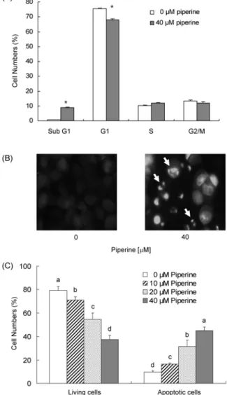
Piperine이 HT-29 세포의 증식을 효과적으로 억제하였으므 로(Fig. 1) piperine의 세포 증식 억제가 세포주기 진행 지연 에 의한 것인지를 확인하기 위해 HT-29 세포의 세포 배양액 에 piperine을 첨가하여 세포를 48시간 배양한 후 핵을 pro- pidium iodide로 염색하여 flow cytometry 방법으로 세포주 기 진행의 변화를 확인하였다. Piperine을 40 μM 농도로 48 시간 처리한 경우 sub G1기에 머무른 세포수가 증가하였고 G1기에 머무른 세포 수는 감소하였으며 S기와 G2/M기에 머무른 세포 수는 유의적인 차이가 나타나지 않았다(Fig.
2A). Sub G1기에 머무른 세포수의 증가는 세포사멸의 특징 으로 간주되므로(32) piperine이 세포사멸을 유도하는 것으 로 생각되어 본 연구에서는 piperine이 세포사멸에 미치는 영향을 조사하였다. HT-29 세포의 세포 배양액에 piperine 을 첨가하여 세포를 72시간 배양한 후 Hoechst H 33258로 염색하여 세포의 형태학적인 변화를 관찰하였다. Fig. 2B에 서 보는 바와 같이 piperine을 40 μM로 처리한 경우 세포사 멸이 일어나면 나타나는 apoptotic bodies가 관찰되었다.
Piperine에 의해 유도된 세포사멸을 정량하기 위해 HT-29 세포의 세포 배양액에 piperine을 다양한 농도로 첨가하여 72시간 배양한 후 Annexin V와 7-AAD로 염색하여 flow cytometry 방법으로 early apoptotic 세포 수를 측정하였다.
Piperine 처리 농도가 증가할수록 살아있는 세포 수는 현저 히 감소하였고, 세포사멸 세포수는 농도 의존적으로 증가하 였다(Fig. 2C). 이상의 결과를 통해 piperine에 의한 세포증 식 억제 효과가 세포사멸 유도에 기인함을 알 수 있으며,
Fig. 2. Effect of piperine on the cell cycle progression and apoptosis of HT-29 cells. Cells were plated and treated with piperine as described in Fig. 1. (A) Flow cytometry analysis of cell cycle distribution. Cells were treated with 0 or 40
μ
M piperine for 48 hr. Cells were trypsinzed, fixed, and treated RNase. Cellular DNA was then stained with propidium iodide. The percentage of cells in sub G1, G1, S, and G2/M phase of the cell cycle was analyzed by flow cytometry. Each bar represents the mean±
SE (n=6). Means without a common letter differ, p<0.05. (B) Hoechst H 33258 staining. Cells were treated with 0 or 40μ
M piperine for 72 hr. Cell were fixed and stained with a DNA specific dye, Hoechst H 33258. Images were obtained using a fluorescence microscope. Microphotographs are representative of three in- dependent experiments. Magnification,×
200. (C) Fluorescence- activated cell sorting analysis. Cells were treated with piperine for 72 hr. Cells were trypsinzed, stained with 7-amino-actino- mycin D and Annexin V, and then analyzed by flow cytometry.The number of living cells and early apoptotic cells is expressed as a percentage of total cell number. Each bar represents the mean
±
SE (n=6). Means without a common letter differ, p<0.05.piperine이 항암제로서 개발 가능성을 제시한다.
Piperine이 Bcl-2 family 단백질 수준 및 미토콘드리아 막 투과성에 미치는 영향
Bcl-2 family 단백질은 미토콘드리아 막 투과성을 조절하 는 단백질로, 미토콘드리아 막에 존재하거나 세포사멸 유도
신호에 의해 미토콘드리아 막으로 이동하여 세포사멸을 조 절하는 중요한 조절인자이다. Bcl-2 family 단백질은 아미노 산 서열의 유사성과 단백질의 기능에 따라 anti-apoptotic 단백질, pro-apoptotic 단백질 그리고 Bcl-2 homology do- main(BH)3-only 단백질로 구분된다. Pro-apoptotic Bcl-2 family 단백질과 BH3-only Bcl-2 family 단백질은 미토콘 드리아의 막 바깥쪽으로 이동하여 막 투과성을 증가시킴으 로써 cytochrome c의 방출을 촉진하여 세포사멸을 유도한 다. 반면 anti-apoptotic Bcl-2 family 단백질은 미토콘드리 아 막의 탈분극을 억제하여 막 투과성을 감소시켜 cyto- chrome c의 방출을 억제하여 세포사멸을 억제한다(17).
Piperine이 Bcl-2 family 단백질 수준에 미치는 영향을 알아 보기 위해 HT-29 세포에 piperine을 처리하여 세포를 배양 한 후 total cell lysate를 만들어 Western blot analysis를 수행하였다. HT-29 세포에서 anti-apoptotic 단백질인 Bcl-xL, pro-apoptotic 단백질인 Bax와 Bak, BH3-only 단 백질인 Bad와 Bmf 등의 단백질 수준은 piperine 처리에 의 해 변하지 않았다(data not shown). 그러나 piperine 처리에 의해 anti-apoptotic 단백질인 Bcl-2와 Mcl-1 단백질 수준 은 감소하였고, BH3-only 단백질인 Bid 단백질 수준은 감소 하였으며, Bik 단백질 수준은 증가하였다(Fig. 3). Bid는 불 활성 전구체로 세포질에 존재하며, 활성화된 caspase-8에
Fig. 3. Effect of piperine on the levels of Bcl-2 family pro- teins in HT-29 cells. Cells were plated and treated with various concentration of piperine for 72 hr as described in Fig. 1. Cell lysates were analyzed by Western blotting with the indicated antibodies. Photograph of chemiluminescent detection of the blots, which were representative of three independent experiments, are shown. The relative abundance of each band to its own
β
-actin was quantified and the control levels were set at 1. The adjusted mean±
SE (n=3) of each band is shown above each blot. Means without a common letter differ, p<0.05.의해 분절되어 활성형인 truncated Bid(t-Bid)가 된다.
T-Bid는 미토콘드리아 막으로 이동하여 Bax와 결합하고, Bax의 구조적 변화를 초래하여 미토콘드리아 막의 투과성 을 증가시켜 cytochrome c의 세포질로의 방출을 유도한다 (33). Piperine에 의해 t-Bid의 증가를 확인할 수 없었으나 분자량 22 kDa인 full length Bid가 감소하였으므로(Fig. 3) piperine에 의해 Bid의 분절화가 증가하였을 것으로 사료된다.
Piperine에 의해 Bcl-2 family 단백질의 변화가 초래되었 으므로 piperine이 미토콘드리아의 막 투과성에 영향을 미쳤 는지 조사하기 위해 piperine을 처리한 세포를 JC-1로 염색 하여 미토콘드리아 막의 탈분극 정도를 측정하였다.
Piperine 처리에 의해 red-fluorescent를 띠는 세포 수는 감 소하였고 green-fluorescent를 띠는 세포 수는 증가하였다.
이는 piperine에 의해 미토콘드리아 막 투과성이 증가하였음 을 나타낸다. Piperine에 의한 미토콘드리아 막 투과성 증가 는 piperine 처리 농도에 따라 변하지는 않았다(Fig. 4A).
Piperine에 의해 미토콘드리아 막 투과성이 증가하였으므로, piperine을 처리하여 세포를 배양한 후 세포질을 분리하여 Western blot을 실시하여 세포질로 방출된 cytochrome c를 측정하였다. Fig. 4B에 나타난 바와 같이 piperine 처리에 의해 세포질로 방출된 cytochrome c의 양은 유의적으로 증 가하였다. 이상의 결과는 piperine이 Bcl-2 family 단백질의 변화를 초래하여 미토콘드리아 막의 투과성을 증가하고 세 포질로 cytochrome c의 방출을 증가함을 나타낸다. 암세포 의 세포사멸을 유도한 성분들은 Bcl-2 family 단백질의 변화 를 초래하고 미토콘드리아 막의 투과성을 증가하여 세포사 멸을 유도한다고 보고되고 있으므로(23-26,29), piperine에 의한 Bcl-2 family 단백질의 변화, 미토콘드리아 막의 투과 성 증가가 piperine에 의해 유도된 HT-29 세포의 세포사멸 의 유도 기전 중의 하나로 사료된다.
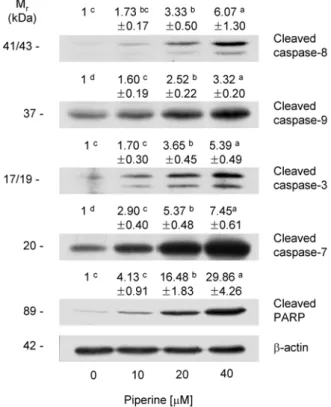
Piperine이 caspase 활성과 PARP 분절에 미치는 영향 Cysteine aspartic acid proteases인 caspase은 세포사멸 을 조절하는 주요한 조절인자로 세포내에서 불활성 형태인 proenzyme으로 합성된 후 스스로 또는 다른 caspase에 의해 분절되어 활성화된다. Caspase는 initiator caspase와 effec- tor caspase로 구분되며, initiator caspase는 death-inducing signal에 의해 활성화되어 effector caspase를 활성화하고 활 성화된 effector caspase는 lamin A, α-fodrin, DNA frag- mentation factor와 PARP 등의 단백질을 분해하여 세포사 멸을 유도한다(16). 여러 연구에서 다양한 항암 성분은 cas- pase의 활성을 조절하여 암세포의 세포사멸을 유도한다고 보고되고 있어(23-26) 본 연구에서는 piperine이 caspase 활 성에 미치는 영향을 조사하기 위해 caspase의 활성화된 형 태인 cleaved caspase의 단백질 수준을 조사하였다. Fig. 5에 나타난 바와 같이 initiator caspase인 caspase-8, -9의 활성 형인 cleaved caspase-8과 -9 단백질 수준이 piperine 처리 에 의해 농도 의존적으로 증가하였다. 또한 effector caspase
Fig. 4. Effect of piperine on the mitochondrial membrane permeability in HT-29 cells. (A) Cells were plated and treated with piperine for 48 hr as described in Fig. 1. Cells were loaded with JC-1 and then analyzed by flow cytometry. The number of cells with normal polarized mitochondrial membrane (red) and with depolarized mitochondrial membrane (green) is expressed as a percentage of total cell number. Each bar represents the mean
±
SE (n=6). Means without a common letter differ, p<0.05.(B) Cells were treated with various concentrations of piperine for 48 hr and subjected to subcellular fractionation. The resulting cy- tosolic fractions were analyzed by Western blotting with the in- dicated antibodies. Photograph of chemiluminescent detection of the blots, which are representative of three independent experi- ments, are shown. The relative abundance of each band was quantified and the control levels were set at 1. The adjusted mean
±
SE (n=3) of each band is shown above each blot. Means without a common letter differ, p<0.05.인 caspase-3과 -7의 활성형인 cleaved caspase-3, -7 단백 질 수준도 piperine 처리에 의해 현저히 증가하였다(Fig. 5).
핵에 존재하고, DNA 수선에 관여하여 세포의 생존 유지 에 중요한 역할을 담당하는 PARP는 caspase-3의 주요한 표적 단백질로 caspase-3에 의해 분절되면 불활성화 되어 세포의 분해를 촉진하여 세포사멸을 야기한다(34). Piperine 에 의해 caspase-3의 활성이 증가하였으므로 piperine이 PARP의 분절에 미치는 영향을 조사하였다. Piperine 처리 농도가 증가할수록 PARP의 불활성 형태인 cleaved PARP 단백질 수준이 현저히 증가하였고 piperine을 40 μM로 처리 한 경우 piperine을 처리하지 않은 군에 비해 cleaved PARP 수준이 29.9배 증가하였다(Fig. 5).
Initiator caspase인 caspase-8과 caspase-9은 서로 다른 경로를 통해 활성화된다. Caspase-8은 세포막에 존재하는
Fig. 5. Effect of piperine on the levels of cleaved caspases and cleaved PARP in HT-29 cells. Cells were plated and treat- ed with various concentration of piperine for 72 hr as described in Fig. 1. Total cell lysates were analyzed by Western blotting with the indicated antibodies. Photograph of chemiluminescent detection of the blots, which are representative of three in- dependent experiments, are shown. The relative abundance of each band to its own
β
-actin was quantified and the control levels were set at 1. The adjusted mean±
SE (n=3) of each band is shown above each blot. Means without a common letter differ, p<0.05.Fas 등의 cell death receptor에 cell death receptor ligands 가 결합하면 활성화되는 외적 경로에 의해 활성화된다. 반 면 caspase-9은 미토콘드리아 막 투과성 증가에 의해 세포 질로 방출된 cytochrome c에 의한 내적 경로에 의해 활성화 된다(35-38). Piperine은 caspase-9의 활성을 증가하였는데 (Fig. 5) 이는 piperine에 의해 Bcl-2 family 단백질의 변화 가 초래되고(Fig. 3) 미토콘드리아 막의 투과성이 증가하여 세포질로 방출된 cytochrome c(Fig. 4B)에 의해 caspase-9 이 활성화된 것으로 사료된다. Piperine에 의해 caspase-8 의 활성이 증가하였으므로(Fig. 5), piperine이 cell death receptor인 Fas와 그 ligand인 Fas ligand(FasL)에 미치는 영향을 조사하였다. HT-29 세포에서 membrane-bound FasL은 Western blot analysis에 의해 검출되지 않았으나 분자량 42 kDa인 Fas는 검출되었다. Piperine 처리에 의해 Fas 단백질 수준은 유의적으로 증가하였다(Fig. 6). 이는 HT-29 세포에서 piperine이 Fas를 통한 외적 경로에 의해 caspase-8을 활성화하고 이를 통해 세포사멸을 유도함을 나타낸다.
Fig. 6. Effect of piperine on the protein levels of Fas in HT-29 cells. Cells were plated and treated with various concen- tration of piperine for 72 hr as described in Fig. 1. Total cell ly- sates were analyzed by Western blotting with an antibody raised against Fas. Photograph of chemiluminescent detection of the blots, which were representative of three independent experi- ments, are shown. The relative abundance of each band to its own
β
-actin was quantified and the control levels were set at 1. The adjusted mean±
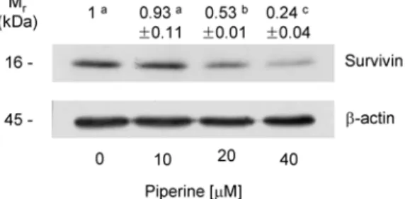
SE (n=3) of each band is shown above each blot. Means without a common letter differ, p<0.05.Piperine이 세포사멸억제단백질인 survivin에 미치는 영향
XIAP, c-IAP, livin 및 survivin 등의 세포사멸억제단백질 (inhibitors of apoptosis protein, IAP)은 effector caspase인 caspase-7과 caspase-3에 직접적으로 작용하여 caspase의 활성을 억제하여 세포사멸을 억제한다. 세포사멸억제단백 질 중 survivin은 다른 세포사멸억제단백질과는 달리 태아 기에 발현이 증가하다가 정상 성인에서는 발현이 거의 되지 않지만 여러 악성 종양에서 현저하게 재발현이 되고 암 환자 에 있어 생존기간, 예후, 치료에 대한 저항성 및 암 재발 등에 관여하는 것으로 알려져 있다(18,19). Piperine이 세포사멸 억제단백질인 survivin 단백질 발현에 미치는 영향을 조사 하였다. Fig. 7에 나타난 바와 같이 survivin 단백질 수준은 piperine 처리에 의해 현저히 감소하였으며, piperine을 40 μM 농도로 처리한 경우 piperine을 처리하지 않은 군에 비해 survivin 단백질 수준이 76% 감소하였다(Fig. 7). Piperine은
Fig. 7. Effect of piperine on the levels of survivin in HT-29 cells. Cells were plated and treated with various concentration of piperine for 72 hr as described in Fig. 1. Total cell lysates were analyzed by Western blotting with the indicated antibodies.
Photograph of chemiluminescent detection of the blots, which were representative of three independent experiments, are shown.
The relative abundance of each band to its own
β
-actin was quantified. And the control levels were set at 1. The adjusted mean±
SE (n=3) of each band is shown above each blot. Means without a common letter differ, p<0.05.Fig. 8. A shematic representation of a possible mechanism of piperine-induced apoptosis in HT-29 cells.
caspase-3과 -7의 활성을 억제하는 survivin의 단백질 수준 을 감소시켜 HT-29 세포에서 세포사멸을 초래하였을 것으 로 사료된다.
이상의 결과들은 후추의 주요 성분인 piperine이 대장암 세포인 HT-29 세포에 세포사멸을 유도하여 암세포의 증식 을 억제함을 나타낸다. HT-29 세포에서 piperine은 세포사 멸의 주요한 조절인자인 Bcl-2 family 단백질 발현 변화에 의한 미토콘드리아 막 투과성 증가와 cytochrome c 방출 증가, caspase 활성 증가, 세포사멸억제단백질인 survivin 단백질 발현을 억제하여 세포사멸을 유도하는 것으로 사료 되며 이를 Fig. 8에 나타내었다. 다른 연구들에 의해 piperine 의 암예방 가능성이 제시되었고(10-13), 본 연구를 통해 pi- perine이 대장암에 강한 항암 효과가 있음을 밝혔으나 향후 암예방 및 암치료제로서 piperine을 활용하기 위해서는 다양 한 동물실험 및 임상실험을 수행해야 할 것으로 사료된다.
요 약
후추의 주요 성분인 piperine은 다양한 생리활성을 나타 내고 있으며, 특히 암예방 효과가 있는 것으로 생각되고 있 다. 본 연구에서는 piperine의 항암 효과를 밝히기 위해 pi- perine이 인간의 대장에서 유래한 암세포인 HT-29 세포의 증식에 미치는 영향과 작용 기전을 연구하였다. Piperine을 HT-29 세포 배양액에 여러 농도(0~40 μM)로 첨가하여 세 포를 배양한 경우 piperine 처리 농도가 증가할수록 세포의 증식이 감소하였고, 세포사멸이 증가하였다. 이는 piperine 이 HT-29 세포의 세포사멸을 유도하여 세포 증식을 억제함 을 제시한다. Piperine의 세포사멸 기전을 조사하기 위해 세 포사멸 조절인자의 변화를 조사하였다. Piperine에 의해 an- ti-apoptotic Bcl-2 family 단백질인 Bcl-2와 Mcl-1 단백질 수준은 감소하였고, BH3-only 단백질인 Bid 단백질 수준은
감소하였으나, Bik 단백질 수준은 증가하였다. 또한 piperine 에 의해 미토콘드리아 막의 투과성이 증가하였고, cyto- chrome c의 세포질로의 방출이 증가하였다. 또한 piperine 처리에 의해 caspase의 활성형인 cleaved caspase-8, -9, -7, -3 단백질 수준이 증가하였고, PARP의 불활성형인 cleaved PARP 수준이 증가하였다. Caspase의 활성을 저해하는 세 포사멸억제단백질 중의 하나인 survivin 단백질 발현이 pi- perine에 의해 감소하였다. 이 결과로부터 대장암세포인 HT-29 세포에서 piperine이 Bcl-2 family 단백질 발현 변화 를 초래하여 미토콘드리아 막 투과성 증가시키고 cyto- chrome c 방출을 증가시키고, caspase 활성을 증가시키고 survivin 단백질 발현을 억제하여 세포사멸을 유도하여 항 암 효과를 나타냄을 알 수 있다. 본 연구는 piperine이 대장암 에 강한 항암 효과가 있음을 밝혔으나 향후 암예방 및 암치 료제로서 piperine을 활용하기 위해서는 동물실험 및 임상실 험 등 다양한 추가 실험이 필요할 것으로 보인다.
감사의 글
본 연구는 교육과학부 한국과학재단 바이오식품소재기반 기술개발사업과 지식경제부 지역혁신센터사업(한림대 식 의약품의 효능평가 및 기능성소재개발센터)의 지원에 의해 수행되었으며 이에 감사드립니다.
문 헌
1. Jemal A, Siegel R, Ward E, Murray T, Xu J, Thun MJ.
2007. Cancer statistics, 2007. CA Cancer J Clin 57: 43-66.
2. Kim WS, Lee RA, Hwang DY, Hong YJ, Hong SI. 2004.
Histoculture drug response assay in colorectal cancer specimen. J Korean Surg Soc 66: 109-115.
3. Maroun JA, Anthony LB, Blais N, Burkes R, Dowden SD, Dranitsaris G, Samson B, Shah A, Thirlwell MP, Vincent MD, Wong R. 2007. Prevention and management of chem- ptherapy-induced diarrhea in patients with colorectal can- cer: a consensus statement by the canadian working group on chemotherapy-induced diarrhea. Curr Oncol 14: 13-20.
4. de Gramount A, Figer A, Seymour M, Homerin M, Hmissi A, Cassidy J, Boni C, Cortes-Funes H, Cervantes A, Freyer G, Papamichael D, Le Bail N, Louvet C, Hendler D, de Braud F, Wilson C, Morvan F, Bonetti A. 2000. Leucovorin and fluorouracil with or without oxaliplatin as first-line treat- met in advanced colorectal cancer. J Clin Oncol 18:
2938-2947.
5. Parmar VS, Jain SC, Bisht KS, Jain R, Taneja P, Jha A, Tyagi OD, Prasad AK, Wengel J, Olsen CE, Boll PE. 1997.
Phytochemistry of the genus Piper. Phytochemistry 46:
597-673.
6. Mittal R, Gupta RI. 2000. In vitro antioxidant activity of piperine. Methods Find Exp Clin Pharmacol 22: 271-274.
7. Singh J, Reen RK, Wiebel FJ. 1994. Piperine, a major in- gredient of black and long peppers, protects against AFB1-induced cytotoxicity and micronucliei formation in H4IIEC3 rat hepatoma cells. Cancer Lett 86: 195-200.
8. Lee CS, Han ES, Kim YK. 2006. Piperine inhibition of
1-methyl-4-phenylpyridinium-induced mitochondrial dys- function and cell death in PC12 cells. Eur J Pharmacol 537:
37-44.
9. Pradeep CR, Kuttan G. 2004. Piperine is a potent inhibitor of nuclear factor-kappaB (NF-kappaB), s-Fos, CREP, ATF-2 and proinflammatory cytokine gene expression in B16F-10 melanoma cells. Int Immunopharmacol 4: 1795- 1803.
10. Selvendiran K, Banu SM, Sakthisekaran D. 2004. Protective effect of piperine on benzo(a)pyrene-induced lung carcino- genesis in Swiss albino mice. Clin Chim Acta 350: 73-78.
11. Selvendiran K, Singh JPV, Sakthisekaran D. 2006. In vivo effect of piperine on serum and tissue glycoprotein levels in benzo(a)pyrene induced lung carcinogenesis in Swiss al- bino mice. Pulm Pharmacol Ther 19: 107-111.
12. Pradeep CR, Kuttan G. 2002. Effect of piperine on the in- hibition of lung metastasis induced B16F-10 melanoma cells in mice. Clin Exp Metastasis 19: 703-708.
13. Bezerra DP, Castro FO, Alves AP, Pessoa C, Moraes MO, Silveira ER, Lima MA, Elmiro FJ, Costa-Lotufo LV. 2006.
In vivo growth-inhibition of Sarcoma 180 by piplartine and piperine, two alkaloid amides from piper. Braz J Med Biol
Res 39: 801-807.
14. Duessel S, Heuertz RM, Ezekiel UR. 2008. Growth in- hibition of human colon cancer cells by plant compounds.
Clin Lab Sci 21: 151-157.
15. Hengarther MO. 2000. The biochemistry of apoptosis.
Nature 407: 770-776.
16. Jin Z, El-Deiry WS. 2005. Overview of cell death signaling pathways. Cancer Biol Ther 4: 139-163.
17. Yao J, Jiang Z, Duan W, Huanq J, Zhanq L, He L, Li F, Xiao Y, Shu B, Lin C. 2008. Involvement of mitochondrial pathway in triptolide-induced cytotoxicity in human normal liver L-02 cells. Biol Pharm Bull 31: 592-597.
18. Ambrosini G, Adida C, Altieri DA. 1997. A novel anti-apop- tosis gene, survivin, expressed in cancer and lymphoma.
Nat Med 3: 917-921.
19. Altieri DC, Marchisio PC. 1999. Survivin apoptosis: an in- terloper between cell death and cell proliferation in cancer.
Lab Invest 79: 1327-1333.
20. Reed JC. 2004. Apoptosis mechanisms: implications for cancer drug discovery. Oncology 18: 10-20.
21. Frankfurt OS, Krishan A. 2003. Apoptosis-based drug screening and detection of selective toxicity to cancer cells.
Anticancer Drugs 14: 555-561.
22. Lowe SW, Lin AW. 2000. Apoptosis in cancer. Carcinogen-
esis 21: 485-495.
23. Kim EJ, Park SY, Hong J, Shin M, Lim SS, Shin HK, Park JHY. 2007. Inhibitory effect of the methanolic extract of
Symphyocladia latiuscula on the growth of HT-29 human
colon cancer cells. J Korean Soc Food Sci Nutr 36: 431- 438.24. Kim EJ, Park H, Lim SS, Kim JS, Shin HK, Park JHY. 2008.
Effect of the hexane extracts of Saussure lappa on the growth of HT-29 human colon cancer cells. Korean J Food
Sci Technol 40: 207-214.
25. Kim EJ, Park SY, Shin HK, Kwon DY, Surh YJ, Park JHY.
2007. Activation of caspase-8 contributes to 3,3'-diindoly- methane-induced apoptosis in colon cancer cells. J Nutr 137: 1-6.
26. Kim EJ, Lee YJ, Shin HK, Park JHY. 2006. A study on the mechanisms by which the aqueous extract of Inonotus
obliquus induces apoptosis and inhibits proliferation in
HT-29 human colon cancer cells. J Korean Soc Food SciNutr 35: 516-523.
27. Denizot F, Lang R. 1986. Rapid colorimetric assay for cell growth and survival modifications to the tetrazolium dye procedure giving improved sensitivity and reliability. J
Immunol Methods 89: 271-277.
28. Kim EJ, Shin HK, Cho JS, Lee SK, Won MH, Kim HW, Park JHY. 2006. Trans-10, cis-12 conjugated linoleic acid inhibits the G1-S cell cycle progression in DU145 human prostate carcinoma cells. J Med Food 9: 293-299.
29. Jung JI, Lim SS, Choi HJ, Cho HJ, Shin HK, Kim EJ, Chung WY, Park KK, Park JH. 2006. Isoliquiritigenin induces apoptosis by depolarizing mitochondrial membranes in prostate cancer cells. J Nutr Biochem 17: 689-696.
30. Eguchi Y, Srinivasan A, Tomaselli KJ, Shimizu S, Tsujimoto Y. 1999. ATP-dependent steps in apoptotic sig- nal transduction. Cancer Res 59: 2174-2181.
31. Sunila ES, Kuttan G. 2004. Immunomodulatory and anti- tumor activity of Pier longum Linn. and piperine. J
Ethnopharmacol 90: 339-346.
32. Yu Z, Li W. 2006. Induction of apoptosis by puerarin in co- lon cancer HT-29 cells. Cancer Lett 238: 53-60.
33. Luo X, Budihardjo I, Zou H, Slaughter C, Wang X. 1998.
Bid, a Bcl2 interacting protein, mediates cytochrome c re- lease from mitochondria in response to activation of cell surface death receptors. Cell 94: 481-490.
34. Oliver FJ, de la Rubia G, Rolli V, Ruiz-Ruiz MC, de Murcia G, Murcia JM. 1998. Importance of poly(ADP-ribose) poly- merase and its cleavage in apoptosis. Lesson from an un- cleavable mutant. J Biol Chem 273: 33533-33539.
35. Budihardjo I, Oliver H, Lutter M, Wang X. 1999. Biochem- ical pathways of caspase activation during apoptosis. Annu
Rev Cell Dev Biol 15: 269-290.
36. Ashkenazi A, Dixit VM. 1998. Death receptor: signaling and modulation. Science 281: 1305-1308.
37. Nijhawan LP, Budihardjo D, Srinivasula I, Ahmad SM, Alnemri M, Wang X. 1997. Cytochrome c and dATP-de- pendent formation of Apaf-1/caspase-9 complex initiates an apoptotic protease cascade. Cell 91: 479-489.
38. Baker SJ, Reddy EP. 1998. Modulation of life and death by the TNF receptor superfamily. Oncogene 17: 3261-3270.
(2009년 2월 23일 접수; 2009년 4월 2일 채택)