Inhibition of Migration and Invasion of LNCap Human Prostate Carcinoma Cells by Doxorubicin through Inhibition of Matrix Metalloproteinase Activity and
Tightening of Tight Junctions
Yung Hyun Choi
1*, Dong Yeok Shin
2and Wun-Jae Kim
31
Department of Biochemistry, Dongeui University College of Oriental Medicine, Busan 614-052 and Anti-Aging Research Center & Blue-Bio Industry RIC, Dongeui University, Busan 614-714, Korea
2
Dongnam Institute of Radiological & Medicine Sciences, Busan 619-953, Korea
3
Department of Urology, Chungbuk National University College of Medicine, Cheongju 361-763, Korea
Received March 9, 2014 /Revised March 26, 2014 /Accepted March 26, 2014Doxorubicin (trade name adriamycin), an anthracycline antibiotic, is commonly used in the treatment of a wide range of cancers, including hematological malignancies, many types of carcinoma, and soft tissue sarcomas. It is closely related to the natural product daunomycin, and like all anthracyclines, it works by intercalating DNA. Its most serious adverse effect is life-threatening heart damage. Its an- ti-metastatic mechanisms in human prostate carcinomas are not fully understood. In this study, we used LNCap human prostate carcinoma cells to investigate the inhibitory effects of doxorubicin on cell motility and invasion, two critical cellular processes that are often deregulated during metastasis.
Doxorubicin treatment inhibited cell migration and invasiveness of LNCap cells without showing any toxicity. Doxorubicin treatment also suppressed the activity and expression of matrix metal- loproteinase (MMP)-2 and MMP-9, which were associated with up-regulated expression of tissue in- hibitor of metalloproteinase (TIMP)-1 and TIMP-2 in LNCap cells. Doxorubicin treatment also attenu- ated the expression levels of claudin family members (claudin-1, -2,-3 and -4), major components of tightening of tight junctions (TJs) and increased the tightening of TJs, as demonstrated by an increase in transepithelial electrical resistance. The present findings demonstrate that doxorubicin reduces the migration and invasion of prostate carcinomas LNCap cells by modulating the activity of TJs and MMPs.
Key words : Claudin, doxorubicin, invasion, matrix metalloproteinase (MMP), tight junctions
*Corresponding author
*Tel : +82-51-850-7413, Fax : +82-51-853-4036
*E-mail : [email protected]
This is an Open-Access article distributed under the terms of the Creative Commons Attribution Non-Commercial License (http://creativecommons.org/licenses/by-nc/3.0) which permits unrestricted non-commercial use, distribution, and reproduction in any medium, provided the original work is properly cited.
ISSN (Online) 2287-3406 Journal of Life Science 2014 Vol. 24. No. 6. 700~706 DOI : http://dx.doi.org/10.5352/JLS.2014.24.6.700
서 론
Doxorubicin은 Streptomyces peucetius 균주가 생산하는 an- thracycline 계열 항생제의 일종으로 혈액학상의 악성종양 (hematological malignancy), 다양한 종류의 암종(carcinoma) 및 연부 조직 육종 (soft tissue sarcoma) 등의 치료 목적으로 가장 널리 사용 중인 항암제이다 [7, 8]. Doxorubicin은 DNA 염기 서열 사이에 삽입되어 DNA 구조 손상을 유발하여 세포 분열을 차단시킴으로서 강력한 항암 활성을 나타내지만 , 백혈 구 감소증 , 빈혈, 심장 기능 상실 등의 부작용이 나타나기도 한다 . Doxorubicin은 단일 항암제로도 널리 사용이 되지만, 항암활성의 상승과 부작용의 최소화를 위하여 다른 항암제와
복합 처리하여 사용되기도 한다 [10]. Doxorubicin이 암세포의 증식 억제 측면에서 세포주기 교란 [18, 30] 및 세포사멸 유도 [6, 28]를 포함한 전이 억제 효과[5, 14, 27] 등은 이미 잘 알려진 사실이지만 , 전립선 암세포에서의 전이 억제 관련 기전 연구 는 여전히 미비한 실정이다 .
암에 의한 사망률의 주요 요인인 전이는 암이 발병 부위로
부터 암세포가 인체의 다른 곳으로 옮겨서 증식하는 현상이
다 . 특히 상피세포에는 세포들 사이에는 gap junction, tight
junction (TJ), adherens junction 및 desmosome과 같은 특화
된 세포간의 구조를 가지는데 , 그중 TJ는 세포 사이의 치밀한
이음부로서 다양한 분자와 이온의 이동 조절에 중요한 역할을
한다 [19, 22]. 그러나 상피조직 세포에서 TJ의 붕괴로 인한 상
피 전기 저항성 (transepithelial electrical resistance, TER)으로
대변되는 TJ의 전기적 저항의 감소나 상피 경세포측 삼투성
(transepithelial paracellular permeability, TPP)의 증가는 정
상세포의 암화 과정에 동반되는 현상이다 . 최근 연구들에 의
하면 상피세포 유래 암세포에서 비정상적으로 높게 발현되며
TJ의 근간을 이루는 claudin family에 속하는 주요 필수 막단
백질들이 세포사이 유동성 조절에 중요한 역할을 하고 있음이
알려졌다 [16, 23, 24]. 따라서 TJ 복합체에서 이들 단백질들의
- Note -
A
B
C
Time(hr)
Fig. 1. Effects of doxorubicin on cell motility and invasion in LNCap cells. (A) Chemical structure of doxorubicin. (B) LNCap cells were grown to confluency on 30-mm cell culture dishes; a scratch was then made through the cell layer using a pipette tip. After washing with PBS, serum free media containing either vehicle or doxorubicin (0.4 μM) was added for different times. Photographs of the wounded area were taken for evaluation of cell move- ment into the wounded area. (C) The cells pretreated with the indicated concentration of doxorubicin for 6 h were plated onto the apical side of matrigel coated filters in serum-free medium containing either vehicle or doxorubicin. Medium containing 20% FBS was placed in the basolateral chamber to act as a chemoattractant.
After 48 h, cells on the apical side were wiped off using a Q-tip. Next, cells on the bottom of the filter were stained using hematoxylin and Eosin Y, and then counted. Data are shown as the mean of triplicate sam- ples and represent invasive cell numbers compared with those of control cells. *
p
<0.05 versus untreated control.역할 규명은 암의 증식 뿐 만 아니라 전이를 제어할 수 있는 주요 수단으로 인식되어지고 있다 . Zinc-dependent endo- peptidase family에 속하는 matrix metalloproteinases (MMPs)는 암세포의 전이에 핵심적인 역할을 하는데, 침윤성 암세포는 이들 효소를 사용하여 세포외 기질 (extracellular matrix, ECM)의 분해와 전이 동안 기저체 막의 붕괴를 촉진시 킨다 [9, 26]. MMPs 중, 특히 MMP-2와 -9가 다양한 암세포에서 활성이 비정상적으로 높게 관찰되며 , 암세포의 침윤과 전이에 중요한 역할을 하는 것으로 알려져 있다 [3, 15]. 한편 MMPs의 억제인자로 알려진 tissue inhibitors of metalloproteinases (TIMPs)는 활성화된 MMPs에 직접 결합하여 그들의 효소적 활성을 저해함으로서 ECM의 파괴를 억제하는 것으로 알려져 있다 [11, 25]. 따라서 TJ의 견고성과 연관된 MMPs의 활성 저 해는 암세포의 전이를 차단할 수 있는 표적 인자가 될 수 있다 . 본 연구에서는 doxorubicin의 암세포 전이 억제 효능에 관 한 추가적인 기전 해석의 일환으로 LNCap 인체 전립선 암세 포를 이용하여 이들 세포의 이동성과 침윤성에 미치는 효과와 TJ 및 MMPs 활성 조절 관련성을 조사하였다.
재료 및 방법 세포배양 및 doxorubicin 처리
LNCap 전립선 암세포는 American Type Culture Collec- tion (Rockville, MD, USA)에서 구입하였으며, 10% fetal bo- vine serum (FBS, Gibco BRL, Gaithersburg, MD, USA) 및 1% penicillin–streptomycin이 함유된 RPMI 1640 배지를 사 용하여 37℃, 5% CO
2조건에서 배양하였다 . Doxorubicin (Fig.
1A)은 Sigma-Aldrich Chemical Co. (St Louis, MO, USA)에서 구입하였으며 , DMSO에 1 mM 농도로 녹여 적정 농도로 희석 하여 처리하였다 .
Wound healing migration assay
20 μg/ml의 rat tail collagen이 코팅된 dish (BD Bioscien- ces, Bedford, MA, USA)에 LNCap 세포를 적정 시간 동안 배양한 후 , 파이펫 팁을 이용한 scraping으로 wounded 영역 을 만들고 정상 배지로 2~3회 수세하였다. 다시 1% FBS가 함 유된 배지로 교체 후 적정 농도의 doxorubicin을 48시간 처리 후 wounded 영역으로의 세포 이동성의 정도를 비교 관찰하 였다 .
In vitro invasiveness assay
Matrigel invasion assay를 이용한 doxorubicin의 침윤성 억제 효과를 관찰하기 위하여 적정 농도의 doxorubicin이 6 시간 동안 처리된 LNCap 세포를 doxorubicin이 함유된 FBS-free 배지를 함유한 Matrigel-coated filter의 apical side 에 분주하였다 . 이때 basolateral chamber에는 20% FBS가 함 유된 배지를 분주하였으며 , 48시간 후 filter의 하단부로 이동
한 세포를 hematoxylin 및 Eosin Y 염색액으로 염색하고 계수 하였다 .
RNA의 분리 및 reverse transcription-polymerase chain reaction (PCR)
Doxorubicin이 처리된 적정 조건에서 배양된 세포들의
RNA를 RNeasy kit (Qiagen, La Jolla, CA, USA)를 이용하여
분리한 후 , AMV reverse transcriptase (Amersham Corp., Arlington Heights, IL, USA)를 이용하여 cDNA를 합성하였 다 . 특정 유전자에 적절한 primer를 이용한 PCR은 Mastercy- cler (Eppendorf, Hamburg, Germany)를 사용하여 수행하였 으며 , PCR 반응 산물을 1% agarose을 이용하여 분리한 후, ethidium bromide (EtBr) 염색을 실시한 후 발현의 정도를 비 교하였다 .
단백질의 분리와 Western blot analysis
준비된 세포에서 총 단백질을 lysis buffer (25 mM Tris–Cl (pH 7.5), 250 mM NaCl, 5 mM ethylendiaminetetra acetic acid, 1% nonidet P-40, 0.1 mM sodium orthovanadate, 2 μg/
ml leupeptin 및 100 μg/ml phenylmethylsulfonyl fluoride)를 이용하여 분리하고 정량하였다 . Western blot analysis를 위해 동량의 단백질들을 sodium dodecyl sulfate (SDS)–poly- acrylamide gel을 이용하여 전기영동하고 nitrocellulose membrane (Schleicher & Schuell, Keene, NH, USA)으로 전이 시켰다 . 각각의 membrane을 적정 항체 및 enhanced chem- iluminescence (ECL, Amersham Corp.) 용액을 이용하여 단 백질들의 발현 변화를 조사하였다 . 본 실험에 사용된 1차 항체 들은 Santa Cruz Biotechnology Inc. (Santa Cruz, CA, USA) 및 Calbiochem (Cambridge, MA, USA)에서 구입하였으며, 2 차 항체들은 Amersham Corp.에서 구입하였다.
TER의 측정
TER 값의 변화 측정은 STX-2 chopstick electrode가 짝으로 형성된 EVOM Epithelial Tissue Voltohmmeter (World Precision Instruments, FL, USA)를 이용하여 조사하였다. 이 를 위하여 LNCap 세포를 Transwell® (Corning Costar Corp., NY, USA)의 8.0 μm pore size insert (upper chamber)로 seed- ing한 후 100% confluence를 이룰 때까지 배양하였다. 이 후 doxorubicin을 48시간 동안 처리하고, upper 및 lower cham- ber에 electrode를 넣어 전기 저항도를 측정하였다[4].
In vitro MMPs 활성 측정
세포 배양액을 이용한 MMPs 활성 측정을 위하여 MMP Gelatinase Activity Assay Kit (Chemicon International Inc., Temecula, CA, USA)를 사용하였다. 이를 위하여 배양액에 bi- otinylated gelatinase substrate를 MMP-2 및 -9의 기질로 첨가 하였고 , 분해 산물을 biotin이 결합된 96-well plate 분주 후, 실온에서 30분간 반응시켰다. 분해는 되었으나 결합되어 있지 않은 기질은 제거한 후 streptavidin–enzyme complex를 첨 가하고 microplate reader (Molecular Devices, Palo Alto, CA, USA)를 이용하여 540 nm 파장에서 흡광도를 측정하였다.
통계 처리
모든 실험결과는 평균 ±표준편차로 표시하였고 SigmaPlot을 이용하여 Student t-test를 이용하여 통계적 유의성을 얻었다.
결과 및 고찰
Doxorubicin에 의한 LNCap 세포의 이동성 및 침윤성 억제 암세포의 전이능을 결정짓는 요소로 작용하는 이동성과 침 윤성의 억제 여부는 항암활성을 지니는 물질들의 항전이능을 평가하는 중요한 기준으로 사용되고 있다 . 최근 비정상적인 상피세포의 극성 소실과 삼투성의 변화가 상피세포 암화 및 전이 단계의 특징임이 밝혀지면서 , 이들 활성 유지에 중요한 역할을 하는 TJ와 암세포 전이와의 연관성이 주요 관심 분야로 대두되고 있다 [19, 21]. 이는 많은 선행 연구에서 항암활성을 가지는 물질들의 암세포 이동성 및 침윤성 차단이 TPP의 상승 과 연관되어 있다는 점에서 지지를 받고 있다 . 본 연구에서는 doxorubicin에 의한 LNCap 전립선 암세포의 이동성과 침윤 성의 억제 여부를 조사하기 위하여 Wound healing migration 및 matrigel invasion assay를 실시하였다. Fig. 1B 및 C의 결과 에서 알 수 있듯이 정상배지에 배양된 세포에 비하여 doxor- ubicin이 처리된 세포에서 처리 농도 의존적으로 LNCap 세포 의 이동성이 현저하게 억제되었고 (Fig. 1B), doxorubicin 처리 농도 증가에 따라 침윤성 또한 유의적으로 억제되었다 (Fig.
1C). 그러나 동일 조건에서 유의적인 증식억제 효과는 관찰되 지 않아 (data not shwon), 이러한 이동성 및 침윤성의 억제가 doxorubicin의 세포독성 효과에 의한 것은 아님을 알 수 있었다.
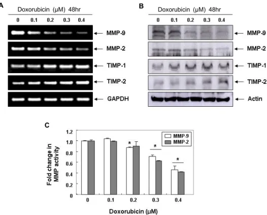
Doxorubicin에 의한 LNCap 세포의 MMPs 발현 및 활 성 억제
ECM의 분해에 결정적인 역할을 하는 MMPs는 정상 기관
의 발생과 조직 재생에 필수적인 효소이지만 , 암세포의 전이
를 위한 침윤성 증대에 핵심적인 역할을 담당하고 있다 . 특히
MMP-2와 -9는 암세포의 침윤과 암조직 주변의 혈관신생에
필수적이므로 , 이들의 합성과 활성 차단은 종양 전이 및 증식
억제를 위한 필수적인 방법이다 [3, 15]. 한편 MMPs의 활성은
TIMPs의 발현 증대에 따라 유의적으로 억제될 수 있으므로
MMPs에 대한 TIMPs의 상대적인 발현 증가는 암세포 침윤성
의 억제를 측정하는 주요 인자로 활용이 가능하다 [11, 25]. 따
라서 doxorubicin에 의한 LNCap 세포의 침윤성 억제가 이들
효소의 발현 및 활성 억제와 연관성을 가지는지를 조사한 결
과 , MMP-2와 -9의 mRNA 및 단백질의 발현뿐만 아니라, 그들
의 효소적 활성도 유의적으로 억제되었음을 알 수 있었다 . 그
러나 doxorubicin은 조사된 두 종류의 TIMPs 발현을 처리 농
도 의존적으로 매우 증가시켰다 (Fig. 2). 따라서 doxorubicin의
TIMPs 발현 증가와 동반된 MMPs의 생성 및 활성 억제는
ECM의 분해를 차단함으로서 LNCap 세포의 이동성과 침윤
A B
C
Doxorubicin (μM) 48hr Doxorubicin (μM) 48hr
Fig. 2. Inhibition of MMPs expression and their activities, and induction of TIMPs expression by doxorubicin in LNCap cells. (A) LNCap cells were treated with various concentrations of doxorubicin for 48 hr. Total RNA was isolated from cells treated with or without various concentrations of doxorubicin for 48 hr, and reverse-transcribed. Resulting cDNAs were then subjected to PCR. The reaction products were subjected to electrophoresis in a 1% agarose gel and visualized by EtBr staining. GAPDH was used as an internal control. (B) Cells grown under the same conditions as (A) were sampled, lysed, and 50 μg of proteins were separated by electrophoresis on SDS-polyacrylamide gels. Western blotting was then performed using the indicated antibodies, and an ECL detection system. Actin was used as an internal control. (C)
In vitro
activity of MMP-2 and -9 in cell culture supernatant was measured using a MMP-2 and -9 gelatinase activity assay kit. The biotinylated gelatinase substrates were cleaved by active MMPs in the samples, and the fragments were added to a biotin-binding plate. The digested but unbound fragments were removed by washing. Data are mean ± SD from three independent experiments and are presented as fold change compared with vector control (*p
<0.05 versus untreated control).성을 억제하였을 것으로 생각된다 .
Claudin family의 발현 및 TER에 미치는 doxorubicin 의 영향
한편 TJ 구성 단백질들의 비정상적인 발현과 수반된 TJ의 붕괴는 암세포의 침윤과 전이의 초기 단계에 나타나는 현상이 다 [21, 23].
그 중, claudin family에 속하는 단백질들은 TJ를 통한 TPP의 조절뿐 뿐만 아니라 세포와 세포사이의 접착성 조절에 중요한 인자로 작용한다 [2, 16, 25]. 그 예로서, 간세포 의 침윤성 획득에 claudin-1이 중요한 역할을 함이 밝혀졌는 데 , 이는 MMP-2의 발현 및 활성 증가를 유도하였으며, clau- din-1의 발현을 인위적으로 차단할 경우 간암세포의 침윤성 이 완벽하게 차단되었음이 보고된 바 있다 [29]. 또한 clau- din-3과 -4는 유방암과 난소암을 포함한 다양한 종양조직에 서 과발현되어 있으며 , 이들 단백질의 발현을 차단할 경우 암 세포의 침윤성이 억제되는 것으로 나타났다 [1, 17]. 그러나 최
근 몇몇 결과에 따르면 어떤 종류의 claudins 발현 증가는 암
세포의 증식과 전이를 촉진시킬 수 있음이 보고되고 있다 . 예
를 들어 , claudin-3 또는 claudin-4의 발현을 억제하였을 경우
ovarian cancer의 증식과 전이뿐만 아니라 epithelial-to-mes-
enchymal transition이 촉진되었으며[12, 20], TER의 유의적
인 변화는 없었지만 , β-catenin signaling 또는 phosphatidyli-
nositol 3-kinase (PI3K)/Akt signaling의 활성 증가가 동반되
었다 . 또한 유방암세포에서 basic helix-loop-helix transcrip-
tional regulator인 differentiated embryo-chondrocyte ex-
pressed gene 1 (DEC1)을 knockdown 시킨 경우 invasive ca-
pacity가 증가되었으며, 이는 claudin-1의 전사활성 증가와
연관성이 있음이 보고된 바 있다 [13]. 따라서 doxorubicin의
LNCap 세포 이동성 및 침윤성의 저해 효과가 TJ의 기능 변화
와 관련이 있는지를 조사하기 위하여 TJ 구성 요소로서 중요한
역할을 하는 claudin family 인자들의 발현과 TJ의 견고성을
나타내는 TER의 값[4]에 미치는 doxorubicin의 영향을 조사
A B
C
Doxorubicin (μM) 48hr Doxorubicin (μM) 48hr
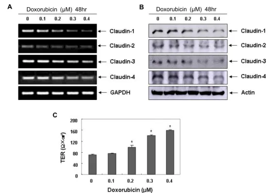
Fig. 3. Effects of doxorubicin on expression of claudins and values of TER in LNCap cells. (A) LNCap cells were treated with the indicated concentrations of doxorubicin for 48 hr. Total RNA was isolated and reverse-transcribed using the indicated primers. Resulting cDNAs were then subjected to PCR and the reaction products were subjected to electrophoresis in a 1% agarose gel and visualized by EtBr staining. GAPDH was used as an internal control. (B) The cells were sampled, lysed, and 50 μg of proteins were separated by electrophoresis on SDS-polyacrylamide gels. Western blotting was then performed using the indicated antibodies, and an ECL detection system. Actin was used as an internal control. (C) After 48 hr, TER values were measured using an EVOM Epithelial Tissue Voltohmmeter, as described in the Materials and Methods section.
Results are shown as the mean ± SD of three independent experiments. A Student’s t-test was used for determination of significance (*,
p
<0.05 versus untreated control).하였다 . 본 연구의 결과에 의하면, doxorubicin 처리 농도 의존 적으로 조사된 4종류의 claudin family 인자들(claudin-1, -2, -3, 및 -4)이 전사 및 번역수준에서의 모두 감소하였으며, TER 의 값은 상대적으로 증가하였음을 알 수 있었다 (Fig. 3).
이상의 결과는 세포독성이 없는 범위에서 doxorubicin에 의한 LNCap 인체 전립선 암세포의 이동성과 침윤성의 차단 은 TIMPs의 발현 증가에 의한 MMPs의 발현과 활성 억제에 의한 것임을 보여주는 것이다 . 아울러 TER의 증가는 doxor- ubicin이 TJ의 누출을 억제하거나 역전시켰음을 의미하는 것 으로서 최소한 TJ의 견고성을 증가시킴으로서 항전이 활성을 나타낸다고 할 수 있다 . 물론 추가적인 기전 연구의 보완과 동물 실험의 검증이 필요하지만 , 본 연구의 결과는 doxor- ubicin이 항전이 인자로서 항암활성을 나타낸다는 기초적 실 험 증거로서 활용될 것으로 생각한다 .
감사의 글
이 논문은 정부 (미래창조과학부)의 재원으로 한국연구재단 의 지원을 받아 수행된 연구임 (No. 2008-0062611).
References
1. Agarwal, R., D'Souza, T. and Morin, P. J. 2005. Claudin-3 and claudin-4 expression in ovarian epithelial cells enhances invasion and is associated with increased matrix metal- loproteinase-2 activity.
Cancer Res
65, 7378-7385.2. Angelow, S. and Yu, A. S. 2007. Claudins and paracellular transport: an update.
Curr Opin Nephrol Hypertens
16, 459-464.3. Gibbs, D. F., Warner, R. L., Weiss, S. J., Johnson, K. J. and Varani, J. 1999. Characterization of matrix metal- loproteinases produced by rat alveolar macrophages.
Am J Respir Cell Mol Biol
20, 1136-1144.4. Grant-Tschudy, K. S. and Wira, C. R. 2005. Effect of oestra- diol on mouse uterine epithelial cell tumour necrosis fac- tor-alpha release is mediated through uterine stromal cells.
Immunology
115, 99-107.5. Grenader, T., Goldberg, A., Hadas-Halperin, I. and Gabizon, A. 2009. Long-term response to pegylated liposomal doxor- ubicin in patients with metastatic soft tissue sarcomas.
Anticancer Drugs
20, 15-20.6. Huang, J., Yang, J., Maity, B., Mayuzumi, D. and Fisher,
R. A. 2011. Regulator of G protein signaling 6 mediates dox- orubicin-induced ATM and p53 activation by a reactive oxy- gen species-dependent mechanism.
Cancer Res
71, 6310-6319.7. Hutchinson, C. R. and Colombo, A. L. 1999. Genetic en- gineering of doxorubicin production in
Streptomyces peuce- tius
: a review.J Ind Microbiol Biotechnol
23, 647-652.8. Hutchinson, C. R. 1995. Anthracyclines.
Biotechnology
128, 331-357.9. John, A. and Tuszynski, G. 2001. The role of matrix metal- loproteinases in tumor angiogenesis and tumor metastasis.
Pathol Oncol Res
7, 14-23.10. Lal, S., Mahajan, A., Chen, W. N. and Chowbay, B. 2010.
Pharmacogenetics of target genes across doxorubicin dis- position pathway: a review.
Curr Drug Metab
11, 115-128.11. Lambert, E., Dasse, E., Haye, B. and Petitfrere, E. 2004.
TIMPs as multifacial proteins.
Crit Rev Oncol Hematol
49, 187-198.12. Lin, X., Shang, X., Manorek, G. and Howell, S. B. 2013.
Regulation of the epithelial-mesenchymal transition by clau- din-3 and claudin-4.
PLoSOne
8, e67496.13. Liu, Y., Miao, Y., Wang, J., Lin, X., Wang, L., Xu, H. T. and Wang, E. H. 2013. DEC1 is positively associated with the malignant phenotype of invasive breast cancers and neg- atively correlated with the expression of claudin-1.
Int J Mol Med
31, 855-860.14. Minisini, A. M., Andreetta, C., Fasola, G. and Puglisi, F.
2008. Pegylated liposomal doxorubicin in elderly patients with metastatic breast cancer.
Expert Rev Anticancer Ther
8, 331-342.15. Mook, O. R., Frederiks, W. M. and Van Noorden, C. J. 2004.
The role of gelatinases in colorectal cancer progression and metastasis.
Biochim Biophys Acta
1705, 69-89.16. Morin, P. J. 2005. Claudin proteins in human cancer: promis- ing new targets for diagnosis and therapy.
Cancer Res
65, 9603-9606.17. Rangel, L. B., Agarwal, R., D'Souza, T., Pizer, E. S., Alò, P. L., Lancaster, W. D., Gregoire, L., Schwartz, D. R., Cho, K. R. and Morin, P. J. 2003. Tight junction proteins claudin-3 and claudin-4 are frequently overexpressed in ovarian can- cer but not in ovarian cystadenomas.
Clin Cancer Res
9, 2567-2575.18. Rathos, M. J., Khanwalkar, H., Joshi, K., Manohar, S. M.
and Joshi, K. S. 2013. Potentiation of
in vitro
andin vivo
antitumor efficacy of doxorubicin by cyclin-dependent kin- ase inhibitor P276-00 in human non-small cell lung cancercells.
BMC Cancer
13, 29.19. Schneeberger, E. E. and Lynch, R. D. 2004. The tight junc- tion: a multifunctional complex.
Am J Physiol Cell Physiol
286, C1213-1228.20. Shang, X., Lin, X., Alvarez, E., Manorek, G. and Howell, S. B. 2012. Tight junction proteins claudin-3 and claudin-4 control tumor growth and metastases.
Neoplasia
14, 974-985.21. Singh, A. B., Sharma, A. and Dhawan, P. 2010. Claudin fam- ily of proteins and cancer: an overview.
J Oncol
2010, 541957.22. Soler, A. P., Miller, R. D., Laughlin, K. V., Carp, N. Z., Klurfeld, D. M. and Mullin, J. M. 1999. Increased tight junc- tional permeability is associated with the development of colon cancer.
Carcinogenesis
20, 1425-1431.23. Turksen, K. and Troy, T. C. 2011. Junctions gone bad: clau- dins and loss of the barrier in cancer.
Biochim Biophys Acta
1816, 73-79.24. Utech, M., Brüwer, M. and Nusrat, A. 2006. Tight junctions and cell-cell interactions.
Methods Mol Biol
341, 185-195.25. Uzui, H., Harpf, A., Liu, M., Doherty, T. M., Shukla, A. and Chai, N. 2002. Increased expression of membrane type 3-ma- trix metalloproteinase in human atherosclerotic plaque: role of activated macrophages and inflammatory cytokines.
Circulation
106, 3024-3030.26. Vihinen, P., Ala-aho, R. and Kähäri, V. M. 2005. Matrix met- alloproteinases as therapeutic targets in cancer.
Curr Cancer Drug Targets
5, 203-220.27. Wang, Z., Yu, Y., Dai, W., Lu, J., Cui, J., Wu, H., Yuan, L., Zhang, H., Wang, X., Wang, J., Zhang, X. and Zhang, Q. 2012. The use of a tumor metastasis targeting peptide to deliver doxorubicin-containing liposomes to highly meta- static cancer.
Biomaterials
33, 8451-8460.28. Weber, T. G., Pöschinger, T., Galbán, S., Rehemtulla, A. and Scheuer, W. 2013. Noninvasive monitoring of pharmacody- namics and kinetics of a death receptor 5 antibody and its enhanced apoptosis induction in sequential application with Doxorubicin.
Neoplasia
15, 863-874.29. Yoon, C. H., Kim, M. J., Park, M. J., Park, I. C., Hwang, S. G., An, S., Choi, Y. H., Yoon, G. and Lee, S. J. 2010.
Claudin-1 acts through c-Abl-protein kinase Cdelta (PKCd) signaling and has a causal role in the acquisition of invasive capacity in human liver cells.
J Biol Chem
285, 226-233.30. Zuryń, A., Litwiniec, A., Gackowska, L., Pawlik, A., Grzanka, A. A. and Grzanka, A. 2012. Expression of cyclin A, B1 and D1 after induction of cell cycle arrest in the Jurkat cell line exposed to doxorubicin.
Cell Biol Int
36, 1129-1135.초록:Doxorubicin에 의한 치밀결합 강화 및 MMPs의 활성 억제를 통한 LNCap 전립선 암세포의 이동성 및 침윤성의 억제
최영현
1*․신동역
2․김원재
3(
1동의대학교 한의과대학 생화학교실 , 항노화연구소 및 블루바이오소재개발센터,
2동남권원자력의학원 ,
3