Toxicology Reports 7 (2020) 1443–1447
Available online 17 October 2020
2214-7500/© 2020 The Authors. Published by Elsevier B.V. This is an open access article under the CC BY-NC-ND license
(http://creativecommons.org/licenses/by-nc-nd/4.0/).
Alterations in telomere length and mitochondrial DNA copy number in human lymphocytes on short-term exposure to moderate hypoxia
Mohammad Rizwan Alama, Dae-Kwang Kima,b,*
aDepartment of Medical Genetics, School of Medicine, Keimyung University, Daegu, Republic of Korea
bHanvit Institute for Medical Genetics, Daegu, Republic of Korea
A R T I C L E I N F O Keywords:
Moderate hypoxia Human lymphocyte Telomere length elongation Mitochondrial DNA copy number Genomic instability
A B S T R A C T
Hypoxia is related to a variety of diseases, such as cardiovascular and inflammatory diseases and various cancers.
Telomere length (TL) may vary according to the hypoxia level and cell types. To the best of our knowledge, no study has investigated the effect of moderate hypoxia on TL and mitochondrial DNA copy number (mtDNAcn) in human lymphocytes. Therefore, in this study, we analyzed the effect of moderate hypoxia on TL in correlation with mtDNAcn. This study included 32 healthy male nonsmoker’s subjects; in this cohort, we had previously studied sister chromatid exchange and microsatellite instability. Blood samples from each subject were divided into three groups: a control group and two experimental groups exposed to moderate hypoxia for 12 or 24 h.
Relative TL and mtDNAcn were measured by a quantitative real-time polymerase chain reaction. The TL in the control group did not significantly differ from that in the experimental group subjected to hypoxia for 12 h;
however, the TL in the 24 h hypoxia–treated experimental group was significantly higher than that in the control group. The correlation between TL and mtDNAcn was not statistically significant in the two hypoxic states. The increase in TL was observed on exposure to hypoxia for 24 h and not for 12 h; thus, the findings suggest that telomere elongation is related to hypoxia exposure duration. The mtDNAcn in the two experimental groups did not significantly differ from that in the control group. These observations suggest that mtDNAcn alterations show more genetic stability than TL alterations. To the best of our knowledge, this is the first in vitro study on human lymphocytes reporting an increase in TL and no alteration in mtDNAcn after short-time exposure to moderate hypoxia.
1. Introduction
The ends of eukaryotic chromosomes are protected by telomeres, which are cap-like nucleoprotein structures [1,2]. Telomeres are re- petitive nucleotide sequences (5′-TTAGGG-3′), coated with a protein complex termed shelterin, which consists of a group of six proteins, including telomere repeat-binding factor 1(TRF1), telomere repeat-binding factor 2 (TRF2), protection of telomeres protein 1 (POT1), TERF1-interacting nuclear factor 2 (TIN2), repressor/activator protein 1(Rap1) and tripeptidyl-peptidase 1 (TPP1) [3]. Telomeres are stabilized by telomerase enzyme through the addition of telomeric re- peats at the ends of chromosomes [4]. Telomerase contains telomerase RNA (TER), which serves as the template, and telomerase reverse transcriptase (TERT), which elongates telomeric DNA. In many human tissues, age-related telomere shortening has been observed because of repeated cell division in the absence of TERT [5]. Telomere shortening
can cause cellular senescence or permanent growth arrest [6]. Previous research has also shown a correlation between telomere length (TL) and drug abuse, resulting in premature biological aging [7]. A recent study in human peripheral blood mononuclear cells in vitro has shown that specific natural compounds activate telomerase [8]. A previous study has also shown that the nutraceutical supplements administration in healthy adults may sustain the TL [9].
In addition to aging, telomere shortening is affected by the stress that cells are subjected to because of factors like disease conditions, imbal- ance in oxidative stress, noxious stimuli, energy limitations [10–13];
and the disturbance in telomere homeostasis is an etiological factor for many human diseases [14]. The oxidative stress pathway was disturbed in many diseases while approaching a balanced diet, lifestyle, and melatonin supplementation can reduce oxidative stress [15–20]. Studies have also shown an association with TL and infertility [21]. Several studies have also looked at telomere shortening in many diseases and
* Corresponding author at: Department of Medical Genetics, School of Medicine, Keimyung University, Daegu, Republic of Korea.
E-mail address: [email protected] (D.-K. Kim).
Contents lists available at ScienceDirect
Toxicology Reports
journal homepage: www.elsevier.com/locate/toxrep
https://doi.org/10.1016/j.toxrep.2020.10.011
Received 13 May 2020; Received in revised form 11 October 2020; Accepted 13 October 2020
age-related diseases, such as diabetes, cardiovascular diseases (CAD), rheumatoid arthritis, cancer, and psychiatric disorders [3,14,22–24].
Hypoxia is related to a wide range of conditions, such as cardiovas- cular diseases, inflammatory diseases, and different cancer types. Telo- merase activity is modulated by hypoxia for cell growth and survival [25]. One study reported that various transcription factors could be induced by oxygen depletion [26]. The study showed that in response to hypoxia, different cell types respond differently [27]. Hypoxia is a hallmark of acute and chronic diseases; it can be beneficial or harmful to organ recovery, depending on the level and exposure time [28]. Hypo- baric hypoxia due to high altitude alters oxygen homeostasis, stimulates oxidative stress, and affects the human body. A study reported that the leukocyte TL of Wistar rats significantly increased at moderate but not high altitudes and suggested that a mild hypoxic state may increase TL [29]. Some studies have shown that the presence of one or a few dysfunctional telomeres in cells is sufficient to trigger a DNA damage response, which can progress to a pathological process [30,31]. Un- derstanding the mechanism by which healthy cells and organisms sur- vive under hypoxic conditions would provide a concept that can be utilized for study for other pathological conditions and tumor model systems.
The defective functioning of mitochondria in relation to hypoxia has been studied in various complex disease conditions, such as type 2 diabetes [32,33], Alzheimer’s disease [34], tissue inflammation [35], and cancer [36]. The mitochondrial genome is circular and double-stranded; it contains 37 genes, which encode 13 polypeptides involved in ATP synthesis [37,38]. The number and function of mito- chondria may vary depending on metabolic requirements and cell type [39]. Hypoxia-inducible factor (HIF)-1 signaling influences mitochon- dria and leads to a decrease in mitochondrial mass under prolonged hypoxic conditions. Mutation in mitochondrial DNA (mtDNA) is asso- ciated with cancer development [40]. Some studies revealed that the expression of nuclear and mitochondrial-encoded proteins was down- regulated under hypoxia and that hypoxia also damaged mtDNA [41, 42]. Some studies showed that changes in mitochondrial DNA copy number (mtDNAcn) in different tissues could act as a biomarker of dysfunctional mitochondria and a risk factor for cardiometabolic dis- eases, neurodegenerative diseases, and cancers [43–45].
Nuclear DNA senescence affects mitochondrial integrity and bio- energetics because of the relationship between telomere shortening and repression of peroxisome proliferator-activated gamma coactivator (PGC)-1α [46]. A study showed that mitochondrial dysfunction is related to telomere shortening and mtDNAcn is related to lymphocyte TL [47]. In many previous studies, alterations in TL and mtDNAcn in relation to age-related or disease conditions have shown contradictory results [48–50]. TL was found to vary depending on the hypoxia level and cell type [51–53].
In our previous study, we analyzed the genetic instability of human lymphocytes exposed to hypoxia by using sister chromatid exchange (SCE); we found that the DNA repair system was intact because micro- satellite instability (MSI) was not induced by hypoxia [54]. Our previous study’s outcome about SCE and MSI led us to investigate further the role of hypoxia at the molecular level and whether it affects telomeres and mtDNA. Other previous studies have reported changes in TL in human lymphocytes in relation to different conditions, but, to our knowledge, there is a lack of studies on the effect of moderate hypoxia on TL and mtDNAcn in human lymphocytes. Therefore, in this study, we analyzed TL and mtDNAcn in blood lymphocytes subjected to moderate hypoxia to elucidate the effect of short-term hypoxia treatment on TL in correlation with mtDNAcn.
2. Materials and methods
2.1. Participants and hypoxic conditions
This study included 32 healthy male nonsmokers (age 21–28 years)
previously recruited for SCE and MSI study [54]. Briefly, all the subjects had no history of exposure to any genotoxic agent and did not have any disease conditions. Whole blood samples were collected in heparinized tubes for all the participants. The institutional review committee approved this study, and informed consent was obtained from the study subjects. The blood samples collected from each subject (32 samples) were divided into three groups: a control group and two experimental groups, with 32 subjects in each group.
Cell culture and hypoxic conditions were maintained according to the criteria described in our previously published study [54]. Briefly, 1.0 mL heparinized whole blood was added to 9.0 mL Roswell Park Me- morial Institute (RPMI) 1640 medium with 10 % fetal bovine serum (FBS), and lymphocytes were stimulated using phytohemagglutinin (PHA). Normoxic conditions were maintained for the control group at 37
◦C in a humidified atmosphere of 95 % air/5% CO2. For the hypoxic experimental groups, the samples were kept in a controlled incubator wherein 92 % N2/5% CO2/3% O2 was maintained for the last 12 or 24 h, and the pH was maintained between 7.2 and 7.4.
2.2. Measurement of TL and mtDNAcn
Genomic DNA was isolated from cultured cells using a DNA extrac- tion kit (BioSewoom, Seoul, Korea). The DNA extracted was quantified using Nanodrop. To analyze TL and mtDNAcn, ß-globulin was used as a single-copy reference gene; primers for specific amplification of telo- mere repeats and cytochrome c oxidase I (COX I, an mtDNA gene) were selected from previous studies [55,56]. The primer sequences used in this study have been provided in Table 1. Quantitative real-time poly- merase chain reaction (qPCR) was performed with a Takara TP850/TP870 Thermal Cycler Dice™ Real Time System (Takara, USA), using a 25 μL reaction mixture volume consisting of 12.5 μL of 2 × TB Green™ Premix Ex Taq™ II (Tli RNaseH Plus), 1 μL of genomic DNA (5 ng/μl), 1 μL (10 pmol) of each forward and reverse primer, and 9.5 μL of ultrapure water. The PCR thermal profile was as follows: initial dena- turation at 95 ◦C for 30 s, followed by 40 cycles each at 95 ◦C for 10 s, 59
◦C for 30 s, and 72 ◦C for 10 s. The T/S ratio for telomeres and mtDNAcn was determined based on the Ct value for each gene and was approxi- mately 2−ΔCt, where ΔCt is the difference between telomere/COX I and ß-globulin (reference gene) [57]. The relative T/S ratio of one sample relative to T/S of another sample was 2− (ΔCt1−ΔCt2) =2−ΔΔCt. All the samples were analyzed in triplicate, and mean values were used for the calculations.
2.3. Statistical analysis
GraphPad Prism version 5.02 was used to analyze the data. Unpaired one-way analysis of variance (ANOVA), followed by a post-hoc Tukey’s test, was used to analyze the statistically significant differences among groups. Pearson correlation analysis was used for correlation analysis. A P-value of < 0.05 was considered significant.
Table 1
Primer sequences used in this study.
Primer Annealing
temperature β-globin
Forward 5′-TGTGCTGGCCCATCACTTTG-3’
Reverse 5′-ACCAGCCACCACTTTCTGATAGG-3’
Telomere Forward 5′-
CGGTTTGTTTGGGTTTGGGTTTGGGTTTGGGTTTGGGTT-3’ 59 ◦C Reverse 5′-
GGCTTGCCTTACCCTTACCCTTACCCTTACCCTTACCCT-3’
COX I
Forward 5′- CCC CAC ATT AGG CTT AAA AAC AGA T -3’
Reverse 5′- TAT ACC CCC GGT CGT GTA GC- -3’
3. Results 3.1. TL
Human peripheral lymphocytes were exposed in vitro to moderate hypoxia for 12 or 24 h to determine its impact on TL by comparing the findings to those of the control group. The TLs of the three groups have been compared in Fig. 1. The relative TL (mean ± SD) values tended to increase on 12 or 24 h of exposure to hypoxia and were found to be 1.31
±0.73 and 1.47 ± 0.69, respectively. Cells exposed to moderate hypoxia for 24 h had significantly greater TL than cells under normoxia (P = 0.008). The TL in cells treated with moderate hypoxia for 12 h did not significantly differ from that in the control group. The TL also did not significantly differ between the two experimental groups subjected to hypoxia for 12 or 24 h.
3.2. mtDNAcn
The mtDNAcn (mean ± SD) for the 12-h and 24-h hypoxia groups was 0.98 ± 0.33 and 1.06 ± 0.48, respectively, and was not significantly different from that in the control group. Furthermore, the mtDNAcn did not significantly differ between the two experimental groups (Fig. 2).
3.3. Correlation between TL and mtDNAcn
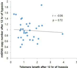
A negative correlation was observed between TL and mtDNAcn under both hypoxic conditions, i.e., 12 h (r = -0.06, P = 0.72, Fig. 3) and 24 h (r = -0.27, P = 0.13, Fig. 4). This correlation was not statistically significant under both hypoxic conditions.
4. Discussion
Alterations in TL can cause cell death and genomic instability.
Genome instability can cause temporary or permanent genomic alter- ations. Previous studies have shown that aging and inadequate lifestyle are connected with inflammation, altered oxidative stress levels, and decreased telomerase activity [58–60]. Hypoxia and cellular senescence determine oxidative stress level, which leads to molecular changes and can eventually cause cell death via apoptosis [61,62]. The oxidative stresses induced by hypoxia can damage cell apparatus and trigger specific signaling pathways. Therefore, to obtain more in-depth infor- mation on the effect of hypoxia, we studied whether moderate hypoxia can cause genomic instability in healthy human lymphocytes by
Fig. 1. Comparison of telomere length among three groups (Control, 12 h hypoxia, and 24 h hypoxia). Significant changes were marked by an asterisk (**P = 0.008), NS- Non-significant.
Fig. 2. Comparison of mitochondrial copy number among three groups (Con- trol, 12 h hypoxia, and 24 h hypoxia). NS- Non-significant.
Fig. 3. Association between mitochondrial DNA copy number and telomere length. Telomere length and mitochondrial DNA copy number in the group exposed to 12 h hypoxia.
Fig. 4. Association between mitochondrial DNA copy number and telomere length. Telomere length and mitochondrial DNA copy number in the group exposed to 24 h hypoxia.
analyzing TL and mtDNAcn. We noted increased TL in lymphocytes exposed to moderate hypoxia for 12 or 24 h. The TL in cells exposed to moderate hypoxia for 24 h was significantly greater than that in the normoxic control group. In cells exposed to moderate hypoxia for 12 h, the increase in TL was not statistically significant. Therefore, we concluded that telomere elongation is related to the duration of expo- sure to moderate hypoxia.
A study involving Wistar rats reported a significant increase in TL at moderate altitude in comparison to that at sea level or simulated high altitude and showed that HIF-1α and TERT levels and TL increased on the first day of exposure to hypoxia and then decreased to a stable level with further exposure [29]. Our result agrees with previous research indicating that TL is induced by hypoxia [25,63,64]. Some studies also observed that hypoxia triggers the adaptive cellular response through HIF-1 and telomerase activation [25,63]. However, to our knowledge, this is the first study to report an increase in TL in human lymphocytes exposed to moderate hypoxia in vitro. A study reported that telomeres could influence mitochondrial function and metabolism [46]. Telomere dysfunction can induce the p53 pathway, which acts as a link between telomere dysfunction and mitochondrial compromise [46]. In the cur- rent study, the mtDNAcn in the experimental groups exposed to mod- erate hypoxic conditions for 12 and 24 h did not significantly differ from that in the control group; the TL showed significant alterations, but the mtDNAcn did not. Therefore, we suggest that mtDNAcn is more stable than telomere length on exposure to moderate hypoxic conditions. The finding that TL changed but mtDNAcn did not can be explained as fol- lows. In humans, there are 46 chromosomes and 92 telomeres because there is a telomere at both ends of each chromosome. During mitosis, cells replicate all of their content and therefore the number of telomeres increases to 184 and telomeres contain thousands of repeats of the six-nucleotide sequence 5′-TTAGGG-3′; therefore, the presence of numerous repeat sequences in the telomeres may easily lead to genetic instability.
Mitochondrial dysfunction due to mtDNA mutation is not observed until a certain threshold of heteroplasmy is reached. While some studies have reported that TL and mtDNAcn are positively correlated in healthy individuals [47,65], others have reported that low lymphocyte mtDNAcn correlates with mitochondrion-related metabolic disorders [66–68]. Previous studies have reported conflicting results related to mtDNAcn in response to hypoxia; some studies reported an increase in mtDNAcn under hypoxic conditions [69–71], whereas others reported that, in some cell types, hypoxia could reduce mitochondrial biogenesis and mtDNA content [72–74]. Although in the current study, the corre- lations between TL and mtDNAcn under both moderate hypoxic condi- tions for 12 and 24 h showed negative trends, the correlations were not statistically significant. These results suggested that TL and mtDNA are not correlated in the case of short-time exposure to moderate hypoxia.
In our previous study, we noted an increase in SCE frequency and no change in MSI after exposure to moderate hypoxia for 12 or 24 h, which suggested that the increase in SCE frequency indicated a degree of chromosomal instability and the lack of change in MSI indicated sta- bility at the nucleotide level [54]. Our current results showed that the increase in TL on exposure to moderate hypoxia for 24 h indicated ge- netic instability at the nucleotide level rather than the chromosomal level. There were no significant changes in mtDNAcn, which could indicate genetic stability at the mitochondrial level, upon exposure to moderate hypoxia for 12 or 24 h. These findings indicated that, for moderate hypoxia, the genetic stability threshold increases in the following order: SCE, TL, and mtDNAcn.
Telomere gene therapy plays a beneficial role, but some studies re- ported that constitutive telomerase activation could lead to oncogenesis [75,76]. In the current study, an increase in TL was noted after exposure to moderate hypoxia, and further research is required to determine whether telomere elongation is beneficial or harmful. The moderate hypoxia conditions used in this study could be linked to the hypobaric hypoxia experienced by people living at high altitudes; therefore, this
study design may be utilized to study the other effects of hypoxia at high altitudes.
There are limitations to this study. First, the HIF-1α, TERT, and oxidative stress marker expression levels were not measured. A study reported that increased levels of reactive oxygen species downregulated HIF-1α expression [77]. Some other studies stated that oxidative stress could be a significant cause of telomere shortening via telomerase ac- tivity inhibition and damage to telomere DNA [78–81]. Second, we studied short-term hypoxia exposure for only 12 and 24 h; further studies are required on the effects of prolonged exposure to moderate hypoxia with respect to oxidative imbalance, telomere damage, and the effects of telomere elongation.
In conclusion, an increase in TL was observed upon exposure to moderate hypoxic conditions for 24 h and not after exposure for 12 h, suggesting that telomere elongation is related to the duration of expo- sure to moderate hypoxia. On short-term exposure to moderate hypoxia, the mtDNAcn in the experimental groups did not significantly differ from that in the control group. These results suggested that mtDNAcn is a more stable marker than TL alterations in the case of short-term moderate hypoxia. Further studies that use a larger sample size and also analyze the levels of oxidative stress markers, including TERT and HIF-1α, are required to make conclusive remarks regarding the role of moderate hypoxia in terms of its correlation with telomere elongation to unveil the underlying mechanism.
Contributions
KDK conceptualized and designed this study. MRA performed the experiment. MRA prepared the manuscript. KDK critically revised the manuscript. The authors proofread and approved the final manuscript.
Declaration of Competing Interest There is no conflict of interest to declare.
Acknowledgments
This work was supported by the National Research Foundation of Korea (NRF) grant funded by the Korea government (Nos.
2018R1D1A1B07040673, 2014R1A5A2010008).
Appendix A. Supplementary data
Supplementary material related to this article can be found, in the online version, at doi:https://doi.org/10.1016/j.toxrep.2020.10.011.
References
[1] S. Adnot, et al., Telomere dysfunction and cell senescence in chronic lung diseases:
therapeutic potential, Pharmacol. Ther. 153 (2015) 125–134.
[2] M. Armanios, Telomeres and age-related disease: how telomere biology informs clinical paradigms, J. Clin. Invest. 123 (3) (2013) 996–1002.
[3] E. Vakonaki, et al., Common mental disorders and association with telomere length, Biomed. Rep. 8 (2) (2018) 111–116.
[4] C.W. Greider, Telomere length regulation, Annu. Rev. Biochem. 65 (1) (1996) 337–365.
[5] P.J. Hornsby, Cell proliferation in mammalian aging, in: Handbook of the Biology of Aging, 5, 2001, pp. 207–266.
[6] J.W. Shay, W.E. Wright, Hallmarks of telomeres in ageing research, J. Pathol. 211 (2) (2007) 114–123.
[7] E. Vakonaki, et al., Effect of chronic and heavy drug abuse on biological aging, World Acad. Sci. J. 1 (2) (2019) 67–73.
[8] D. Tsoukalas, et al., Discovery of potent telomerase activators: unfolding new therapeutic and anti-aging perspectives, Mol. Med. Rep. 20 (4) (2019) 3701–3708.
[9] D. Tsoukalas, et al., Association of nutraceutical supplements with longer telomere length, Int. J. Mol. Med. 44 (1) (2019) 218–226.
[10] M. Hau, et al., Repeated stressors in adulthood increase the rate of biological ageing, Front. Zool. 12 (1) (2015) 4.
[11] A. Zalli, et al., Shorter telomeres with high telomerase activity are associated with raised allostatic load and impoverished psychosocial resources, Proc. Natl. Acad.
Sci. 111 (12) (2014) 4519–4524.
[12] M. Asghar, et al., Hidden costs of infection: chronic malaria accelerates telomere degradation and senescence in wild birds, Science 347 (6220) (2015) 436–438.
[13] E.S. Epel, et al., Accelerated telomere shortening in response to life stress, Proc.
Natl. Acad. Sci. 101 (49) (2004) 17312–17315.
[14] R.T. Calado, N.S. Young, Telomere diseases, N. Engl. J. Med. 361 (24) (2009) 2353–2365.
[15] D. Tsoukalas, et al., Metabolic fingerprint of chronic obstructive lung diseases: a new diagnostic perspective, Metabolites 9 (12) (2019) 290.
[16] D. Tsoukalas, et al., Targeted metabolomic analysis of serum fatty acids for the prediction of autoimmune diseases, Front. Mol. Biosci. 6 (2019) 120.
[17] Y.A. Hajam, et al., Combined administration of exogenous melatonin and insulin ameliorates streptozotocin induced toxic alteration on hematological parameters in diabetic male Wistar rats, Toxicol. Rep. 7 (2020) 353–359.
[18] C. Chiara, et al., MicroRNAs alteration as early biomarkers for cancer and neurodegenerative diseases: new challenges in pesticides exposure, Toxicol. Rep.
(2020).
[19] M. Sharifi-Rad, et al., Lifestyle, oxidative stress, and antioxidants: back and forth in the pathophysiology of chronic diseases, Front. Physiol. 11 (2020) 694.
[20] J. Sharifi-Rad, et al., Diet, lifestyle and cardiovascular diseases: linking pathophysiology to cardioprotective effects of natural bioactive compounds, Int. J.
Environ. Res. Public Health 17 (7) (2020) 2326.
[21] E. Vasilopoulos, et al., The association of female and male infertility with telomere length, Int. J. Mol. Med. 44 (2) (2019) 375–389.
[22] H. Fujii, et al., Telomerase insufficiency in rheumatoid arthritis, Proc. Natl. Acad.
Sci. 106 (11) (2009) 4360–4365.
[23] X. Wu, et al., Telomere dysfunction: a potential cancer predisposition factor, J. Natl. Cancer Inst. 95 (16) (2003) 1211–1218.
[24] P. Willeit, et al., Leucocyte telomere length and risk of type 2 diabetes mellitus:
new prospective cohort study and literature-based meta-analysis, PLoS One 9 (11) (2014) e112483.
[25] T. Minamino, S. Mitsialis, S. Kourembanas, Hypoxia extends the life span of vascular smooth muscle cells through telomerase activation, Mol. Cell. Biol. 21 (10) (2001) 3336–3342.
[26] G.L. Semenza, Oxygen sensing, hypoxia-inducible factors, and disease pathophysiology, Annu. Rev. Pathol. Mech. Dis. 9 (2014) 47–71.
[27] E. Krzywinska, C. Stockmann, Hypoxia, metabolism and immune cell function, Biomedicines 6 (2) (2018) 56.
[28] N.R. Prabhakar, G.L. Semenza, Oxygen sensing and homeostasis, Physiology 30 (5) (2015) 340–348.
[29] Y. Wang, et al., Telomeres are elongated in rats exposed to moderate altitude, J. Physiol. Anthropol. 33 (1) (2014) 19.
[30] M.T. Hemann, et al., The shortest telomere, not average telomere length, is critical for cell viability and chromosome stability, Cell 107 (1) (2001) 67–77.
[31] L.-Y. Hao, et al., Short telomeres, even in the presence of telomerase, limit tissue renewal capacity, Cell 123 (6) (2005) 1121–1131.
[32] M.N. Sack, Type 2 diabetes, mitochondrial biology and the heart, J. Mol. Cell.
Cardiol. 46 (6) (2009) 842–849.
[33] S.-B. Catrina, et al., Hyperglycemia regulates hypoxia-inducible factor-1α protein stability and function, Diabetes 53 (12) (2004) 3226–3232.
[34] C. Peers, H.A. Pearson, J.P. Boyle, Hypoxia and Alzheimer’s disease, Essays Biochem. 43 (2007) 153–164.
[35] V. Nizet, R.S. Johnson, Interdependence of hypoxic and innate immune responses, Nat. Rev. Immunol. 9 (9) (2009) 609.
[36] F. Weinberg, N.S. Chandel, Mitochondrial metabolism and cancer, Ann. N. Y. Acad.
Sci. 1177 (1) (2009) 66–73.
[37] E.A. Dominic, et al., Mitochondrial cytopathies and cardiovascular disease, Heart 100 (8) (2014) 611–618.
[38] M. Saraste, Oxidative phosphorylation at the fin de siecle, Science 283 (5407) (1999) 1488–1493.
[39] A.N. Sferruzzi-Perri, E.J. Camm, The programming power of the placenta, Front.
Physiol. 7 (2016) 33.
[40] M. Yu, Generation, function and diagnostic value of mitochondrial DNA copy number alterations in human cancers, Life Sci. 89 (3–4) (2011) 65–71.
[41] C. Vijayasarathy, et al., Adaptive changes in the expression of nuclear and mitochondrial encoded subunits of cytochrome c oxidase and the catalytic activity during hypoxia, Eur. J. Biochem. 270 (5) (2003) 871–879.
[42] E.W. Englander, et al., Hypoxia-induced mitochondrial and nuclear DNA damage in the rat brain, J. Neurosci. Res. 58 (2) (1999) 262–269.
[43] R. Stark, M. Roden, Mitochondrial function and endocrine diseases, Eur. J. Clin.
Invest. 37 (4) (2007) 236–248.
[44] P. Jeˇzek, et al., Distinctions and similarities of cell bioenergetics and the role of mitochondria in hypoxia, cancer, and embryonic development, Int. J. Biochem.
Cell Biol. 42 (5) (2010) 604–622.
[45] A. Pyle, et al., Reduced mitochondrial DNA copy number is a biomarker of Parkinson’s disease, Neurobiol. Aging 38 (2016) 216, e7-216. e10.
[46] E. Sahin, et al., Telomere dysfunction induces metabolic and mitochondrial compromise, Nature 470 (7334) (2011) 359.
[47] J.-H. Kim, et al., The relationship between leukocyte mitochondrial DNA copy number and telomere length in community-dwelling elderly women, PLoS One 8 (6) (2013) e67227.
[48] D. Gilley, H. Tanaka, B.-S. Herbert, Telomere dysfunction in aging and cancer, Int.
J. Biochem. Cell Biol. 37 (5) (2005) 1000–1013.
[49] M. Picard, B.S. McEwen, Psychological stress and mitochondria: a systematic review, Psychosom. Med. 80 (2) (2018) 141.
[50] N. Monroy-Jaramillo, E. Dyukova, C. Walss-Bass, Telomere length in psychiatric disorders: Is it more than an ageing marker? World J. Biol. Psychiatry 19 (sup2) (2018) S2–S20.
[51] J.-Z. Guan, et al., Different levels of hypoxia regulate telomere length and telomerase activity, Aging Clin. Exp. Res. 24 (3) (2012) 213–217.
[52] J.-Z. Guan, et al., Alteration of telomere length and subtelomeric methylation in human endothelial cell under different levels of hypoxia, Arch. Med. Res. 43 (1) (2012) 15–20.
[53] P. Davy, R. Allsopp, Hypoxia: are stem cells in it for the long run? Cell Cycle 10 (2) (2011) 206–211.
[54] J.-H. Lee, et al., Genetic instability in the human lymphocyte exposed to hypoxia, Cancer Genet. Cytogenet. 196 (1) (2010) 83–88.
[55] N. Kim, et al., Association between internet gaming addiction and leukocyte telomere length in Korean male adolescents, Soc. Sci. Med. 222 (2019) 84–90.
[56] J.-H. Lee, D.-K. Kim, Association between mitochondrial D-loop polymorphism and copy number, Korean J. Phys. Anthropol. 27 (3) (2014) 131–136.
[57] R.M. Cawthon, Telomere measurement by quantitative PCR, Nucleic Acids Res. 30 (10) (2002) e47.
[58] M. Gleeson, et al., The anti-inflammatory effects of exercise: mechanisms and implications for the prevention and treatment of disease, Nat. Rev. Immunol. 11 (9) (2011) 607.
[59] C.V. de Sousa, et al., The antioxidant effect of exercise: a systematic review and meta-analysis, Sport. Med. 47 (2) (2017) 277–293.
[60] E.H. Blackburn, E.S. Epel, J. Lin, Human telomere biology: a contributory and interactive factor in aging, disease risks, and protection, Science 350 (6265) (2015) 1193–1198.
[61] M.J. McEachern, A. Krauskopf, E.H. Blackburn, Telomeres and their control, Annu.
Rev. Genet. 34 (1) (2000) 331–358.
[62] T. Von Zglinicki, Oxidative stress shortens telomeres, Trends Biochem. Sci. 27 (7) (2002) 339–344.
[63] N. Mouraret, et al., Role for telomerase in pulmonary hypertension, Circulation 131 (8) (2015) 742–755.
[64] S.-l. Xu, et al., Effects of hypoxia on promoter of telomerase reverse transcriptase and cell cycle distribution in neonatal rat cardiac myocytes, Chin. Med. J. 118 (5) (2005) 409–411.
[65] A.R. Tyrka, et al., Association of telomere length and mitochondrial DNA copy number in a community sample of healthy adults, Exp. Gerontol. 66 (2015) 17–20.
[66] J. Song, et al., Peripheral blood mitochondrial DNA content is related to insulin sensitivity in offspring of type 2 diabetic patients, Diabetes Care 24 (5) (2001) 865–869.
[67] H. Lee, et al., Decreased mitochondrial DNA content in peripheral blood precedes the development of non-insulin-dependent diabetes mellitus, Diabetes Res. Clin.
Pract. 42 (3) (1998) 161–167.
[68] S.-W. Weng, et al., Peripheral blood mitochondrial DNA content and dysregulation of glucose metabolism, Diabetes Res. Clin. Pract. 83 (1) (2009) 94–99.
[69] J. Carabelli, et al., High fat diet-induced liver steatosis promotes an increase in liver mitochondrial biogenesis in response to hypoxia, J. Cell. Mol. Med. 15 (6) (2011) 1329–1338.
[70] Y. Luo, et al., Altitude can alter the mtDNA copy number and nDNA integrity in sperm, J. Assist. Reprod. Genet. 28 (10) (2011) 951–956.
[71] Y. Luo, et al., Long-term cycles of hypoxia and normoxia increase the contents of liver mitochondrial DNA in rats, Eur. J. Appl. Physiol. 113 (1) (2013) 223–232.
[72] P.H. Oliveira, et al., Impact of hypoxia and long-term cultivation on the genomic stability and mitochondrial performance of ex vivo expanded human stem/stromal cells, Stem Cell Res. 9 (3) (2012) 225–236.
[73] H. Hoppeler, et al., Response of skeletal muscle mitochondria to hypoxia, Exp.
Physiol. 88 (1) (2003) 109–119.
[74] H. Zhang, et al., HIF-1 inhibits mitochondrial biogenesis and cellular respiration in VHL-deficient renal cell carcinoma by repression of C-MYC activity, Cancer Cell 11 (5) (2007) 407–420.
[75] K. Okamoto, H. Seimiya, Revisiting telomere shortening in cancer, Cells 8 (2) (2019) 107.
[76] S. Cacchione, A. Biroccio, A. Rizzo, Emerging roles of telomeric chromatin alterations in cancer, J. Exp. Clin. Cancer Res. 38 (1) (2019) 1–12.
[77] Z.-Z. Yang, et al., Redox regulation of HIF-1α levels and HO-1 expression in renal medullary interstitial cells, Am. J. Physiol. Renal Physiol. 284 (6) (2003) F1207–F1215.
[78] J. Haendeler, et al., Hydrogen peroxide triggers nuclear export of telomerase reverse transcriptase via Src kinase family-dependent phosphorylation of tyrosine 707, Mol. Cell. Biol. 23 (13) (2003) 4598–4610.
[79] J. Haendeler, et al., Antioxidants inhibit nuclear export of telomerase reverse transcriptase and delay replicative senescence of endothelial cells, Circ. Res. 94 (6) (2004) 768–775.
[80] T. Richter, T. von Zglinicki, A continuous correlation between oxidative stress and telomere shortening in fibroblasts, Exp. Gerontol. 42 (11) (2007) 1039–1042.
[81] M. Satoh, et al., Association between oxidative DNA damage and telomere shortening in circulating endothelial progenitor cells obtained from metabolic syndrome patients with coronary artery disease, Atherosclerosis 198 (2) (2008) 347–353.