70
http://dx.doi.org/10.4046/trd.2013.74.2.70 ISSN: 1738-3536(Print)/2005-6184(Online) Tuberc Respir Dis 2013;74:70-73
CopyrightⒸ2013. The Korean Academy of Tuberculosis and Respiratory Diseases. All rights reserved.
Interstitial Lung Disease in a Patient with Dyskeratosis Congenita
Hyun Jung Kim, M.D.1,2, Kyu Jin Kim, M.D.1,2, Kwan Ho Lee, M.D., Ph.D.1,2, Kyeong-Cheol Shin, M.D., Ph.D.1,2, Jin Hong Chung, M.D., Ph.D.1,2, Myung Soo Hyun, M.D., Ph.D.1, Ki-Hong Kim, M.D., Ph.D.3
1Department of Internal Medicine, Yeungnam University College of Medicine, 2Regional Center for Respiratory Disease, Yeungnam University Medical Center, 3Department of Dermatology, Yeungnam University College of Medicine, Daegu, Korea
Dyskeratosis congenita is a rare congenital disorder characterized by a triad of reticular pigmentation of the skin, dystrophic nails, and leukoplakia of the mucous membrane. Sometimes it is associated with bone marrow failure, secondary malignancy and interstitial lung disease. Though it is rare, Dyskeratosis congenita is diagnosed relatively easily when clinicians suspect it. It can be diagnosed just by gross inspection with care. Dyskeratosis congenita should be considered as one cause associated with interstitial lung disease. In Korea, interstitial lung disease with dyskeratosis congenita has not been reported. We report a case and review the literature.
Key Words: Lung Diseases, Interstitial; Dyskeratosis Congenita; Anemia, Aplastic
Address for correspondence: Jin Hong Chung, M.D., Ph.D.
Department of Internal Medicine, Yeungnam University Medical Center, Yeungnam University College of Medicine, 170, Hyeonchung-ro, Nam-gu, Daegu 705-717, Korea Phone: 82-53-620-3840, Fax: 82-53-654-8386
E-mail: [email protected] Received: Jun. 12, 2012 Revised: Jun. 21, 2012 Accepted: Jul. 17, 2012
CCIt is identical to the Creative Commons Attribution Non-Commercial License (http://creativecommons.org/licenses/by-nc/3.0/).
Introduction
Dyskeratosis congenita is a rare hereditary disorder characterized by dystrophic nails, abnormal skin pig- mentation, and mucous leukoplakia. Non-mucocuta- neous features include bone marrow failure, pulmonary fibrosis, and predisposing malignancy
1. The prevalence of classic dyskeratosis congenita is approximately 1/
1,000,000 individuals
2. The primary causes of death in patients with dyskeratosis congenita are bone marrow failure and immunodeficiency (60–70%), pulmonary complications (10–15%), and malignancy (10%)
3. While dyskeratosis congenita has been reported in the Korean literature, to our knowledge, pulmonary in- volvement has not been mentioned
4-8. Since pulmonary involvement is considered to be an important complica-
tion of dyskeratosis congenita, we report a case and re- view the literature.
Case Report
A 29-year-old Korean man who had smoked a fourth of a pack daily for twelve years, visited the hospital be- cause of easy fatigue. His height was 172 cm and weight was 49 kg. He has never been exposed to occu- pational noxious gas like asbestos, chemical fumes, or organic dusts. He has taken no medications.
Recently, he suffered from dry cough and dyspnea on exertion. He felt easily exhausted but his weight or appetite was not changed. He denied febrile sensation or chills, and peripheral edema was not noted. His skin seemed mottled with pigmentation on the anterior chest and neck, but he didn't have photosensitivity or skin allergy. Also, leukoplakia was noted at his tongue and almost all of his finger and toe nails were markedly atrophic and dystrophic (Figure 1). However, we couldn't find joint swelling or deformity.
Auscultation of the lungs revealed bilateral diffuse fine crackles without wheezes or rhonchi. The heart beat sounded regular without murmur. On the day of his visit, his blood pressure was 105/54 mm Hg, pulse
Case Report
Tuberculosis and Respiratory Diseases Vol. 74. No. 2, Feb. 2013
71 Figure 1. Mucocutaneous triad of abnormal skin pig- mentation, nail dystrophy, and leukoplakia. (A) Abnor- mal fine reticular pigmenta- tion around the neck. (B) Leukoplakia on the tongue.
(C, D) Markedly atrophic and dystrophic finger and toe nails.
rate was 79/min, respiratory rate was 17/min, and body temperature was 36.4
oC.
He took a pulmonary function test and chest X-ray.
The results showed that forced vital capacity (FVC) was decreased to 3.24 L (66% of predicted value), forced ex- piratory volume 1 second (FEV
1) was 3.00 L (77% of predicted value). It showed a typical restrictive pulmo- nary disease pattern. Diffusion capacity of the lung for carbon monoxide (DLCO) was 37% of predicted value.
Chest X-ray revealed fibrostreaky and reticular at- tended infiltration in both lungs. His electrocardiogram showed normal sinus rhythm. Hemoglobin was 12.4 g/dL and hematocrit was 36.3%. Anti-nuclear antibody and rheumatic factor were negative.
We began to review his hospital record and noticed that he has been admitted several times. When he was 17 years old, he had neurosurgery because of a trau- matic subdural hematoma injury from a bicycle acci- dent. At that time, thrombocytopenia was noted. During
his hospital stay, he received a liver scan, bone marrow study, chromosomal analysis and other tests. His bone marrow biopsy revealed hypocellularity without dys- plasia. He was not a candidate for bone marrow trans- plantation because marrow suppression was not that severe. Aplastic anemia without dysplasia was diag- nosed throughout hematologic evaluation. Chromoso- mal analysis and intelligence quotient were normal.
Viral marker showed no specific viral infection like par- vovirus, hepatitis B or C that could cause pancytopenia.
Liver scan showed splenomegaly with increased uptake at bone marrow.
During a previous admission period, the physician was curious about his unusual skin and dystrophied nails. The physician consulted regarding his skin lesion with a dermatologist. The dermatologist diagnosed his disease as dyskeratosis congenita.
His family includes two sisters who had no skin pig-
mentation or nail dystrophy. Only his mother's left sec-
HJ Kim et al: Interstitial lung disease in a patient with dyskeratosis congenita
72
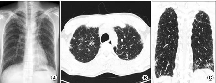
Figure 2. Chest X-ray and computed tomography taken on the first visit. (A) Fibrostreaky and reticular patterned infiltration at both lung fields. (B, C) Interstitial thickening along the subpleural area.
Figure 3. Chest X-ray and computed tomography 2 years later. (A) Increased in the bilateral basal reticulolinear shadows.
(B, C) Progression of interstitial fibrosis at both lung subpleural areas.
ond nail was dystrophied. He has two children, one son and one daughter. Presently they also have no symp- toms of dyskeratosis congenita.
His disease was reported to The Korean Journal of Hematology
5. He was the first case of dyskeratosis con- genita with pancytopenia in Korea. But he didn't return after discharge because he had no further symptoms.
Ten years later, he visited the department of pulmo- nology because of easy fatigue with a tentative diag- nosis of pulmonary tuberculosis. But chest computed tomography (CT) showed an interstitial lung disease pattern (Figure 2). His symptoms and laboratory find- ings didn't support the possibility of autoimmune
disorder. We diagnosed interstitial lung disease related to dyskeratosis congenita.
He was treated with prednisolone 20 mg but was ad- mitted because of gastric ulcer bleeding. He has spleno- megaly and thrombocytopenia, so bleeding risk was high. Prednisolone was stopped and he was followed up with supportive care. His symptom, dyspnea on ex- ertion became aggravated. After 2 years, we reassessed his status by pulmonary function test and chest CT and found it had worsened. FVC was much declined to 2.57 L (52% of predicted value) and FEV
1was 2.32 L (59%
of predicted value). The ratio FEV
1of FVC was 90%,
but DLCO was 17%, DLCO/alveolar volume was 25%.
Tuberculosis and Respiratory Diseases Vol. 74. No. 2, Feb. 2013