Background: To evaluate the demographics, clinical and radiographic features of calcific tendinitis of the shoulder in the Korean popula- tion, specifically focusing on the incidence of coexisting rotator cuff tear.
Methods: Between October 2014 and January 2015, we performed a prospective multicenter study with 506 patients from 11 training hos- pitals in Korea. We collected data of demographics and radiographic analysis based on simple radiographs, clinical assessments based on visual analog scale (VAS) and the American Shoulder Elbow Surgeons (ASES) score, and treatment modalities that are used currently. We also evaluated coexisting rotator cuff tear by ultrasonography (US) or magnetic resonance imaging (MRI) images.
Results: There were 402 female patients (79%) with mean age of 55 years (range, 31–87 years). Mean duration of symptoms was 16 months.
Mean size of calcific materials was 11.4 mm (range, 0–35 mm). Mean value of VAS and ASES scores were 6.5 (range, 1–10) and 47 (range, 8–95), respectively. Of 383 patients (76%), 59 (15%) had rotator cuff tear including 15 full-thickness tears on US or MRI. Patients with rota- tor cuff tears were significantly associated with older age, recurrent symptoms, menstrual disorders in females, and having undergone calci- fication removal surgery and rotator cuff repair (all p<0.05).
Conclusions: This study reported demographic, radiographic, and clinical features of calcific tendinitis of the shoulder in Korean popula- tion, which were not different from those of Western population. Coexisting rotator cuff tear was found with 15% incidence in this large se- ries, suggesting that further radiographic study to evaluate rotator cuff tear might be needed in some calcific tendinitis patients of older age and presenting with recurrent symptoms.
Keywords: Calcific tendinitis; Epidemiology; Rotator cuff Clin Shoulder Elbow 2021;24(1):21-26
https://doi.org/10.5397/cise.2020.00010
Calcific tendinitis of the shoulder in the Korean population:
demographics and its relation with coexisting rotator cuff tear
Yon-Sik Yoo
1, Jin-Young Park
2, Myung-sun Kim
3, Nam-Su Cho
4, Yong-Beom Lee
5, Seung-Hyun Cho
6, Kyoung Jin Park
7, Chul-Hyun Cho
8, Bong Gun Lee
9, Dong Joo Shin
10, Han-Hoon Kim
11, Tae Kang Lim
111Department of Orthopedic Surgery, Hallym University Dongtan Sacred Heart Hospital, Hwaseong, Korea
2Global Center for Shoulder, Elbow and Sports at Neon Orthopaedic Clinic, Seoul, Korea
3Department of Orthopaedics, Chonnam National University College of Medicine, Gwangju, Korea
4Department of Orthopedic Surgery, Kyung Hee University Hospital at Gangdong, Seoul, Korea
5Department of Orthopaedic Surgery, Hallym University Sacred Heart Hospital, Anyang, Korea
6Seoul Metropolitan Seonam Hospital, Seoul, Korea
7Goodsam Orthopaedic Clinic, Cheongju, Korea
8Department of Orthopedic Surgery, Keimyung University Dongsan Medical Center, Keimyung University School of Medicine, Daegu, Korea
9Department of Orthopaedics, Hanyang University College of Medicine, Seoul, Korea
10Department of Orthopaedics, Daegu Fatima Hospital, Daegu, Korea
11Department of Orthopedic Surgery, Nowon Eulji Medical Center, Eulji University School of Medicine, Seoul, Korea
IRB approval: Nowon Eulji Medical Center (No. EMCIRB 19-126).
Financial support: None.
Conflict of interest: None.
Received: January 14, 2020 Revised: December 4, 2020 Accepted: December 6, 2020 Correspondence to: Tae Kang Lim
Department of Orthopedic Surgery, Nowon Eulji Medical Center, Eulji University School of Medicine, 68 Hangeulbiseong-ro, Nowon-gu, Seoul 01830, Korea
Tel: +82-2-970-8036, Fax: +82-2-973-3024, E-mail: [email protected], ORCID: https://orcid.org/0000-0001-8752-3987
eISSN 2288-8721
Copyright© 2021 Korean Shoulder and Elbow Society. All Rights Reserved.
This is an Open Access article distributed under the terms of the Creative Commons Attribution Non-Commercial License (http://creativecommons.org/licenses/by-nc/4.0/) which permits unrestricted non-commercial use, distribution, and reproduction in any medium, provided the original work is properly cited.
INTRODUCTION
Calcific tendinitis is one of the common causes of shoulder pain and discomfort, and an incidence rate of 2.7%–20% has been re- ported [1]. Several hypotheses have been raised as to the cause of calcific tendinitis, including active cell-mediated process, rotator cuff tendon degeneration, overuse and microinjury, genetic pre- disposition, local metabolic or hemodynamic abnormalities, and subacromial impingements, but the exact pathophysiology re- mains unclear [2]. The calcific tendinitis of the shoulder is preva- lent in the dominant and more frequently used arm [1]. It is highly correlated to old age, diabetes, or smoking and frequently occurs in patients who raise their arms overhead [2].
Demographic factors can be related to the pathogenesis of dis- ease and also affect its prognosis. In calcific tendinitis, it has been reported that being female, bilateral morbidity, and having multi- ple calcific deposits are associated with poor prognosis in long- term clinical results [3]. Contrary to the existing belief that the treatment of calcific tendinitis is benign and self-limited, de Witte et al. [3] recently reported that pain and discomfort of the shoulder still persisted in more than 50% of patients even after 14 years of follow-up. Considering these outcomes, studies of demo- graphic and radiographic features to determine treatment op- tions and assess prognosis at the initial diagnosis of calcific ten- dinitis are clinically highly significant.
Most of the existing studies [4] on demographic factors, radio- graphic characteristics, long-term course, and prognostic factors of the disease are limited by small numbers of patients. Studies involving larger numbers of patients are needed. Besides, studies [3,5] that included relatively large numbers of research subjects were all conducted outside the country. No studies have been re- ported investigating demographic factors and characteristics as- sociated with calcific tendinitis of the shoulder in the Korean population.
One of the controversies related to calcific tendinitis of the shoulder is whether it is related to the rotator cuff tear. Since cal- cific tendinitis can be easily diagnosed with only a simple radio- graphic examination, it is common to first perform conservative treatment without first performing expensive imaging tests such as magnetic resonance imaging (MRI) [5]. In patients who do not respond to conservative treatment, however, a common clin- ical occurrence is that accompanying rotator cuff tear is diag- nosed when additional imaging tests are performed. These clini- cal experiences occur in patients with calcific tendinitis of the shoulder diagnosed by simple radiographic examination. The question remains as to whether there is a need to diagnose early the presence of rotator cuff tear. Indeed, according to a study by
Jim et al. [4], rotator cuff rupture was reported in 22 of 81 pa- tients with calcified tendinitis. However, this study is limited in the number of patients, and because it is a study performed through simple radiographs of arthrography after administration of a contrast agent, diagnostic accuracy is limited. In addition to this study, the presence of rotator cuff tear in calcified tendinitis of the shoulder has been reported rarely.
Therefore, the purpose of this study was to investigate the de- mographic, clinical, and radiological factors of calcific tendinitis of the shoulder joint in a large number of Koreans and to investi- gate the prevalence of rotator cuff tear associated with calcific tendinitis.
METHODS
Materials
From November 2014 to January 2015, a retrospective study was conducted on 506 patients diagnosed with calcific tendinitis of the shoulder at 11 major training hospitals nationwide. In this study, the inclusion criteria were patients that visited the hospital for shoulder pain and a simple radiographic examination with observed calcium deposits around the shoulder. Patients with in- fections or previous fracture or surgery in the same area of the shoulder joint were excluded from the study. Informed consent was waived due to retrospective nature of this study.
Demographic and Clinical Evaluation
Demographic characteristics of patient age, sex, dominant arm,
bilateral arm, trauma history, occupation, regular exercise, hob-
bies using shoulders, and smoking were investigated. Occupa-
tions were categorized into housework, labor, office work, and
others. Labor and office work were classified according to wheth-
er arms were used in vocational activities. Regular exercise was
defined as occurring at least twice a week (ex. swimming or jog-
ging, etc.). Smoking was defined as only current smokers. Diabe-
tes, thyroid disorder, and rheumatoid arthritis were investigated
as comorbid diseases, and the history of menopause and obstet-
rics and gynecological disease was investigated in females. Dis-
eases that affect estrogen metabolism such as endometriosis,
ovarian cysts, polycystic ovary syndrome, and infertility treat-
ment history were investigated. At the first visit, the duration of
symptoms, the presence of nocturnal pain, visual analog scale
(VAS), and American Shoulder Elbow Surgeons (ASES) scores
were measured for clinical evaluation. The investigation classi-
fied treatments into nonsurgical such as extracorporeal shock
wave treatment or surgical.
Radiological Evaluation
The location of calcium was evaluated in the standard radio- graphs of anteroposterior, axial and supraspinatus outlet views.
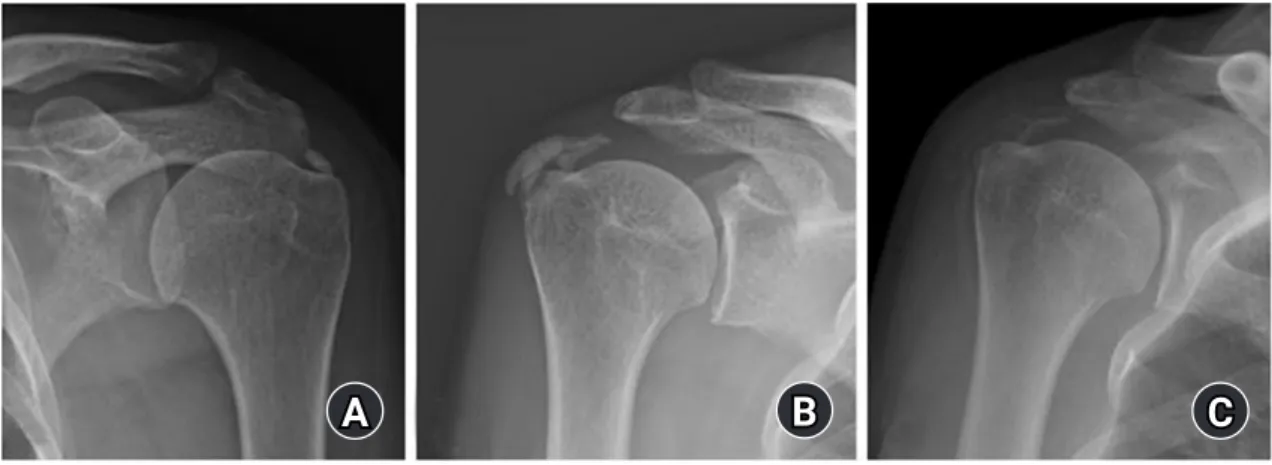
Calcium located in the anterior of the scapular spine and over the greater tuberosity was particularly noted as calcium deposit in the supraspinatus tendon. Deposits in the posterior of the scapu- lar spine and the greater tuberosity were considered as in the in- fraspinatus tendon. Calcium originating from the subscapularis was determined as the case in front of the lesser tuberosity in the outlet and axial view. The size of calcium deposit was measured on picture archiving and communicating system (PACS) and its characteristic was classified by Gartner method [6] (Fig. 1). Fur- thermore, ultrasound or MRI was performed to investigate the presence or absence of accompanying rotator cuff tear.
Statistical Analysis
Based on these results, the relationship between demographic variables such as age, sex, presence of comorbid diseases, clinical symptoms, radiographic findings, and surgical treatment was statistically analyzed. The clinical relationship between symp- toms and surgery was statistically analyzed, particularly the rela- tion between rotator cuff tear and demographic factors. A chi- square test was performed for nominal variable analysis, and an independent t-test was performed for continuous variable analy- sis. Cases where the p-value was less than 0.05 were judged to have statistical significance.
RESULTS
Demographic Factor
Of the total 506 patients, there were 402 females (79%) and 104 males (21%). Of the total 506 patients, there were 402 females (79%) and 104 males (21%) and the average age was 55 years
(range, 31–87 years). By age group, the 50s had the highest inci- dence at 45%, followed by 23% in 60s, 21% in 40s, 6% in 70s, and 5% in 30s (Fig. 2). Among the total patients, 404 (79%) were con- firmed as being affected in the dominant arm, and 98 (19%) cas- es were bilaterally affected at the time of diagnosis. There were 64 patients (12.6%) who had a history of trauma before the onset of symptoms.
As for the occupation, unemployed, including housekeeping, was the most common, with 187 (37%), followed by laborers with 108 (21%), office workers with 94 (19%), and others with 87 (17%). One hundred and six patients (21%) answered that they were exercising regularly, and 77 (15%) were exercising using their shoulders. The current smokers included 42 (8%). As for systemic and medical diseases, diabetes was present in 62 (12%), thyroid disease in 32 (6%), and rheumatic disease in 13 (3%).
Among 377 female patients, 179 (47%) had achieved menopause,
B
A C
Fig. 1. Gartner classification of calcific tendinitis. (A) Gartner type 1 has a clear, uniform, and dense shape. (B) Gartner type 2 has a clear boundary but is uneven inside or a uniform inside but an uneven boundary. (C) Gartner type 3, with no borders, transparent or cloud-like.
250
200
150
100
50
0
Age
Number
25 104
228
114
32 3 30s 40s 50s 60s 70s 80s
Fig. 2. Distribution of calcific tendinitis by age group. The 50s had the highest incidence at 45%, 60s were 23%, 40s at 21%, 70s at 6%, and 30s at 5% by age group.
and 39 (10%) had a history of obstetric diseases (Table 1).
When comparing clinical characteristics according to gender, the frequency of night pain in females was statistically signifi- cantly higher (73% for women and 61% for men, p=0.015). Oth-
er demographic and clinical characteristic differences between men and women were not observed (Table 2).
Clinical Features
The average symptom duration was 16 months (range, 1–180 months), the average pain score (VAS) at the time of visit was 6.6 (range, 1–10), and the ASES score was 47 (range, 8–95). Previ- ously, 102 (20%) were diagnosed with calcific tendinitis and had recurrence of symptoms after a symptom-free period. Sleep dis- turbance due to night pain was observed in 358 (70.7%). The most common treatment for patients was the use of oral analge- sics (93%), steroid injection therapy (53%), physical therapy (42%), extracorporeal shock wave therapy (16%), and surgical arthroscopic calcium removal and rotator cuff repair (6%).
Radiological Results
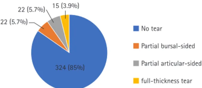
The average size of calcium deposit was 11.6 mm (range, 0.9–35 mm) on simple radiographic examination. The location of oc- currence was supraspinatus: 322 cases (64%), Infraspinatus 125 cases (25%), subscapularis 79 cases (16%), and Teres Minor 5 cases (1%). According to Gartner classification [6], there were 171 cases of type 1 (34%), 177 cases of type 2 (35%), and 120 cas- es of type 3 (24%). Ultrasound or MRI was performed in 383 out of 506 cases (76%), of which 59 (15%) were diagnosed with rota- tor cuff tear. Partial-thickness tear was more common with 44 cases (11%), but patients with full-thickness tear (Fig. 3) were also observed in 15 cases (4%). (Fig. 4)
When comparing calcific tendinitis patients with rotator cuff tear against those without rotator cuff tear on ultrasound or MRI, the average age of patients with rotator cuff tear was 58 years,
Table 1. Demographics and clinical outcomesVariable Value
Age (yr) 55 (31–87)
Female sex 402 (79)
Dominant side 404 (79)
Bilateral 98 (19)
Symptom duration (mo) 16 (0–180)
Recurrent symptom 102 (20)
Trauma history 64 (13)
Night pain 358 (71)
Occupation
Housework 187 (39)
Manual labor 108 (21)
Office work 94 (19)
Other 87 (17)
Regular exercise 106 (21)
Exercise using shoulder 77 (15)
Smoking 42 (8)
Diabetes 62 (12)
Thyroid disease 32 (6)
Rheumatoid disease 13 (3)
Menopause 179 (47)
Obstetric disease 39 (10)
Body mass index (kg/m2) 23.6 (16.42–41.67)
Size of calcification (mm) 11.4 (0–35)
Gartner classification
Type I 171 (34)
Type II 177 (35)
Type III 120 (24)
Coexisting rotator cuff tear 59 (15)
Visual analog scale score 6.5 (1–10)
American Shoulder Elbow Surgeons score 47 (8–95)
Operation 30 (6)
Values are presented as mean (range) or number (%).
Table 2. Clinical characteristics by sex
Variable Male
(n=104) Female
(n=401) p-value
Age (yr) 56.7±10.4 55.1±8.9 0.124
Recurrent symptom 22 (21) 102 (25) 0.443
Night pain 63 (61) 295 (73) 0.015
Trauma history 13 (13) 50 (13) 0.993
Visual analogue scale score 6.2±1.9 6.6±2.0 0.067 Size of calcification (mm) 11.4±7.2 11.4±6.1 0.972 Values are presented as mean±standard deviation or number (%).
B
A C
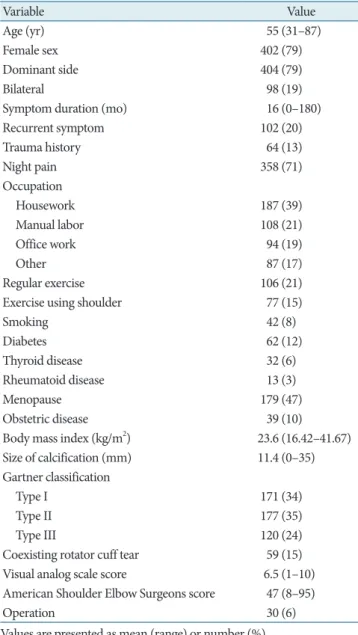
Fig. 3. A 59-year-old female patient with calcific tendinitis with rota- tor cuff tear. (A) Radiation examination of the shoulder joint exit showed calcium in the inferior tendon. (B, C) Ultrasound examina- tion showed rotator cuff tear (solid arrows) and calcium deposition at the same time (dotted arrow).
of night pain. Among treatment modalities, oral analgesics (93%) were the most common, followed by steroid injection therapy (53%).
According to Harvie et al.'s study [7] on the relationship be- tween the natural course of calcific tendinitis and endocrine dis- eases, the prevalence of endocrine diseases such as hypothyroid- ism was high in patients with calcific tendinitis. The proportion of female patients with calcific tendinitis was high, suggesting that estrogen might play a role in the pathogenesis. In this study's results, the prevalence of females was at 79%, diabetes was con- firmed in 12% of patients, and thyroid disease was also con- firmed in 6% of patients. Among female patients with calcific tendinitis, previous studies reported that menstrual irregularities and hysterectomy rates were higher than those of the general population [7] and that finding corresponded to a high propor- tion of menstrual irregularities (47%) and obstetric diseases (10%) found in this study.
According to the results of a long-term follow-up study by de Witte et al. [3], long-term prognosis was poor when calcific ten- dinitis invaded the dominant arm or bilateral arms. It is more common for calcific tendinitis to invade the dominant arm [1]
because the rotator cuff contracts during the lifting motion, mak- ing it hemodynamically susceptible to local ischemia [2]. In this study, it was confirmed that the dominant arm invading patients accounted for a large proportion (79%) of cases, and it was more frequent in houseworkers and laborers than in office workers. As such, a patient's dominant arm activities in occupation and hob- bies is related to the cause of the disease and the prognosis, and so it must be carefully checked before treatment.
According to previous studies, the radiological size and Gart- ner classification of calcium cannot accurately predict patients' prognosis. Still, radiographic improvement (reduction in size or
Fig. 4. Rotator cuff tear associated with calcific tendinitis. Rotatorcuff tear accompanied in 15% of cases: partial rupture of the mucous sac was present in 5.7%, partial rupture of the joint in 5.7%, and 3.9%
showed full rupture.
Table 3. Clinical characteristics comparing groups with and without coexisting rotator cuff tear
Variable No rotator cuff tear (n=324) Coexisting rotator cuff tear (n=59) p-value
Age (yr) 55.2±8.9 58.0±8.6 0.025
Female sex 241(74) 42 (71) 0.157
Recurrent symptom 76 (23) 24 (41) 0.012
Night pain 250 (68) 48 (83) 0.042
Trauma history 48 (15) 10 (17) 0.297
Diabetes 30 (9) 2 (6) 0.748
Thyroid diseases 26 (8) 4 (7) 0.556
Menopause 113 (35) 29 (50) 0.014
Obstetric disease 30 (9) 7 (12) 0.087
Rheumatoid disease 12 (4) 2 (3) 0.66
Size of calcification (mm) 11.1±6.0 12.3±7.2 0.332
Operation 31 (10) 19 (32) <0.001
Values are presented as mean±standard deviation or number (%).
significantly higher than the 55 year average age of patients with- out (p=0.025). In addition, the associated frequency of rotator cuff tear in patients with recurring symptoms (41%) was signifi- cantly higher (p=0.012) than in the other group (23%). The fre- quency (83%) was significantly higher than that of the other group (68%) (p=0.042). In patients who had menopause, the as- sociated frequency of rotator cuff tears (50%) was significantly higher than in the group without menopause (35%, p=0.014).
The associated frequency of rotator cuff tear in the surgical group (33%) was also significantly higher than in the nonoperative group (10%, p<0.001) (Table 3).
DISCUSSION
This study is a multicenter epidemiologic investigation of the de- mographics, imaging, and clinical features of calcific tendinitis in Koreans. The most important finding was that 15% of patients with calcific tendinitis had a rotator cuff tear. The average symp- tom age of calcific tendinitis was 55 years, and it tended to be more prevalent in females. About 71% of patients had a high rate
22 (5.7%)
22 (5.7%) 15 (3.9%)
324 (85%)
■ No tear
■ Partial bursal-sided
■ Partial articular-sided
■ full-thickness tear