Ultrasound Dimensions of the Rotator Cuff and Other Associated Structures in Korean Healthy Adults
In evaluating patients complaining of shoulder pain, ultrasonography is an emerging imaging tool due to convenience, low cost, high sensitivity and specificity. However, normative values of ultrasound dimensions of the shoulder to be compared with pathologic findings in Korean adults are not provided yet. We evaluated the ultrasound dimensions of the rotator cuff, long head of biceps tendon, deltoid muscle and acromioclavicular joint in Korean healthy adults. Shoulder ultrasonography was performed on 200 shoulders from 100 healthy adults. The dimensions of the thickness of rotator cuff (supraspinatus, infraspinatus, subscapularis tendon), deltoid muscle, long head of biceps tendon, subacromial subdeltoid bursa, and acromioclavicular joint interval were measured in a standardized manner. Differences in measurements among sex, age, and dominant arms were compared. The thickness of rotator cuff tendons (supraspinatus, infraspinatus, subscapularis) and deltoid muscle were significantly different between men and women.
The thickness of subacromial subdeltoid bursa was significantly different between men and women for non-dominant side. In rotator cuff tendon measurements, the differences between dominant and non-dominant shoulders were not significant, which means the asymptomatic contralateral shoulder can be used to estimate the normal reference values.
When stratified by age divided by 10 years, the measurements of supraspinatus, subscapularis and deltoid thickness showed tendency of increase with the age. The acromioclavicular joint interval, on the other hand, revealed decreasing tendency. This report suggests normative values of ultrasound dimensions of healthy Korean population with varying age, and can be useful as reference values in evaluating shoulder pathology, especially in rotator cuff tendon pathology.
Keywords: Reference Values; Rotator Cuff; Shoulder; Ultrasonography Kyeongwon Kim, Hong Geum Kim,
Daeheon Song, Jung Yoon Yoon, and Myung Eun Chung
Department of Rehabilitation Medicine, St. Paul’s Hospital, College of Medicine, The Catholic University of Korea, Seoul, Korea Received: 31 October 2015 Accepted: 29 May 2016 Address for Correspondence:
Myung Eun Chung, MD
Department of Rehabilitation Medicine, St. Paul’s Hospital, College of Medicine, The Catholic University of Korea, 180 Wangsan-ro, Dongdaemun-gu, Seoul 02559, Korea E-mail: [email protected]
http://dx.doi.org/10.3346/jkms.2016.31.9.1472 • J Korean Med Sci 2016; 31: 1472-1478
INTRODUCTION
Rotator cuff lesions are the most common causes of painful shoulder in the elderly, accounting for up to 70% of cases (1).
Although magnetic resonance (MR) arthrography is reported to be the most sensitive and specific technique for diagnosing ro- tator cuff tears, ultrasound (US) is widely used since it is a cost- effective and easily assessable tool, providing dynamic imaging studies (2-4). Ultrasound is reported to be accurate, with a sen- sitivity range from 92.4% to 96% and a specificity range from 93% to 94.4% for diagnosing full thickness tear and a sensitivity range from 66.7% to 84% and a specificity range from 89% to 93.5% for partial thickness tear (2,5). Measuring the tendon thickness by ultrasound is proven to be a valid method in diag- nosing enthesopathy of plantar fascia, distal Achilles tendon, patellar ligament, distal quadriceps and brachial triceps tendon in spondyloarthropathy patients (6). In addition, ultrasound di- mension of Achilles tendon thickness can be used as one of the diagnostic criteria in Achilles tendon pathology (7). In diagnos-
ing rotator cuff lesions, tendon thickness should be compared with normal dimensions of the rotator cuff. There have been re- searches suggesting diagnostic cut off values of rotator cuff tear or supraspinatus tendinopathy (5,8). However, there is no defi- nite reference providing normal ultrasound dimensions of the shoulder with a wide range of age groups, especially in the Ko- rean population. In this study, the ultrasound dimensions of the rotator cuff, long head of biceps tendon, deltoid muscle, subacromial subdeltoid bursa and acromioclavicular joint in healthy Korean adults were measured and the possible vari- ability among different sex, dominant hands, and ages were as- sessed to provide accurate references for rotator cuff measure- ments.
MATERIALS AND METHODS Subjects
A total of 100 subjects were recruited in the study via open invi- tation. All of the subjects enrolled in the study were recruited Musculoskeletal Disorders
from volunteers who visited our institution, from April 2014 to February 2015.
Healthy adults aged between 20 and 70 years who had no shoulder problems were included. Exclusion criteria were the following: Subjects with 1) shoulder pain, 2) history of shoulder instability or dislocation, 3) shoulder pathology such as rotator cuff injury, impingement syndrome, biceps tendinopathy, ad- hesive capsulitis, subacromial bursitis, or acromioclavicular joint injury, 4) history of surgery involving rotator cuff injury, 5) shoulder weakness due to underlying pathology such as supra- scapular neuropathy, brachial plexopathy, cervical root disor- der, cervical myelopathy and stroke, 6) diabetes mellitus, 7) rheumatic disorders or systemic diseases (renal, hepatic, cardi- ac, etc.). To exclude preexisting shoulder pathology, physical examinations including shoulder range of motion and palpa- tion were performed. In addition, provocative tests to evaluate glenohumeral instability, labral pathology, rotator cuff injury, impingement, acromioclavicular joint pathology, bicep tendon injury were performed. Patients who have pain, tenderness, limited range of motion, or positive findings in at least one of those physical examinations were excluded.
Data collection
Demographic details including age, sex, and hand dominance were collected. Ultrasound examination was performed by a single physiatrist, who is a formal member of Korean Academy of Rehabilitation Medicine, with experience in musculoskeletal
ultrasound scanning for more than 10 years. Both shoulders were evaluated in each individual. Acuson Sequoia 512 (Sie- mens, Germany) ultrasound scanner with an 8-15 MHz linear array probe was used. The axial spatial resolution for this probe was 0.280 mm. Ultrasonographic scanning was performed ac- cording to the protocol recommended by the European Society of Musculoskeletal Radiology.
The measurements of following structures were evaluated (Fig. 1).
1. The thickness of long head of biceps tendon 2. The thickness of subscapularis tendon 3. The thickness of supraspinatus tendon
4. The thickness of subacromial subdeltoid (SASD) bursa 5. The interval of acromioclavicular joint (AC joint) 6. The thickness of infraspinatus tendon
7. The thickness of deltoid muscle
The tendon of long head of biceps was measured in the transverse view at the highest point of the groove with the sub- ject having neutral shoulder and elbow flexed 90’ with palm up position. The probe was tilted to be positioned perpendicular to the direction of the tendon. Then, with the probe fixed in the same position, the subject was instructed to externally rotate the arm, fixing the elbow on the lateral chest. When the sub- scapularis tendon emerged inferior to the coracoid process, the probe was slightly moved to find the site of insertion to the less- er tuberosity and the thickness of subscapularis tendon was measured at just medial to the insertion site (Fig. 2A). The thickness of supraspinatus tendon was measured on the coro-
Fig. 1. Schematic figures of measuring thickness of subscapularis, supraspinatus, and infraspinatus tendons. SSc, subscapularis; SST, supraspinatus; IST, infraspinatus.
SSc
SST IST
Deltoid
Coracoid Lesser tuberosity
Greater tuberosity
Humeral head
Fig. 2. Ultrasound dimensions of subscapularis, supraspinatus and infraspinatus tendon. (A) The thickness of subscapularis tendon was measured at just medial to the site of insertion to the lesser tuberosity. (B) The thickness of supraspinatus tendon was measured on the coronal view at the sulcus located between greater tuberosity and articular cartilage. (C) The thickness of infraspinatus tendon was measured at the level of the posterior border of the acromion.
A B C
nal view at the sulcus located between greater tuberosity and articular cartilage with the Modified Crass position (Fig. 2B).
The Modified Crass position means placing the subjects’ arm posteriorly and the palmar side of the hand on the superior as- pect of the iliac wing with the elbow flexed, directed posteriorly.
With this position, the probe was positioned more parallel to the supraspinatus tendon at the insertion site. The reason why we chose the Modified Crass position over the Crass position is that the majority of patients with rotator cuff pathology experi- ence less pain and are able to position closer to the instruction in the former than the latter. The probe was moved anteriorly and posteriorly to precisely observe the insertion of supraspi- natus tendon located anteriorly to the running of the biceps tendon. The thickness of the subacromial subdeltoid bursa and supraspinatus tendon were measured on the coronal view in the same plane. Acromioclavicular joint interval was measured over the top of the shoulder in a coronal plane. Infraspinatus tendon thickness was measured at the level of the posterior border of the acromion with the hand placing on the opposite shoulder (Fig. 2C). Deltoid muscle thickness was measured at the anterolateral edge of acromion with the same position as the infraspinatus tendon thickness.
Considering previous researches of measuring ultrasound dimensions in asymptomatic adults, our sample size was de- cided (8,9). In our study, we assumed ultrasonographic dimen- sions of shoulders would be values of standard deviation of 0.7 and a difference of 0.5 mm in thickness of rotator cuff between males and females would be significant statistically with P value of 5%. The sample size of 43 in each sex group was assumed to be adequate to compare with each other with power of 80%.
The rate of falling out was assumed to be 15%, we planned to enroll 55 patients for each group.
Statistical analysis
Unpaired t-test (or Wilcoxon rank sum test for nonparametric test) was used to determine differences in measurements be- tween males and females. Paired t-test (or Wilcoxon signed rank test for nonparametric test) was used to compare mea-
surements of dominant and non-dominant shoulders. Differ- ences in values according to the age were determined by analy- sis of variance (ANOVA or Kruskal-Wallis for nonparametric tests). Data analyses were performed with SPSS ver. 21.0 for Windows (SPSS Inc., Chicago, IL, USA).
Ethics statement
This study had been approved by the institutional review board of St. Paul’s Hospital (No. PC14OISI0038). All participants sub- mitted informed consents.
RESULTS
Out of 109 volunteers, 9 subjects were excluded because of pos- itive findings in physical examinations and experiences of shoulder injection to relieve pain. A total of 200 shoulders from 100 subjects (50 males and 50 females, 95 right handed and 5 left handed, age 21 to 69 years) were scanned. Baseline demo- graphic features of the participants are presented on Table 1.
In male subjects, the mean thickness of supraspinatus, infra- spinatus and subscapularis tendon was 5.1 ± 0.8 mm, 4.7 ± 0.6 mm, 4.6 ± 0.8 mm in dominant arm and 5.0 ± 0.7 mm, 4.8 ± 0.7 mm, 4.7 ± 0.7 mm in non-dominant arm, respectively. In fe- male subjects, the mean thickness of supraspinatus, infraspina- tus and subscapularis tendon was 4.6 ± 0.9 mm, 4.0 ± 0.7 mm, 4.1 ± 0.7 mm in dominant arm and 4.4 ± 0.8 mm, 4.1 ± 0.6 mm,
Table 1. Study participant characteristics
Participant’s characteristics Males (n = 50) Females (n = 50)
Mean age, yr 44 45
Age of minimum to maximum, yr 23-69 21-68
Age by decade, yr 20-29 30-39 40-49 50-59 60-69
10 10 10 10 10
10 10 10 10 10 Hand dominance (No.)
Right-handed
Left-handed 48
2 47
3
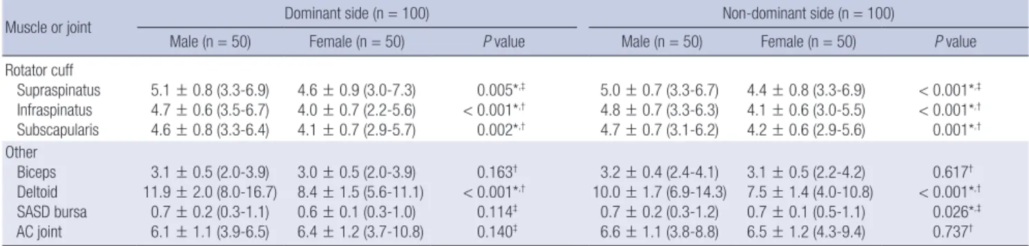
Table 2. Differences between males and females in dominant side and non-dominant side
Muscle or joint Dominant side (n = 100) Non-dominant side (n = 100)
Male (n = 50) Female (n = 50) P value Male (n = 50) Female (n = 50) P value
Rotator cuff Supraspinatus Infraspinatus Subscapularis
5.1 ± 0.8 (3.3-6.9) 4.7 ± 0.6 (3.5-6.7) 4.6 ± 0.8 (3.3-6.4)
4.6 ± 0.9 (3.0-7.3) 4.0 ± 0.7 (2.2-5.6) 4.1 ± 0.7 (2.9-5.7)
0.005*,‡
< 0.001*,†
0.002*,†
5.0 ± 0.7 (3.3-6.7) 4.8 ± 0.7 (3.3-6.3) 4.7 ± 0.7 (3.1-6.2)
4.4 ± 0.8 (3.3-6.9) 4.1 ± 0.6 (3.0-5.5) 4.2 ± 0.6 (2.9-5.6)
< 0.001*,‡
< 0.001*,†
0.001*,†
Other Biceps Deltoid SASD bursa AC joint
3.1 ± 0.5 (2.0-3.9) 11.9 ± 2.0 (8.0-16.7)
0.7 ± 0.2 (0.3-1.1) 6.1 ± 1.1 (3.9-6.5)
3.0 ± 0.5 (2.0-3.9) 8.4 ± 1.5 (5.6-11.1) 0.6 ± 0.1 (0.3-1.0) 6.4 ± 1.2 (3.7-10.8)
0.163†
< 0.001*,†
0.114‡ 0.140‡
3.2 ± 0.4 (2.4-4.1) 10.0 ± 1.7 (6.9-14.3)
0.7 ± 0.2 (0.3-1.2) 6.6 ± 1.1 (3.8-8.8)
3.1 ± 0.5 (2.2-4.2) 7.5 ± 1.4 (4.0-10.8) 0.7 ± 0.1 (0.5-1.1) 6.5 ± 1.2 (4.3-9.4)
0.617†
< 0.001*,†
0.026*,‡
0.737† Data are presented as the mean ± standard deviation. Minimal and maximal values are shown in parenthesis. All data are presented in millimeters.
*P < 0.05; †Unpaired t-test; ‡Wilcoxon rank sum test.
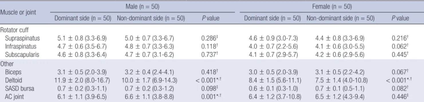
Table 3. Differences between dominant side and non-dominant side in males and females
Muscle or joint Male (n = 50) Female (n = 50)
Dominant side (n = 50) Non-dominant side (n = 50) P value Dominant side (n = 50) Non-dominant side (n = 50) P value Rotator cuff
Supraspinatus Infraspinatus Subscapularis
5.1 ± 0.8 (3.3-6.9) 4.7 ± 0.6 (3.5-6.7) 4.6 ± 0.8 (3.3-6.4)
5.0 ± 0.7 (3.3-6.7) 4.8 ± 0.7 (3.3-6.3) 4.7 ± 0.7 (3.1-6.2)
0.286‡ 0.118† 0.737†
4.6 ± 0.9 (3.0-7.3) 4.0 ± 0.7 (2.2-5.6) 4.1 ± 0.7 (2.9-5.7)
4.4 ± 0.8 (3.3-6.9) 4.1 ± 0.6 (3.0-5.5) 4.2 ± 0.6 (2.9-5.6)
0.216† 0.062† 0.445† Other
Biceps Deltoid SASD bursa AC joint
3.1 ± 0.5 (2.0-3.9) 11.9 ± 2.0 (8.0-16.7)
0.7 ± 0.2 (0.3-1.1) 6.1 ± 1.1 (3.9-6.5)
3.2 ± 0.4 (2.4-4.1) 10.0 ± 1.7 (6.9-14.3)
0.7 ± 0.2 (0.3-1.2) 6.6 ± 1.1 (3.8-8.8)
0.418†
< 0.001*,†
0.098‡ 0.001*,†
3.0 ± 0.5 (2.0-3.9) 8.4 ± 1.5 (5.6-11.1) 0.6 ± 0.1 (0.3-1.0) 6.4 ± 1.2 (3.7-10.8)
3.1 ± 0.5 (2.2-4.2) 7.5 ± 1.4 (4.0-10.8) 0.7 ± 0.1 (0.5-1.1) 6.5 ± 1.2 (4.3-9.4)
0.067†
< 0.001*,†
0.082† 0.446‡ Data are presented as the mean ± standard deviation. Minimal and maximal values are shown in parenthesis. All data are presented in millimeters.
*P < 0.05; †Unpaired t-test; ‡Wilcoxon rank sum test.
Table 4. Differences among different ages by decades
Muscle or joint Age, yr Dominant side (n = 100) Non-dominant side (n = 100)
Mean ± SD Min-Max P value Mean ± SD Min-Max P value
Rotator cuff
Supraspinatus 20-29 (n = 20) 4.3 ± 0.5 3.3-5.1 0.003*,‡ 4.2 ± 0.5 3.3-5.3 0.003*,†
30-39 (n = 20) 4.6 ± 0.7 3.6-5.8 4.7 ± 0.7 3.4-5.8
40-49 (n = 20) 4.9 ± 1.0 3.0-6.9 4.9 ± 0.9 3.7-6.7
50-59 (n = 20) 5.2 ± 1.0 3.5-7.3 4.7 ± 0.8 3.4-6.4
60-69 (n = 20) 5.2 ± 0.8 3.6-6.5 5.1 ± 0.8 3.3-6.9
Infraspinatus 20-29 (n = 20) 4.3 ± 0.8 3.0-6.7 0.306† 4.3 ± 0.6 3.4-5.8 0.290†
30-39 (n = 20) 4.2 ± 0.7 2.6-5.5 4.6 ± 0.7 3.6-6.1
40-49 (n = 20) 4.4 ± 0.8 2.2-5.9 4.3 ± 0.8 3.0-6.3
50-59 (n = 20) 4.5 ± 0.5 3.7-5.7 4.7 ± 0.8 3.5-6.2
60-69 (n = 20) 4.1 ± 0.8 2.6-5.6 4.5 ± 0.8 3.1-5.6
Subscapularis 20-29 (n = 20) 4.0 ± 0.8 2.9-5.6 0.010*,† 4.1 ± 0.6 3.1-5.2 0.034*,†
30-39 (n = 20) 4.5 ± 0.8 3.3-6.2 4.5 ± 0.7 3.4-6.2
40-49 (n = 20) 4.1 ± 0.6 3.3-5.3 4.7 ± 0.6 3.3-6.0
50-59 (n = 20) 4.7 ± 0.7 3.6-6.4 4.3 ± 0.7 2.9-5.7
60-69 (n = 20) 4.6 ± 0.8 3.0-6.0 4.6 ± 0.7 3.3-6.2
Other
Biceps 20-29 (n = 20) 2.8 ± 0.5 2.0-3.4 0.086† 2.9 ± 0.4 2.3-3.6 0.199†
30-39 (n = 20) 3.0 ± 0.5 2.0-3.9 3.1 ± 0.5 2.2-4.1
40-49 (n = 20) 3.1 ± 0.4 2.3-3.9 3.1 ± 0.5 2.2-4.1
50-59 (n = 20) 3.1 ± 0.4 2.4-3.7 3.3 ± 0.4 2.4-4.2
60-69 (n = 20) 3.2 ± 0.5 2.3-3.9 3.2 ± 0.4 2.3-4.1
Deltoid 20-29 (n = 20) 8.7 ± 2.4 5.6-13.7 0.028*,‡ 7.6 ± 1.8 4.0-10.9 0.030*,†
30-39 (n = 20) 11.2 ± 3.1 6.2-16.7 9.2 ± 2.2 5.1-14.3
40-49 (n = 20) 9.7 ± 2.0 6.3-14.1 8.5 ± 1.6 4.9-11.4
50-59 (n = 20) 11.1 ± 2.6 7.0-15.8 9.1 ± 1.9 6.6-14.1
60-69 (n = 20) 10.1 ± 1.5 7.3-12.8 9.4 ± 1.9 6.0-12.9
SASD bursa 20-29 (n = 20) 0.7 ± 0.2 0.4-1.0 0.165† 0.7 ± 0.1 0.5-0.9 0.522†
30-39 (n = 20) 0.6 ± 0.2 0.3-1.1 0.7 ± 0.2 0.3-1.2
40-49 (n = 20) 0.6 ± 0.1 0.3-0.8 0.7 ± 0.1 0.5-1.1
50-59 (n = 20) 0.7 ± 0.1 0.5-1.0 0.7 ± 0.1 0.5-0.9
60-69 (n = 20) 0.7 ± 0.2 0.3-1.1 0.7 ± 0.2 0.5-1.0
AC joint 20-29 (n = 20) 6.5 ± 0.9 4.9-8.1 0.029*,‡ 6.9 ± 1.1 5.1-9.0 0.029*,†
30-39 (n = 20) 6.4 ± 1.2 4.3-8.6 6.8 ± 1.2 5.2-9.4
40-49 (n = 20) 6.7 ± 1.7 3.7-10.8 6.5 ± 1.1 4.2-8.8
50-59 (n = 20) 6.2 ± 0.8 5.0-8.0 6.6 ± 1.0 5.3-8.6
60-69 (n = 20) 5.5 ± 0.9 3.9-7.7 5.9 ± 1.2 3.8-8.3
All data are presented in millimeters.
SD, Standard deviation; Min, minimum; Max, maximum.
*P < 0.05; †ANOVA; ‡Kruskal-Wallis.
4.2 ± 0.6 mm in non-dominant arm, respectively (Table 2). The supraspinatus, infraspinatus, subscapularis tendon and deltoid muscle thickness were significantly different between males and females for dominant and non-dominant arms. The mea- surements of SASD bursa thickness were significantly different between males and females for non-dominant arms.
The differences in measurements of rotator cuff tendons be- tween the dominant and non-dominant arm among males and females showed no statistical significance (Table 3). Only del- toid muscle thickness and AC joint interval in males and del- toid muscle thickness in females were significantly different be- tween dominant and non-dominant arms.
When subjects were stratified by the age groups, divided by ten years, the measurements of supraspinatus tendon thickness showed tendency of increase with the age, whereas the AC joint interval showed decreasing tendency (Table 4). In the other measurements, no significant difference among age groups was found.
DISCUSSION
This study suggests normative reference data of rotator cuff ten- don thickness and acromioclavicular joint interval among Ko- rean population. To make ultrasonographic diagnosis of rotator cuff pathology, especially rotator cuff tear or tendinopathy, the measurements should be compared with reference values to make objective and accurate diagnoses. Although there has been a report suggesting diagnostic criteria of supraspinatus tendinopathy demonstrating maximal thickness of supraspina- tus tendon based on comparison between symptomatic pa- tients and asymptomatic controls, there is no report suggesting normal reference values of rotator cuff dimension (10). This study is in great value for the fact that it is the first report provid- ing normative ultrasound dimensions of the rotator cuff in healthy Korean adults with varying age.
The results of our study possess certain degree of validity since the results show the correlation with prior studies (9,11).
In our study, the supraspinatus, infraspinatus, subscapularis tendon and deltoid muscle thickness were significantly differ- ent between males and females for dominant and non-domi- nant arms. The increasing tendency of rotator cuff thickness in male subjects in our research is assumed to be related to larger strength of the shoulder in males than females. There has been research demonstrating significant correlations between su- praspinatus thickness and external rotation strength, infraspi- natus thickness and internal rotation thickness, subscapularis thickness and internal rotation strength (8). In addition, in oth- er prior research on the rotator cuff dimensions of young adults (aged 18-40 years), the rotator cuff dimensions between males and females were significantly different (9).
The dimensions between dominant and non-dominant
arms were not significantly different in all of the thickness of ro- tator cuff tendon, biceps tendon and subacromial subdeltoid bursa with the exception of AC joint interval and the thickness of deltoid muscle. Our study showed similar results with previ- ous researches, which means the asymptomatic contralateral shoulder can be used to estimate the expected dimension (9,12). However, in the male group, the AC joint interval of dominant arms was significantly lower than non-dominant arms. This result could be associated with arthritic change as a consequence of more usage of dominant arm in males. Further researches regarding association of activities of upper extremi- ties with AC joint interval are needed.
When the measurements were stratified by age, the mea- surements of supraspinatus tendon thickness revealed increas- ing tendency with increasing age groups. This tendency could be related to asymptomatic rotator cuff tendinopathy which shows frequent incidence rate with aging. The prevalence of ro- tator cuff pathology is reported to increase by natural aging process (13). In a research of ultrasonographic findings in as- ymptomatic shoulders, supraspinatus tendon was significantly thicker and demonstrated a lower echogenicity ratio in elderly patients aged more than 60 years and the thickness showed positive correlation with age. This study suggested the increase in thickness of the supraspinatus tendon might be due to chronic tendinopathy by age-related degeneration. In a re- search study of ultrasonographic findings of asymptomatic shoulders, supraspinatus tendinosis was the third most com- mon abnormal finding, accounting for 39%, followed by sub- acromial subdeltoid bursal thickening and acromioclavicular joint osteoarthritis (14). Further studies for clarifying correla- tions between sonographic findings and pathology are needed.
In our data, acromioclavicular joint interval showed the ten- dency of decrease with increasing age. This tendency may be the result of natural degeneration by aging process and similar results are documented in the other studies. Stein et al. (15) re- ported more advanced arthritic changes in acromioclavicular joint were detected in MRI in the over 30 age group. Nicholson et al. (16) reported significant increase in degenerative changes at the acromial facet of the acromioclavicular joint occurred with advancing age.
This study has some limitations to be taken into consider- ation. First, the ultrasound scanning was performed once by a single physiatrist, which means intraobserver and interobserv- er agreement was not assessed. The previous study regarding rotator cuff dimension in young healthy adults showed good intraobserver and interobserver agreement (9). In the study of sonographic evaluation of the painful shoulder, the examiners were in very good agreement for full-thickness rotator cuff tears, supraspinatus tendinosis, abnormalities of the long head of biceps tendon, subacromial bursa abnormalities, acromio- clavicular osteoarthritis and moderate agreement for partial
thickness tear and intratendinous tears (17). The other study revealed good interobserver reliability in grading fatty degener- ation of rotator cuff muscles (18). Although operator depen- dence has been considered a limitation of ultrasonography, good interobserver and intraobserver reliability was reported by previous studies. Although our study could not assess the in- traobserver and interobserver reliability, ultrasonography mea- surements were performed on both arms for each participant and the results of each measurement showed congruence, in- directly increasing the reliability of the single physiatrist’s mea- surement. Second, the number of subjects was not large enough to objectively assess differences among age. However, this is the first report that evaluated the normal dimensions of rotator cuff tendons where at least 10 subjects were recruited within each subgroup of stratified age, having a range of 20 to 70 years. Third, all of the subjects were chosen from one institu- tion and there could be selection bias. However, participants in our study possess diversity in age and gender, which could rep- resent general Korean population. Fourth, subjects’ anthropo- metric factors that can possibly affect the measurements such as height, weight and body mass index (BMI) were not as- sessed. As the previous study showed there were no significant correlation between the height or weight of the subjects and the rotator cuff tendon measurements, we assumed weight and height could be omitted in measuring normal rotator cuff ten- don (9). However, because there has been a study demonstrat- ing obesity is related to increased risk for rotator cuff tendinitis and rotator cuff related surgery, further studies assessing rela- tionship between body mass index and cuff tendon thickness under adjustment of age and gender are needed (19). Fifth, echogenicity that could be useful in diagnosing rotator cuff ten- dinopathy was not measured in our research. Further studies assessing echogenicity as well as rotator cuff tendon thickness in healthy adults would be needed to suggest more accurate normal reference data.
This study has suggested normative reference values of rota- tor cuff dimensions of Korean adults. Further studies with larg- er groups of subjects assessing normal rotator cuff dimensions and defining influencing factors including a wide range of age would be needed to further validate our normative reference values. Furthermore, studies regarding comparison between measurements of normal healthy adults and patients with rota- tor cuff lesion and suggesting the cut-off value of rotator cuff le- sions in Korea would be needed.
DISCLOSURE
The authors have declared no potential conflicts of interest to disclose.
AUTHOR CONTRIBUTION
Study design: Chung ME. Acquisition of clinical data: Kim K, Song D, Kim HG, Yoon JY, Chung ME. Statistical analysis and data analysis: Kim K, Chung ME. Writing and revision of manu- script: Kim K, Song D, Kim HG, Yoon JY, Chung ME. Final ap- proval: all authors.
ORCID
Kyeongwon Kim http://orcid.org/0000-0001-6185-0058 Hong Geum Kim http://orcid.org/0000-0002-3331-7421 Daeheon Song http://orcid.org/0000-0003-0653-8488 Jung Yoon Yoon http://orcid.org/0000-0002-4703-9266 Myung Eun Chung http://orcid.org/0000-0002-7308-2815
REFERENCES
1. Chard MD, Hazleman R, Hazleman BL, King RH, Reiss BB. Shoulder dis- orders in the elderly: a community survey. Arthritis Rheum 1991; 34: 766- 9.
2. de Jesus JO, Parker L, Frangos AJ, Nazarian LN. Accuracy of MRI, MR ar- thrography, and ultrasound in the diagnosis of rotator cuff tears: a meta- analysis. AJR Am J Roentgenol 2009; 192: 1701-7.
3. Lenza M, Buchbinder R, Takwoingi Y, Johnston RV, Hanchard NC, Falop- pa F. Magnetic resonance imaging, magnetic resonance arthrography and ultrasonography for assessing rotator cuff tears in people with shoul- der pain for whom surgery is being considered. Cochrane Database Syst Rev 2013: CD009020.
4. Roy JS, Braën C, Leblond J, Desmeules F, Dionne CE, MacDermid JC, Bu- reau NJ, Frémont P. Diagnostic accuracy of ultrasonography, MRI and MR arthrography in the characterisation of rotator cuff disorders: a sys- tematic review and meta-analysis. Br J Sports Med 2015; 49: 1316-28.
5. Smith TO, Back T, Toms AP, Hing CB. Diagnostic accuracy of ultrasound for rotator cuff tears in adults: a systematic review and meta-analysis.
Clin Radiol 2011; 66: 1036-48.
6. de Miguel E, Cobo T, Muñoz-Fernández S, Naredo E, Usón J, Acebes JC, Andréu JL, Martín-Mola E. Validity of enthesis ultrasound assessment in spondyloarthropathy. Ann Rheum Dis 2009; 68: 169-74.
7. Maffulli N, Regine R, Angelillo M, Capasso G, Filice S. Ultrasound diagnosis of Achilles tendon pathology in runners. Br J Sports Med 1987; 21: 158-62.
8. Bang IK, Lee JP, Kim YJ, Kim C, Kim GH, Reu HW, Oh JK. Tendon diame- ter of rotator cuff and strength of the shoulder external/internal rotator muscles in elite thrower. J Korean Acad Rehabil Med 2007; 31: 730-34.
9. Karthikeyan S, Rai SB, Parsons H, Drew S, Smith CD, Griffin DR. Ultra- sound dimensions of the rotator cuff in young healthy adults. J Shoulder Elbow Surg 2014; 23: 1107-12.
10. Teunis T, Lubberts B, Reilly BT, Ring D. A systematic review and pooled analysis of the prevalence of rotator cuff disease with increasing age. J Shoulder Elbow Surg 2014; 23: 1913-21.
11. Abate M, Schiavone C, Salini V. Sonographic evaluation of the shoulder in asymptomatic elderly subjects with diabetes. BMC Musculoskelet Dis- ord 2010; 11: 278.
12. Cho SH, Cho HL, Lee JS, Kim JW. Ultrasonographic findings of the shoul-
der in asymptomatic high school overhead athletes. J Korean Orthop Ul- trasound Soc 2012; 5: 81-8.
13. Milgrom C, Schaffler M, Gilbert S, van Holsbeeck M. Rotator-cuff chang- es in asymptomatic adults. The effect of age, hand dominance and gen- der. J Bone Joint Surg Br 1995; 77: 296-8.
14. Yu TY, Tsai WC, Cheng JW, Yang YM, Liang FC, Chen CH. The effects of aging on quantitative sonographic features of rotator cuff tendons. J Clin Ultrasound 2012; 40: 471-8.
15. Stein BE, Wiater JM, Pfaff HC, Bigliani LU, Levine WN. Detection of acro- mioclavicular joint pathology in asymptomatic shoulders with magnetic resonance imaging. J Shoulder Elbow Surg 2001; 10: 204-8.
16. Nicholson GP, Goodman DA, Flatow EL, Bigliani LU. The acromion: mor-
phologic condition and age-related changes. A study of 420 scapulas. J Shoulder Elbow Surg 1996; 5: 1-11.
17. Le Corroller T, Cohen M, Aswad R, Pauly V, Champsaur P. Sonography of the painful shoulder: role of the operator’s experience. Skeletal Radiol 2008; 37: 979-86.
18. Wall LB, Teefey SA, Middleton WD, Dahiya N, Steger-May K, Kim HM, Wessell D, Yamaguchi K. Diagnostic performance and reliability of ultra- sonography for fatty degeneration of the rotator cuff muscles. J Bone Joint Surg Am 2012; 94: e83.
19. Wendelboe AM, Hegmann KT, Gren LH, Alder SC, White GL Jr, Lyon JL.
Associations between body-mass index and surgery for rotator cuff ten- dinitis. J Bone Joint Surg Am 2004; 86-A: 743-7.