가천대학교 의과대학 길병원 호흡기내과
김정호, 전효근, 김미경, 경선영, 안창혁, 이상표, 박정웅, 정성환
Particulate Matter from Asian Dust Storms Induces the Expression of Proinflammatory Cytokine in A549 Epithelial Cells
Jung Ho Kim, M.D., Hyo Keun Jeon, M.D., Mi Kyeong Kim, M.D., Sun Yong Kyung, M.D., Chang Hyeok An, M.D., Sang Pyo Lee, M.D., Jung Woong Park, M.D., Sung Hwan Jeong, M.D.
Division of Pulmonary Medicine, Department of Internal Medicine, Gachon Medical School Gil Medical Center, Incheon.
Background: PM10(Particulate matter with a diameter < 10㎛), which is characterized by different environmental conditions, is a complex mixture of organic and inorganic compounds. The Asian dust event caused by meteorological phenomena can also produce unique particulate matter in affected areas. This study investigated the cytokine produced by A549 epithelial cells exposed to particles collected during both the Asian dust pfenomenon and ambient air particles in a non-dusty period.
Method: Air samples were collected using a high volume air sampler(Sibata Model HV500F) with an air flow at 500ℓ /min for at least 6 hours. The cytokine messenger RNA(mRNA) was measured using a reverse transcriptase polymerase chain reaction(RT-PCR). The A549 cells were exposed to 10 to 500㎍/㎖ of a suspension containing PM10 for 24 hours.
Each was compared with those in the non-exposed control cells.
Result: The mRNA levels of interleukin(IL)-1α, IL-Iβ , IL-8, and the granulocyte macrophage colony stimulating factor(GM-CSF) increased after veing exposed to PM10 in the ambient air particles, compared with those in the non-exposed control cells. The increase in IL-1α and IL-8 were dose dependent at a PM10 concentration between 100㎍/㎖
and 500㎍/㎖. The mRNA level of IL-8 in the A549 epithelial cells was higher during the in the Asian dust period(500㎍/
㎖) than during the non dust period.
Conclusion: A549 cells exposed to the PM10 collected during the Asian dust period produce more proinflammatory cytokine than during non-dusty period. This cytokine enhances the local inflammatory response in the airways and can also contribute to the systemic component of this inflammatory process. (Tuberc Respir Dis 2006; 60: 663-672) Key words: Particulate matter, Asian dust, Proinflammatory cytokine.
Address for correspondence : Sung Hwan Jeong, M.D.
Division of Pulmonary Medicine, Department of Internal Medicine, Gachon Medical School
Gil Medical Center, Guwol-1-dong, Namdong-gu, Incheon, 405-760, Korea
Phone : 032-460-3818 Fax : 032-469-4320 E-mail : [email protected], [email protected] Received : Feb. 21. 2006
Accepted : May. 24. 2006
서 론
대기 중의 먼지 현상이란 물이나 얼음입자는 거의 포함되어 있지 않고, 주로 고체 입자들이 대기 중에서 떠다니거나 바람에 의해서 불려 오르는 현상을 말한 다. 먼지 현상의 한 종류인 ‘연무(haze)’는 육안으로 보이지 않는 극히 작고 건조한 고체입자가 대기 중에 떠다니고 있는 현상이다. 특히 바람에 의해서 지면으 로부터 불려 올라간 먼지나 모래의 작은 입자가 대기
중에 떠있는 것이 확실할 때의 현상은 ‘먼지연무(dust haze)’라 하며, ‘황사’는 먼지연무의 일종으로서 주로 아시아 대륙의 건조지대에서 불려 올라간 다량의 흙 먼지가 장거리 수송되어 하늘을 덮고 떠다니며 서서 히 하강하는 현상을 말한다1.
국내에서도 매년 봄철이면 황사가 주기적으로 발 생한다. 황사 현상에서 크기가 큰 입자들은 발원지나 주변에 머물고 국내에 유입되는 황사 입자의 크기는 대개 지름 10㎛이하의 미세먼지(particulate matter 10; PM10)이고, 미세먼지는 흡입시에 하부기관지 및 폐의 가스-교환부분까지 침착하여 호흡기계에 손상 을 일으킬 수 있다고 보고되고 있다2.
기존의 미세먼지의 인체 내 유해성에 대한 연구는 대기오염 연구에서 많이 이루어졌는데, 1930년 벨기 에의 Meuse, 1952년 12월 런던에서 심한 대기오염으 로 많은 사망자가 발생했던 것이 대표적인 대기오염
사례로 잘 알려져 있다3. 임상적으로는 대기오염에 의 하여 천식과 만성 폐쇄성 폐질환과 같은 호흡기 질환 의 유병률 증가 및 급성 악화가 일어날 수 있다는 것 이 여러 연구에서 증명되었으며4-8, 대기오염에 포함 된 미세먼지 중에서 말초호흡기로 침투가 가능한 크 기인 PM10이 천식 발작 및 만성 호흡기 질환의 악화 에 많이 기여하는 것으로 알려져 있다9-11. 국내에서 시행된 연구에서도 대기 오염의 위해성은 임상적, 실 험적으로 밝혀져 있으며12-13, 특수한 자연적 대기오염 의 한 형태인 황사 현상의 경우 이 기간 중에 대기중 의 PM10이 크게 증가하며 이에 따라 기관지 천식 환 자의 호흡기 증상 악화를 유발 할 수 있음이 밝혀진 바 있다14.
미세먼지는 유기물과 무기물의 복합체로 발생시기 와 장소에 따라 물리적 특성과 구성 성분이 매우 다 양하고, 생물학적으로 영향을 줄 수 있는 금속 성분도 함유하고 있다. PM10이 폐장 내에 흡입 시 폐포 대식 세포와 폐 상피세포가 PM10에 직접적으로 노출이 되 고, 특히 폐 상피세포에서 interleukin(IL)-1, IL-6, IL-8, monocyte chemotactic protein(MCP)-1, tumor necrosis factor(TNF)-α, granulocyte macrophage colony stimulating factor(GM-CSF)과 같은 염증성 cytokine의 발현이 증가하는 것으로 알려져 있으며, 이로 인해 호흡기 질환의 악화가 일어나는 것으로 추 측되고 있다15-18. 하지만 황사의 PM10이 폐 상피세포 에서 전신 반응을 일으킬 수 있는 염증성 물질의 생 성에 관한 연구는 아직 많지 않다.
본 연구에서는 황사에 포함된 PM10이 A549 cells에 작용하여 전염증성 cytokine 및 cytokine messenger RNA(mRNA)의 발현에 어떤 영향이 있는지를 관찰 하여 만성 천식 등 호흡기 질환의 증상 악화기전에 미치는 역할을 규명하고자 하였다.
대상 및 방법 1. 미세먼지의 포집
공기 포집은 3일간의 황사 기간과 10일간의 비황사
3월 10일, 3월 30일, 4월 23일 시행하였고 비황사 기 간의 공기 포집은 2004년 4월 21일, 4월 24일, 5월 9 일, 5월 22일, 5월 26일, 6월 3일, 6월 26일, 7월 7일, 7월 31일, 8월 14일 시행하었다.
2. PM10 준비
공기 포집기(HV500F, Sibata, Japan)을 이용하여 도심지인 인천시(인천시 남동구 구월동 가천의대 길 병원 응급센터 옥상)에서 분당 500 liter로 하루 6시간 씩(오전 10시부터 오후 4시까지) 실외의 장소에서 대 기분진을 pore size 0.25㎛ 필터 (Prefilter AP, 124㎜, Milipore, Bedford, MA, USA)에 포집하였다. 포집한 필터를 조각내어 phosphate buffered saline(PBS) buffer 10㎖를 넣은 튜브(tube)에 넣었다. 틈틈이 소 용돌이혼합기(vortex mixer)를 사용하면서 30분간 실온에서 배양했다. 필터를 건져낸 후 10㎛ pore의 필 터(MitexTM membrane filters, Milipore, Bedford, MA, USA)로 여과하였다. 그 후 약 1분간 3번 초음파 파쇠(sonication) 하였다. 1.5㎖ 튜브로 나누어 담아 무게를 측정하고 121℃에서 15분간 멸균한 후 사용 전까지 -20℃에 보관하였다.
3. 세포배양
폐암 상피세포주인 A549 cells(한국세포은행주)을 fetal bovine serum(FBS)이 10% 포함된 RPMI-1640 media(Welgene, Daegu, Korea)에서 배양하였다. 세 포가 90-100%로 배양되었을 때 황사와 비황사 시기 에 포집한 PM10을 각각 10, 100, 500㎍/㎖의 농도로 첨가해 주었다. 그리고 각각을 처리 24시간 후 RNA 를 추출하였다. 대조군은 세포배양만 시행하고 PM10
처리를 하지 않은 것으로 하였다.
4. RNA추출과 역전사중합효소연쇄반응(reverse transcriptase polymerase chain reaction;
RT-PCR)
IL-1α 5'-GTC TCT GAA TCA GAA ATC CTT CTA TC -3' 5'-CAT GTC AAA TTT CAC TGC TTC ATC C -3' (420bp)
IL-1β 5'-AAA CAG ATG AAG TGC TCC TTC CAG G -3' 5'-TGG AGA ACA CCA CTT GTT GCT CCA -3' (388bp)
IL-8 5'- ATG ACT TCC AAG CTG GCC GTG GCT -3' 5'-TCT CAG CCC TCT TCA AAA ACT TCT C-3' (289bp)
GM-CSF 5'- ACA CTG CTG AGA TGA ATG AAA CAG TAG -3'
5'- TGG ACT GGC TCC CAG CAG TCA AAG GGG ATG-3' (172bp)
GAPDH 5'-GTG GAT ATT GTT GCC ATC AAT GAC C-3' 5'-GCC CCA GCC TTC TCC ATG GTG GT-3' (248bp)
GM-CSF; granulocyte macrophage colony stimulating factor, IL; interleukin, RT-PCR; reverse transcriptase polymerase chain reaction
Table 1. RT-PCR에 사용한 primer
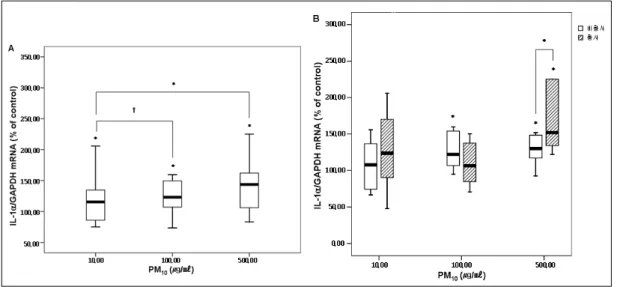
Figure 1. IL-1α mRNA expression in A549 cells after 24 hours incubation with PM10.
A549 cells were exposed to 10 to 500㎍/㎖ of a suspension of ambient particulate matter with a diameter of less than 10㎛(PM10) for 24 hours. Results shown in the box plot graph are the percentage from control(medium alone) values(*p<0.05, † p<0.1) compared with control. The black lines in boxes show median value of IL-1a mRNA expression.
1A. The expression of IL-1α mRNA measured in the supernatant collected at 24 hours increased and were significantly higher than those in the control nonexposed cells (10 to 500㎍/㎖)
1B. The mRNA Levels of IL-1α were increased after exposed to PM10 collected during Asian dust period, compared to those in non-dusty period(500㎍/㎖) and the control(500㎍/㎖)
IL; interleukin, mRNA; cytokine messenger RNA Trizol(Invitrogen, Life Technologies, Inc., Carlsbad, CA, USA)를 넣고 세포를 넣은 후 1.5㎖ 튜브로 옮겼 다. 실온에서 5분간 배양 한 후 chloroform을 첨가하 였고 inverting 15초를 하여 다시 실온에서 3분간 배 양하였다. 12000g 4℃에서 15분간 원심분리한 후
75% 에탄올(ethanol)로 2번 세척하였으며 침전물 (pellet)을 건조시킨 후 diethyl pyrocarbonate(DEPC) 로 처리된 증류수를 넣어 정량하였다.
2㎍의 RNA에 random hexamer(Promega, Madi- son, WI, USA)를 넣어 94℃에 5분, 얼음에 3분간 배
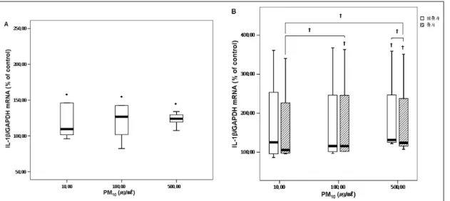
Figure 2. IL-1β mRNA expression in A549 cells after 24 hours incubation with PM10.
A549 cells were exposed to 10 to 500㎍/㎖ of a suspension of ambient particulate matter with a diameter of less than 10㎛(PM10) for 24 hours. Results shown in the box plot graph are the percentage from control(medium alone) values(*p<0.05, † p<0.1) compared with control. The black lines in boxes show median value of IL-1a mRNA expression.
2A. The expression of IL-1β mRNA measured in the supernatant collected at 24 hours increased and were significantly higher than those in the control nonexposed cells(10 to 500㎍/㎖)
2B. The mRNA Levels of IL-1β were increased after exposed to PM10 collected during Asian dust period, compared to those in non-dusty period(500㎍/㎖) and the control(100㎍/㎖, 500㎍/㎖). IL-1β mRNA expression increased after 24 hours of exposure to PM10 in a dose-dependent manner.
양했다. M-MLV(Progma, Madison, WI, USA), RNasin(Progma, Madison, WI, USA), dNTPs(Ta- kara, Otsu, Japan)을 넣어 역전사 반응을 했다(37℃, 1시간). 이 cDNA를 주형 DNA로 이용하고 interleukin(IL)-1α, IL-1β, IL-8, granulocyte macro- phage colony stimulating factor(GM-CSF)의 primer set들을 이용하여 RT-PCR을 시행하였다. 그리고 2% agarose gel에 전기영동 후 각각의 밴드를 농도계 측기(densitometry)을 이용하여 측정하였다(Table 1).
5. 분석 방법
모든 결과는 평균±표준편차로 표시하였다. 두 군간 의 차이는 Wilcoxon signed ranks test로 검정하였다.
분석은 SPSS(Version 13.0)프로그램을 이용하였고 p<0.05인 경우에 통계학적으로 유의 하다고 판단하 였다.
결 과
인천 지역에서 PM10의 연평균 농도는 2001년까지 는 감소하는 추세였으나 그 이후에는 약간의 증가추 세를 보이고 있고 환경부의 보고에 따르면 연구가 수 행된 2004년도에는 연평균 62㎍/㎥, 황사기간을 제외 하면 59㎍/㎥를 보이고 있었다.
본 연구에서 공기 포집이 시행된 2004년도에 인천 지역의 황사 기간은 6일 이었다. 인천 보건환경연구 원 자료에 의하면 황사 기간의 PM10의 평균 농도는 118.67±55.54㎍/㎥이었고 한시간 PM10 최대값은 376.00±164.07㎍/㎥이었다.
1. PM10이 A549 cells에서 IL-1α mRNA발현에 미 치는 영향
A549 cells을 황사기간과 비황사기간을 포함한 전
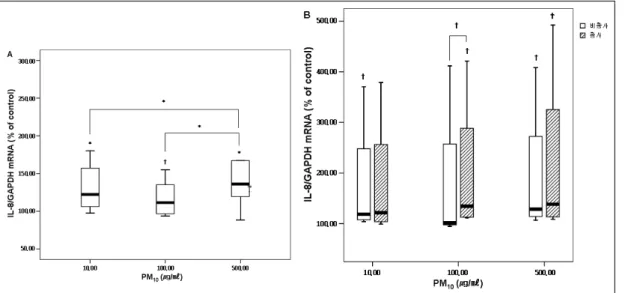
Figure 3. IL-8 mRNA expression in A549 cells after 24 hours incubation with PM10.
A549 cells were exposed to 10 to 500㎍/㎖ of a suspension of ambient particulate matter with a diameter of less than 10㎛(PM10) for 24 hours. Results shown in the box plot graph are the percentage from control(medium alone) values(*p<0.05, † p<0.1) compared with control. The black lines in boxes show median value of IL-1a mRNA expression.
3A. The expression of IL-8 mRNA measured in the supernatant collected at 24 hours increased and were significantly higher than those in the control nonexposed cells(10 to 500㎍/㎖, 100 to 500㎍/㎖).
3B. The mRNA Levels of IL-8 were increased after exposed to PM10 collected during Asian dust period, compared to those in non-dusty period(100㎍/㎖) and the control(100㎍/㎖, 500㎍/㎖)
IL; interleukin, mRNA; cytokine messenger RNA 체 PM10으로 처리하고 24시간 후에 세포배양 상층액 으로 RT-PCR을 시행한 결과를 보면 10, 100, 500㎍/
㎖의 각 농도에서 대조군보다 의미 있게 IL-1α mRNA발현이 증가하였다(PM10 10㎍/㎖ 141.93±88.49%
of control, p=0.039, PM10 100㎍/㎖ 156.03±106.00%
of control, p=0.015, PM10 500㎍/㎖ 171.17±105.95%
of control, p=0.002). PM10 10㎍/㎖보다 100㎍/㎖에서 발현이 증가하는 경향을 보였고(p=0.098), 500㎍/㎖에 서는 유의하게 증가하였다(p=0.016)(Figure 1A). 100
㎍/㎖에서는 황사 기간의 PM10에서는 대조군보다 의 미 있게 mRNA의 발현이 증가하지 않았지만(141.55±
109.12% of control, p=0.400), 비황사기간의 PM10에 서는의미 있게 IL-1α mRNA발현이 증가하였다 (162.19 ±113.34% of control, p=0.035). 비황사 기간 의 PM10 500㎍/㎖에서 대조군보다 의미있게 IL-1α mRNA발현이 증가하였고(156.80±88.39% of control, p=0.025), 황사 시기의 PM10 500 ㎍/㎖에서도 대조군 보다 의미있게 IL-1α mRNA발현이 증가하였다
(205.19±124.73% of control, p=0.012)(Figure 1B).
2. PM10이 A549 cells에서 IL-1β mRNA발현에 미 치는 영향
A549 cells을 PM10으로 황사기간과 비황사기간을 포함한 전체 처리하고 24시간 후에 세포배양 상층액 으로 RT-PCR을 시행한 결과를 보면 10, 100, 500㎍/
㎖의 각 농도에서 대조군보다 의미 있게 IL-1β mRNA발현이 증가하였다(PM10 10㎍/㎖ 163.46±
107.37% of control, p=0.028, PM10 100㎍/㎖ 152.71±
92.19% of control, p=0.008, PM10 500㎍/㎖ 152.02±
87.47% of control, p=0.009)(Figure 2A). 그러나 농도 에 따른 의미 있는 차이는 보이고 있지 않았다. 황사 기간의 PM10 100㎍/㎖와 500㎍/㎖에서는 대조군보다 IL-1β mRNA의 발현이 증가하는 경향을 보였다 (174.02±126.79% of control, p=0.068, 176.63±116.64%
of control, p=0.068). 비황사 기간의 PM10 500㎍/㎖에
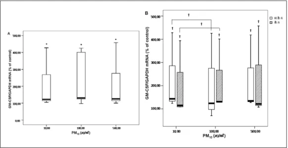
Figure 4. GM-CSF mRNA expression in A549 cells after 24 hours incubation with PM10.
A549 cells were exposed to 10 to 500㎍/㎖ of a suspension of ambient particulate matter with a diameter of less than 10㎛(PM10) for 24 hours. Results shown in the box plot graph are the percentage from control(medium alone) values(*p<0.05, † p<0.1) compared with control. The black lines in boxes show median value of IL-1a mRNA expression.
4A. The expression of GM-CSF mRNA measured in the supernatant collected at 24 hours increased and were significantly higher than those in the control nonexposed cells(10 to 500㎍/㎖).
4B. The mRNA Levels of GM-CSF were increased after exposed to PM10 collected during Asian dust period, compared to those in the control(100㎍/㎖, 500㎍/㎖). GM-CSF mRNA expression increased after 24hours of exposure to PM10 in a dose-dependent manner.
서도 대조군보다 IL-1β mRNA의 발현이 증가하는 경향을 보였다(186.11±115.54% of control, p=0.068).
또한 황사 시기의 PM10에서는 10㎍/㎖에서 보다 100
㎍/㎖와 500㎍/㎖에서 mRNA발현이 증가하는 경향 을 보였다(PM10 10㎍/㎖ 162.00±119.22% of control, PM10 100㎍/㎖ 174.02±126.79% of control, p=0.068, PM10 500㎍/㎖ 176.63±116.64% of control, p=0.068) (Figure 2B).
3. PM10이 A549 cells에서 IL-8 mRNA발현에 미치 는 영향
A549 cells을 황사기간과 비황사기간을 포함한 전 체 PM10으로 처리하고 24시간 후에 세포배양 상층액 으로 RT-PCR을 시행한 결과를 보면 10, 500㎍/㎖의 각 농도에서 대조군보다 의미 있게 IL-8 mRNA발현 이 증가하였다(PM10 10㎍/㎖ 167.17±105.12% of control, p=0.008, PM10 500㎍/㎖ 189.78±132.46% of control,
p=0.006)(Figure 3A). PM10 100 ㎍/㎖에서는 대조군 보다 증가하는 경향을 보이고 있다(PM10 100㎍/㎖
164.52±125.79% of control, p=0.091). PM10 10㎍/㎖보 다 500㎍/㎖에서 의미 있게 IL-8 mRNA발현이 증가 하였고(p=0.041), PM10 100㎍/㎖보다 500㎍/㎖에서도 의미있게 mRNA발현이 증가하였다(p=0.041) (Figure 3A). 비황사 기간의 PM10 에서는 10, 500㎍/㎖에서 (PM10 10㎍/㎖ 177.84±128.59% of control, p=0.068, PM10 500㎍/㎖ 193.23±143.94% of control, p=0.068), 황사 기간의 PM10 에서는 100㎍/㎖, 500㎍/㎖에서 (PM10 100㎍/㎖ 200.50±148.63% of control, p=0.068, PM10 500㎍/㎖ 219.39±183.14% of control, p=0.068) 대조군보다 IL-8 mRNA의 발현이 증가하는 경향을 보였다(p<0.1)(Figure 3B). 그리고 비황사 기간보다 황사기간의 PM10 100㎍/㎖에서 mRNA 의 발현이 증 가하는 경향을 보였다(177.47±156.04% of control, 200.50±148.63% of control, p=0.068)(Figure 3B).
4. PM10이 A549 cells에서 GM-CSF mRNA발현에 미치는 영향
A549 cells을 황사기간과 비황사기간을 포함한 전 체 PM10으로 처리하고 24시간 후에 세포배양 상층액 으로 RT-PCR을 시행한 결과를 보면 10, 100, 500㎍/
㎖의 각 농도에서 대조군보다 의미 있게 GM-CSF mRNA발현이 증가하였다(PM10 10㎍/㎖ 183.08±
130.11% of control, p=0.008, PM10 100㎍/㎖ 218.56±
151.86% of control, p=0.046, PM10 500㎍/㎖ 190.52±
140.97% of control, p=0.008)(Figure 4A). 그러나 농 도에 따른 의미있는 차이는 보이고 있지 않았다. 비황 사 기간의 PM10 10㎍/㎖, 500㎍/㎖과(PM10 10㎍/㎖
209.62±146.03% of control, p=0.066, PM10 500㎍/㎖
203.79±143.02% of control, p=0.066) 황사 기간의 PM10 10㎍/㎖, 100㎍/㎖, 500㎍/㎖에서 (PM10 10㎍/㎖
183.32±140.60% of control, p=0.066, PM10 100㎍/㎖
198.22±135.45% of control, p=0.066, PM10 500㎍/㎖
202.00±170.49% of control, p=0.066) 대조군보다 GM-CSF mRNA발현이 증가하는 경향을 보였다 (p<0.1). 그리고 비황사 기간에는 PM10 10㎍/㎖보다 100㎍/㎖에서 GM-CSF mRNA발현이 증가하는 경 향이 있었다(PM10 10㎍/㎖ 209.62±146.03% of control, PM10 100㎍/㎖ 186.35±161.74% of control, p=0.066)(Figure 4B).
고 찰
본 연구에서 황사기간과 비황사기간을 포함한 전 체 PM10은 A549 cells에서 IL-1α, IL-1β, IL-8, GM-CSF mRNA의 발현을 증가시켰다. IL-1α에서는 500㎍/㎖에서 10㎍/㎖보다 cytokine의 발현이 증가하 였고 IL-8에서는 10㎍/㎖와 100㎍/㎖보다 500㎍/㎖에 서 cytokine의 발현이 증가한 것을 확인할 수 있었다.
또한 IL-1α에서는 500㎍/㎖에서 황사 시기의 PM10이 비황사 시기의 PM10보다 이러한 염증성 cytokine의 발현을 더 많이 유도하고 있었다.
IL-1, IL-8, GM-CSF는 모두 골수에서 백혈구의 방출을 촉진하고 간에서 급성기반응물질(acute pha-
se reactant)를 생산하고 혈관 내피세포를 자극할 수 있는 cytokine들이다19-21. IL-1은 호중구성 및 호산구 성 염증반응에서 다른 종류의 cytokine의 발현 및 합 성을 증가시키는 것을 통해 중요한 역할을 한다18. IL-8은 호중구의 활성화와 화학주성에 가장 중요한 역할을 하며22, 알레르기 질환에서도 중요한 역할을 할 수 있다. GM-CSF는 골수에서 과립백혈구 (granulocyte)와 대식세포(macrophage)의 회전 (turnover)과 유리(release)에 결정적인 역할을 한다
23,24
. 이 cytokine들은 폐 내 국소적인 염증 반응을 야 기할 수 있으며, 전신 순환을 하게 될 경우 전신적 염 증 반응도 유발 할 수 있는 것으로 보고되고 있다25-27. 이로 인해 기관지 천식이나 만성 폐쇄성 폐질환 같은 기도질환의 악화와 나아가 심장 질환의 악화도 유발 할 수 있을 것으로 생각된다.
본 연구에서 황사기간과 비황사기간을 포함한 전 체 PM10은 IL-8의 100 ㎍/㎖의 농도를 제외하고는 10, 100, 500㎍/㎖의 농도에서 IL-1α, IL-1β, IL-8, GM-CSF mRNA의 발현을 대조군에 비하여 유의하 게 증가시켰다. 이러한 결과는 기존의 대기오염 물질 에서 PM10이 전염증성 cytokine의 발현을 유발하고 PM10에 의해 IL-1, IL-8, GM-CSF의 생성이 증가한 다는 결과와 유사하다15,28,29. 하지만 IL-8의 100㎍/㎖
의 농도에서 mRNA발현이 의미있게 증가하지 않은 이유는 정확히 알 수는 없었고 추가 연구가 필요하다 고 사료된다. 본 연구에서는 황사기간과 비황사기간 을 포함한 전체 PM10을 A549 cells에 처리를 해 주었 을 때 고농도에서는 저농도에서보다 IL-1α와 IL-8에 서 mRNA의 발현이 의미 있게 증가하는 양상을 보여 주고 있으나 IL-1β, GM-CSF에서는 의미 있는 차이 는 보여주고 있지 않았다. PM10에 의한 cytokine의 증 가 기전에 대해서는 아직 명확히 규명된 것은 없으나, PM10 입자 자체의 물리적 영향이나 내독소(endo- toxin)에 의해 전염증성 cytokine의 발현이 증가될 수 있으며30,31 최근 PM10에 함유된 철32, 바나듐15, 구리16 같은 금속 성분들이 잠재적으로 산화-환원반응에 관 여하면서 전염증성 cytokine을 증가시킬 수 있다고 보고된 바 있다. 또한 Adamson들은 PM10에 포함된 수용성 금속성분이 쥐의 폐에 염증 반응을 증가시켜
독성을 나타낼 수 있다고 주장하였고33, 박영만들도 A549 cells에서 입자상 물질의 독성에 금속, 특히 철 이 중요하게 관여하고 있다고 보고하였다34.
본 연구에서 황사 시기에 채취한 PM10을 비황사 시기와 비교하였을 때 500㎍/㎖에서는 같은 농도의 PM10인데도 불구하고 IL-1α에서는 황사 시기의 PM10이 비황사 시기의 PM10보다 염증성 cytokine의 발현이 유의하게 증가되었다. 이처럼 비황사 시기의 PM10보다 황사 시기에 같은 장소에서 포집된 PM10이 고농도에서는 일부의 염증성 cytokine의 발현을 증가 시킨다. 이처럼 일부의 cytokine에서 황사 시기의 PM10이 비황사 시기의 PM10에 비해 이러한 세포 염 증반응 유발 효과가 큰 것은 앞서도 언급했듯이 PM10
이 상황에 따라 구성 성분과 물리적 특성이 다양하기 때문일 것으로 추측된다.
황사의 주성분은 광물성 물질로 규소계통의 모래 와 황토, 알루미늄, 칼슘, 철 등이 대부분이지만35 중 국의 급속한 공업화로 인위적인 오염물까지 포함되 어 있을 것으로 예상되고 있다. 본 연구가 진행된 2004년도에는 황사 기간이 총 6일 이었고 이 기간 동 안 PM10의 평균은 118.6 ㎍/㎥로 비황사 기간의 59㎍/
㎥보다 2배 정도 높았다. 구성 성분을 보면, 황사 현 상 발현 시 토양기원 원소인 알루미늄(Al), 칼슘(Ca), 철(Fe), 칼륨(K), 망간(Mn), 스트론튬(Sr) 및 바나듐 (Va)의 대기 중 농도는 비황사 시기보다 4.3배-9.1배 정도(PM10 base)의 고농도를 나타내었고, 이전의 자 료에 의하면 PM10중의 Na+, Mg++, Ca++농도의 황사/
비황사비는 각각 3.3배, 2.7배, 2.6배를 나타내었다36. 이처럼 황사와 비황사 기간의 PM10의 구성 성분은 다 르고 특히 황사 기간에는 금속성분이 많이 포함되어 있는 것을 알 수 있다. 따라서 황사기간의 PM10에서 cytokine mRNA의 발현이 증가하는 원인으로 구성 성분의 차이를 생각해 볼 수 있으며 특히 황사 기간 동안 PM10구성성분 중에 금속 성분이 많이 포함되어 있어 이로 인한 영향이 클 것으로 생각된다.
기존의 보고에 따르면 PM10에 의한 cytokine증가 에 내독소가 영향을 준다는 보고가 있어30,31, 본 연구 에서는 내독소의 영향을 최소화하기 위해 포집된
행하면 내독소의 영향을 다소 줄일 수는 있으나 완전 히 배제할 수는 없으므로 황사 시기와 비황사 시기의 PM10에 의한 영향을 비교함에 있어서 내독소에 의한 영향 등에 대한 추가적인 연구가 필요할 것으로 생각 된다.
본 연구에서 황사기간과 비황사기간을 포함한 전 체 PM10에 노출된 경우 폐 상피세포에서 염증성 cytokine생성이 증가되었고, 또한 IL-1α에서는 고농 도에서 황사 시의 PM10이 비황사 시보다 이러한 영향 이 더 크게 나타났다. 이는 PM10에 의해 폐 내 국소적 염증 반응이 유발 또는 악화 될 수 있으며, 전신적 염 증 반응이 일어날 수 있음을 의미한다. 국내의 특수한 대기오염의 한 형태인 황사 시기에 만성 폐쇄성 폐질 환이나 기관지 천식 등 호흡기 질환의 증상 악화가 반복되며, 이러한 증상 악화를 유발하는 병태생리학 적 기전이 PM10에 의해 폐 상피세포로부터 유도 분비 되는 전염증성 cytokine의 증가와 연관되어 있을 가 능성이 크며, 향후 이에 대한 추가적인 연구가 필요하다.
요 약
연구배경: 미세먼지는 여러 가지 유기물과 무기물 의 복합체로 그 구성 성분이 시간과 장소에 따라 다 르고 모양과 크기도 일정하지 않으며, 특히 지름 10㎛
이하의 미세먼지 (particulate matter 10; PM10)는 흡 입이 가능한 입자의 크기여서 하부기관지 및 폐의 가 스-교환부분까지 침착하여 호흡기계에 손상을 일으 킬 수 있다.
본 연구에서는 황사에 포함된 PM10과 비황사 시기 에 포집된 PM10이 폐상피세포주에 작용하여 전염증 성 사이토카인(proinflammatory cytokine) 및 cyto- kine messenger RNA(mRNA)의 발현에 어떤 영향이 있는지를 관찰하여 기관지 천식과 만성 폐쇄성 폐질 환등 호흡기 질환의 증상 악화기전에 미치는 역할을 규명하고자 하였다.
연구방법: 공기 포집기(HV 500F, sibata model)를 이용하여 황사와 비황사 기간에 하루 6시간씩 실외의 장소에서 대기분진을 membrane filter에 포집한 다
cells(한국세포은행주)에 PM10을 농도에 맞게(10㎍/
㎖, 100㎍/㎖, 500㎍/㎖) 노출시켰다. 각각의 노출된 세포로부터 interleukin(IL)-1α, IL-1β, IL-8, granu- locyte macrophage colony stimulating factor(GM- CSF)의 mRNA를 역전사중합효소연쇄반응(reverse transcriptase polymerase chain reaction; RT-PCR) 방법으로 측정하였다.
결 과: 황사 및 비황사 기간 중 포집된 PM10을 가했을 시 가하지 않은 대조군에 비하여 IL-1α, IL-1 β, IL-8, granulocyte macrophage colony stimulating factor (GM-CSF)의 m`RNA와 cytokine 의 발현이 유의하게 높았으며, 황사 기간의 고농도의 PM10에 노 출된 세포의 IL-1α mRNA는 비황사 기간의 PM10에 노출된 세포의 mRNA보다 증가되어 있었다.
결 론: PM10은 A549 cells에서 전염증성 사이토 카인의 발현을 증가시키고 비황사 기간보다 황사 기 간 중 대기 중에서 채취한 PM10에 노출된 A549 cells 에서 일부의 전염증성 사이토카인의 mRNA발현을 더욱 증가시키는 것을 알 수 있었다. 따라서 황사 기 간의 PM10에 의한 일부의 전염증성 사이토카인의 발 현 증가가 만성 호흡기 질환의 증상 악화기전에 연관 되어 있을 가능성을 시사하였다.
중심단어 ; 미세먼지, 황사, 전염증성 사이토카인
참 고 문 헌
1. Chun YS, Lim JY, Choi BC. The Features of Aerosol in Seoul by Asian Dust and Haze during Springtime from 1998 to 2002. Korean Meteorological Society.
2003;39(4):459-74.
2. Donaldson K, Stone V, Clouter A, Ren wick L, MacNee W. Ultrafine particle. Occup Environ Med 2001;58(3):211-5
3. Logan WP. Mortality in the London fog incident.
1952. Lancet 1953;I:336-8
4. Saric M, Fugas M, Hrustic O. Effects of urban air pollution on school-age children. Arch Envirion Health 1981;36:101-8.
5. Ware JH, Ferris BG, Dockery DW, Spengler JD, Stram DO, Speizer FE. Effects of ambient sulfar dioxides and suspended particles on respiratory health of preadolescent children. Am Rev Respir Dis
1986;133:834-42
6. Euler GL, Abbey DE, Magie AR, Hodgkin JE. Chronic obstructive pulmonary disease symptom effects of longterm cumulative exposure to ambient levels of total suspended particulates and dioxide in California seventh-day adventist resident. Arch Environ Health 1987;42:213-22
7. Ponka A, Virtanen M. Chronic bronchitis, emphysema, and low-level air pollution in Helsinki, 1978-1989. Environ Res 1994;65:207-17.
8. Cohen CA, Hudson AR, Clausen JL, Knelson JH.
Respiratory symptoms, spirometry and oxidant air pollution in non-smoking adults. Am Rev Respir Dis 1972;105:251-61.
9. Pope, C. A., III, M. J. Thun, M. M. Namboodiri, D. W.
Dockery, J. S. Evans, F. E. Speizer, and C. W. Heath, Jr. Particulate air pollution as a predictor of mor- tality in a prospective study of U.S. adults. Am J Respir Crit Care Med 1995;151:669-74.
10. Schwartz J, Slater D, Larson TV, Pierson WE, Koenig JQ. Particulate air pollution and hospital emergency room visits for asthma in Seattle. Am Rev Respir Dis 1993;147:826-31.
11. Dokery DW, Pope CA III. Acute respiratory effects of particulate air pollution. Ann Rev Public Health 1994;15:107-32.
12. Choi KU, Paek DM. Asthma and Air Pollution in Korea. Korean Journal of Epidemiology 1995;17:64-75 13. Leem JH, Lee JT, Kim DG, Shin DC, Roh JH.
Short-term Effects of Air Pollution on Hospital Visits for Respiratory Disease in Seoul. Korean Journal of Occupational and Environmental Medicine 1998;10 (3):333-42.
14. Park JU, Lim YH, Kyung SY, An CH, Lee SP, Jeong SH, Ju YS. Effects of Ambient Particulate Matter (PM10) on Peak Expiratory Flow and Respiratory Symptoms in Subjects with Bronchial Asthma During Yellow Sand Period. Tuberculosis and Respiratory Disease 2003;55(6):570-8.
15. Carter, J. D., A. J. Ghio, J. M. Samet, and R. B.
Devlin. Cytokine production by human airway epi- thelial cells after exposure to an air pollution particle is metal-dependent. Toxicol Appl Pharmacol 1997;146:180-8.
16. Kennedy, T., A. J. Ghio, W. Reed, J. Samet, J.
Zagorski, J. Quay, J. Carter, L. Dailey, J. R. Hoidal, and R. B. Devlin. Copper-dependent inflammation and nuclear factor-kappaB activation by particulate air pollution. Am J Respir Cell Mol Biol 1998;
19:366-78.
17. Becker, S., J. M. Soukup, M. I. Gilmour, and R. B.
Devlin. Stimulation of human and rat alveolar
macrophages by urban air particulates: effects on oxidant radical generation and cytokine production.
Toxicol Appl Pharmacol 1996;141:637-48.
18. Mills, P. R., R. J. Davies, and J. L. Devalia. Airway epithelial cells, cytokines, and pollutants. Am J Respir Crit Care Med 1999;160:S38-43.
19. Platzer, E. Human hemopoietic growth factors. Eur J Haematol 1989;42:1-15.
20. Gabay, C., and I. Kushner. Acute-phase proteins and other systemic responses to inflammation N Engl J Med 1999;340:448-54.
21. Bussolino, F., M. Ziche, J. M. Wang, D. Alessi, L.
Morbidelli, O. Cremona, A. Bosia, P. C. Marchisio, and A. Mantovani. In vitro and in vivo activation of endothelial cells by colony-stimulating factors. J Clin Invest 1991;87:986-95.
22. Keatings, V. M., P. D. Collins, D. M. Scott, and P. J.
Barnes. 1996. Differences in interleukin-8 and tumor necrosis factor-alpha in induced sputum from patients with chronic obstructive pulmonary disease or asthma. Am J Respir Crit Care Med 153:530-34.
23. van Pelt, L. J., M. V. Huisman, R. S. Weening, A. E.
von dem Borne, D. Roos, and R. H. van Oers. A single dose of granulocyte-macrophage colony-stimulating factor induces systemic interleukin-8 release and neutrophil activation in healthy volunteers. Blood 1996;87:5305-13.
24. Lopez, A. F., D. J. Williamson, J. R. Gamble, C. G.
Begley, J. M. Harlan, S. J. Klebanoff, A. Walter- sdorph, G. Wong, S. C. Clark, and M. A. Vadas.
Recombinant human granulocyte-macrophage colony -stimulating factor stimulates in vitro mature human neutrophil and eosinophil function, surface receptor expression, and survival. J Clin Invest 1986;78:
1220-8.
25. Terashima, T, Wiggs B, English D, Hogg JC, van Eeden SF. Phagocytosis of small carbon particles by alveolar macrophages stimulates the release of PMN from the bone marrow. Am J Respir Crit Care Med 1997;155:1441-7.
26. Tan, W. C., D. Qiu, B. L. Liam, T. P. Ng, S. H. Lee, S. F. van Eeden, Y. D’Yachkova, and J. C. Hogg. The human bone marrow response to acute air pollution caused by forest fires. Am J Respir Crit Care Med 2000;161:1213-7.
27. Mukae, H., J. C. Hogg, D. English, R. Vincent, and S.
F. van Eeden. Phagocytosis of particulate air pollu- tants by human alveolar macrophages stimulates the bone marrow. Am J Physiol 2000;279:L924-31.
28. Takeshi Fujii, Shizu Hayashi, James C. Hogg, Renaud Vincent, Stephan F. Van Eeden. Particulate Matter Induces Cytokine Expression in Human Bronchial Epithelial Cells. Am J Respir Cell Mol Biol 2001;25:265-71.
29. Boland, S., A. Baeza-Squiban, T. Fournier, O.
Houcine, M. C. Gendron, M. Chevrier, G. Jouvenot, A.
Coste, M. Aubier, and F. Marano. Diesel exhaust particles are taken up by human airway epithelial cells in vitro and alter cytokine production. Am J Physiol 1999;276:L604-13.
30. Osornio-Vargar AR, Bonner JC, Alfaro-Moreno E, Martinez L, Garcia-Cueller C, Ponce-de-Leon Rosales S, Miranda J, Rosas I. proinflammatory and cytotoxic effects of Mexico City air pollution particulate matter in vitro are dependent on particle size and composition. Environ Health Perspect. 2003;111(10):
1289-93.
31. Long CM, Suh HH, Kobzik L, Catalano PJ, Ning YY, Koutrakis P. A pilot investigation of the relative toxicity of indoor and outdoor fine particles;in vitro effects of endotoxin and other particulate properties.
Environ Health Perspect. 2001;109(10):1019-26 32. van Eeden, S. F., Y. Kitagawa, Y. Sato, and J. C.
Hogg. Polymorphonuclear leukocytes released from the bone marrow and acute lung injury. Chest 1999;116(1 Suppl):S43-6.
33. Adamson, I. Y., H. Prieditis, and R. Vincent. Pul- monary toxicity of an atmospheric particulate sample is due to the soluble fraction. Toxicol Appl Pharmacol 1999;157:43-50.
34. Baak YM, Kim JH, Kim KA, Ro CU, Kim HJ, Lim Y.
Effects of Particulate Matters on A549 and RAW 264.7 Cells. Korean J Prev Med 2001;34:41-46.
35. Chung YS, Kim HS, Park KH, Jhun JG, Chen SJ.
Atmospheric Loadings , Concentrations and Visibility Associated with Yellow Sand Observed in 1997-2000 : Satellite and Meteorological Analysis. Korean Meteorological Society. 2000;36(5):583-600.
36. Kim MY, Cho SJ, Kim KR, Lee MH. The Behaviour of Dust Concentrations During Sand Storm in Seoul Area. Jour. Korean Earth Science Society. 2003;24 (4):315-24.