• 검색 결과가 없습니다.

Isolation and Characterization of Mannanase-Producing Bacillus amyloliquefaciens YJ17 from Spent Mushroom (Flammulina velutipes) Substrates

N/A
N/A
Protected

Academic year: 2021

Share "Isolation and Characterization of Mannanase-Producing Bacillus amyloliquefaciens YJ17 from Spent Mushroom (Flammulina velutipes) Substrates"

Copied!
6
0
0

로드 중.... (전체 텍스트 보기)

전체 글

(1)

21 This is an Open-Access article distributed under the terms of the Creative Commons Attribution Non-Commercial License (http://

creativecommons.org/licenses/by-nc/3.0) which permits unrestricted non-commercial use, distribution, and reproduction in any medium, provided the original work is properly cited.

J. Mushrooms 2016 March, 14(1): 21-26 http://dx.doi.org/10.14480/JM.2016.14.1. 21 Print ISSN 1738-0294, Online ISSN 2288-8853

© The Korean Society of Mushroom Science

*Corresponding author E-mail : [email protected]

Tel : +82-55-751-3397, Fax : +82-55-751-3399 Received March 2, 2016

Revised March 15, 2016 Accepted March 24, 2016

Isolation and Characterization of Mannanase-Producing Bacillus amyloliquefaciens YJ17 from Spent Mushroom (Flammulina velutipes) Substrates

Hye Soo Kim, Chul Hwan Kim, Hyun Sook Kwon

1

and Soo Jeong Cho *

Dept. of Pharmaceutical Engineering, Gyeongnam National University of Science and Technology, Jinju 660-758, Korea

1

Korea Promotion Institute for Traditional Medicine Industry, Gyeongsan, 712-260, Korea

ABSTRACT:

The mannanase-producing bacteria, designated YJ17, was isolated from spent mushroom (Flammulina velutipes) substrates. The isolate YJ17 was a facultative anaerobic and was grown at temperatures ranging from 20

o

C to 50

o

C with an optimal temperature of 40

o

C. The DNA G+C content of the YJ17 was 44 mol%. The major fatty acids were anteiso-15:0 (38.9%), 17:0 (7.6%), and iso-15:0 (36.5%). The 16S rRNA gene sequence similarity between the isolate YJ17 and other Bacillus strains was from 98% to 99%. In the phylogenetic analysis based on these sequences, the isolate YJ17 and Bacillus amyloliquefaciens clustered within a group together and separated from other species of Bacillus. Based on the physiological and molecular properties, the isolate YJ17 was classified within the genus Bacillus as B. amyloliquefaciens YJ17. The optimal pH and temperature for mannanase activity of B. amyloliquefaciens YJ17 were pH 7.0 and 50

o

C, respectively.

KEYWORDS: Bacillus amyloliquefaciens YJ17, Mannanase-producing bacteria, Spent mushroom substrates
Introduction

Mannanes are constructed from the simple sugar mannose as plant polysaccharides and are widely found in nature. Mannan materials include mannan, glucomannan, galactomannan, and galactoglucomannan, which consist of a β-1,4-linked linear backbone of mannose residues that carry other carbohydrates or acid substitutions. In general, there are three known enzymes;endo-1,4- β- mannanase, exo-1,4-β-mannanase, and β-mannosidase, that participate in the complete decomposition and conversion of mannan (Yoon and Lim, 2007). Mannanases are useful enzymes in several processes in the food, feed, pulp and

laundry industries (Admark et al, 1998; McCleary, 1988). In the feed industry, mannanase was commonly used as feed enzyme with xylanase, β-glucanase and phytase to improve the digestion and absorption of dietary nutrients. Because lots of grains are recently used as carbon source for the microbial production of bioenergy, the prices of feed grains have been escalated.

Hence, much attention has been concentrated on feed enzymes to increase the nutritional value of soybean meal, a major component of animal feed (Petty et al, 2002; Zou et al, 2006; Slominski et al, 2006). Despite this numerous applications, the use of mannanase is still limited due to low yields and high-production costs (Zhang et al, 2000).

A number of fungi, yeast, bacteria, and marine algae, as well as from germinating seeds of terrestrial plants, and various invertebrates can produce β-mannanase (Akino et al, 1987; Arisan-Atac et al, 1993; Wozniewski et al, 1992; Yamaura and Matsmoto, 1993). Among these organisms, production of β-mannanases by microorganisms is more promising due to its low cost, high production rate, and readily controlled conditions.

Various microorganisms have been reported as

mannanase producers. The mananases from Streptomyces

sp. (Takahashi et al, 1984), Bacillus subtilis (Mendoza

(2)

et al, 1994; Zakaria et al, 1998), Sclerotium (Athelia) rolfsii (Sachslenhner and Haltrich, 1999), Bacillus stearothermophilus (Zhang et al, 2000), Aspergillus awamori (Kurakake and Komaki, 2001) and Trichoderma harzianum (Ferreria and Filho, 2004) have been purified and characterized, as well as some genes from B. subtilis and B. stearothermophilus encoding mannanases were also cloned, sequenced and expressed (Ethier et al, 1998; Mendoza et al, 1994). Among those, B. subtilis was recommended because of its safety, fast growth and ability to secrete a high level of mannanase into medium (Zakaria et al, 1998; Mendoza et al, 1994b).

The genus Bacillus consists of Gram-positive, rod- shaped, aerobic or facultatively anaerobic, spore-forming bacteria of diverse phenotypic characteristics, including differences with respect to nutritional requirements, growth conditions and DNA base composition (Claus and Berkeley, 1986). The Bacillus activates a variety of defense processes, including sporulation, synthesis of extracellular degradative enzymes and antibiotic production. The Bacillus sp. was easily cultivated in a low-cost culture medium. Their spore-forming characteristics confer better survival in adverse conditions and resistance to many physicochemical factors.

In this study, we isolated and characterized the mannanase-producing strain from freshly spent mushroom substrates (Flammulina velutipes). In addition, the mannanase activity of isolate was investigated under different condition.

Materials and Methods

Isolation of mannanase-producing bacteria from spent mushroom substrates

The mannanase-producing bacteria YJ17 was isolated from freshly spent mushroom (Flammulina velutipes) substrates obtained from the Doremifarm located in Chungnam, Korea. The spent mushroom (Flammulina velutipes) substrates was suspended in the sterilized water and spread on tryptic soy agar (TSA, Difco). The plate was incubated at 40

o

C for 24 h. Screening of mannanase-producing bacteria was performed onto TSA supplemented with 0.5% locust bean gum (Sigma- Aldrich Co., St. Louis, Mo. USA) and 1% trypan blue.

The plate was incubated at 40

o

C for 24 h. Mannanase activity was detected by the appearance of clearing zones around the bacterial colonies. The activity was calculated as the ratio of the diameter of the clearing

zone to the diameter of the colony.

Morphological characteristics

The isolate was grown in Schaeffer's sporulation medium (Schaeffer et al, 1965) at 40

o

C for 12 h. Spores were visualized by staining with malachite green and light microscopy (JEM 1010, Jeol Ltd., Tokyo, Japan).

Cell morphology was examined by light microscopy and transmission electron microscopy. The flagellum type was also examined by transmission electron microscopy after negative stain cells from exponentially growing cultures with 1% phosphotungstic acid. The Gram staining was performed using the BBL

TM

Gram stain kit (Dickinson Co., Sparks, MD. USA) according to the manufacturer's instructions.

Fatty acid methyl ester analysis

The isolate was grown on TSA (Dickinson Co., Sparks, MD. USA) at 40

o

C for 24 h and the cellular fatty acid composition was determined by gas chromatography (model 5890, Hewlett Packard Inc., Santa Clara, CA. USA) fitted with a phenylmethyl silicone fused silica capillary column (25 m, 0.2 mm, Hewlett Packard Inc., Santa Clara, CA. USA). The extraction and the analysis were performed according to the standard manual of the MIDI/Hewlett Packard Microbial Identification System. The profile was compared with the MIDI Microbial Identification database TSBA50, version 5.00 (MIDI Inc., Newark, DE. USA).

Utilization of carbon source

Tests for utilization of substrates as sole carbon source

were performed with BIOLOG automatic identification

system (Biolog, Inc., Hayward, CA. USA). The isolate

was grown on biolog universal growth medium (Biolog

Inc., Hayward, CA. USA) at 40 ℃ for 24 h and

suspended in a sterile solution containing 10% salts. The

cell density was adjusted to an A

590

of 0.34 to 0.39

using a BIOLOG model 21101 photometer. Immediately

after the cells were suspended in the salt solution, the

suspensions were transferred into the BIOLOG GP2

MicroPlates containing 92 different carbon sources and

incubated at 40

o

C for 24 h. The results were read with a

MicroPlate Reader by using MicroLog 4.0 computer

software to perform automated reading. The profile was

compared with the BIOLOG identification database

GN4.01.

(3)

DNA base composition

The DNA G+C content was determined by the thermal melting procedure described by Mandel &

Marmur (Mandel and Marmur, 1968). Escherichia coli DNA with a G+C content of 51 mol% was used for comparison.

Sequencing of 16S rRNA gene

Genomic DNA was isolated and purified using i- genomic BYF DNA extraction kit (iNtRON Biotechnology Inc., Seongnam, Korea). PCR reaction was typically performed in a final volume of 100 ul using 40 pmol of each primer, 50 ng of template, Taq polymerase (Takara), and 1 mM of each dNTP. Reactions comprised 30 cycles of 94

o

C for 1 min, annealing at 55

o

C for 30 sec and extension at 72

o

C for 1 min with a final extension at 72

o

C for 10 min using primers, which designed based on conserved region between nucleotide sequences of 16S rRNA gene of Bacillus sp. The forward primer was 5’-TTCTACGGAGAGTTTGATCC-3’ and the reverse primer was 5’-CACCTTCCGGTACGGCTACC-3’. Amplified 16S rRNA gene fragments were analyzed with an automated DNA sequencer (Applied Biosystems, Foster city, CA. USA).

Sequence comparison and phylogenetic analysis The 16S rRNA sequence was aligned with that of other Bacillus sp. obtained from GenBank database and analyzed using DNAMan analysis system (Lynnon Biosoft, Quebec, Canada). Phylogenetic comparison was inferred by using the neighbor-joining method. The resulting tree was drawn with the DNAMan software and treated as rooted, although the outgroup designation option was included to polarize character states.

Enzyme assays

Mannanase activity was determined by measuring the amount of reducing sugars liberated during the hydrolysis of locust bean gum by the dinitrosalicylic acid (DNS) method (Miller, 1959). The standard assay reaction mixture consisted of 0.5% (w/v) of the locust bean gum supplemented with 50 mM sodium citrate buffer (pH 6.0) and enzyme to make a final volume of 0.3 ml. The reaction mixture was incubated at 50

o

C for 15 min. One unit of enzyme activity was defined as the amount of enzyme that produced 1 µmol of reducing sugar per min. The optimal pH of mannanase activity was examined at pH 3.0-10.0 under standard assay

conditions using 50 mM of buffers. The effect of temperature on mannanase activity was determined by incubating the enzyme with the substrate at temperatures ranging from 10

o

C to 80

o

C in 50 mM sodium citrate buffer at pH 6.0.

Results and Discussion

Screening of mannan-degrading bacteria from from spent mushroom substrates

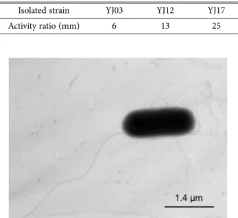
The eighteen bacterial strains were originally isolated from freshly spent mushroom (Flammulina velutipes) substrates. Among them, three isolates were able to hydrolyze mannanase during growth on TSA containing 0.5% locust bean gum and 1% trypan blue, zones of different sizes were cleared around the isolated bacteria.

Therefore, the strain with highest mannanase activity ratio, designated YJ17, was selected for further studies (Table 1). The mannanase activity has been reported in a wide variety of Bacillus sp., but few data are available on mannanase of B. amyloliquefaciens.

Morphological and physiological characteristics The isolate YJ17 was facultative anaerobic and was grown at temperature ranging from 20

o

C to 50

o

C with

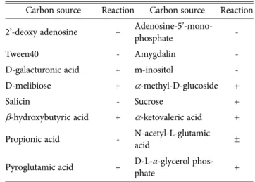
Fig. 1. Transmission electron micrograph of the isolate YJ17.

Exponentially growing culture was observed by negative stain with 1% phosphotungstic acid. Scale bar, 1.4 um.

Table 1. Mannanase activity of the isolated strains from spent mushroom substrate. The activity ratio was calculated from clear zones formed on TSA plate containing 0.5%

locust bean gum as substrate and incubated at 40

o

C for 24 h

Isolated strain YJ03 YJ12 YJ17

Activity ratio (mm) 6 13 25

(4)

an optimal temperature of 40

o

C. After incubation for 24 h on TSA, colonies of the isolate YJ17 were smooth, convex and wrinkled with approximately 2.5 ±0.1 mm in diameter. Cells were Gram-positive, rod-shaped and the measured size by transmission electron microscopy was 1.4×0.7 µm in 36 h culture grown at 40

o

C (Fig. 1).

The isolate YJ17 was identified using BIOLOG automatic identification system. The BIOLOG data of the isolate YJ17 allows Bacillus strain to be classified according to the ability to ferment 92 different carbohydrates. The substrate utilization as carbon source of the isolate YJ17 is presented in Table 2. Analysis based entirely on substrate utilization profiles showed that the isolate YJ17 belonged to B. amyloliquefaciens and was different from Bacillus subgroup: B. subtilis, B.

lincheniformis, B. mojavensis, and B. atrophaeus.

The DNA G+C content of the isolate YJ17 was 44 mol%. This data also show that isolate YJ17 belong to B. amyloliquefaciens in the B. subtilis group and should not be classified as a strain or variety of B. subtilis. The mol% G+C of the DNA of B. amyloliquefaciens (43.5- 44.9) is slightly higher than for B. subtilis (42-43) (Logan and Berkeley, 1984).

Chemotaxonomy

The whole-cell fatty acid composition was determined for the isolate YJ17 and other members of Bacillus closely related to the B. subtilis subgroup. Fatty acid analysis showed that the isolate YJ17 gave matches with B. subtilis with 0.36 similarity indices. Although comparison of bacterial fatty acid profiles revealed

‘closest match’ species, the similarity indices of these Table 2. Biochemical characteristics of isolate YJ17

Carbon source Reaction Carbon source Reaction

L-arabinose + Glycogen +

α-D-lactose - D-fuctose +

β-methyl-D-glucoside - D-mannitol +

D-tagatose - D-rafinose -

Lactomide - D-xylose -

L-alaninamide - L-malic acid +

Adenosine + L-asparagine +

Mannan - Uridine -

D-galactose + N-acetyl- β-D-man-

nosamine +

D-melezitose + α-D-glucose +

D-Ribose + 3-methyl glucose +

α-hydroxybutyric acid - Starchyose +

Succinic acid mono-

methyl ester + α-ketoglutaric acid +

Glycyl-L-glutamic acid - Succinic acid + Thymidine-5’-mono-

phosphate + 2, 3-butanediol +

Dextrin + β-cyclodextrin +

D-cellobiose + Arbutin -

Maltotriose + Maltose +

D-psicose + Palatinose +

Xylitol - Turanose +

D-malic acid - L-lactic Acid +

L-alanyl-glycine - L-alanine ±

Thymine + Inosine +

N-acetyl-D-glucosamine + Tween 80 +

D-gluconic acid + Gentiobiose -

β-methyl-D-galactoside + α-methyl-D-galacto-

side -

D-Sorbitol + Sedoheptulosan -

p-hydroxy-phenyl acetic

acid - γ-hydroxybutyl acid +

Succine amic acid + Pyruvic acid +

Putrescine + L-serine -

α-D-glucose-1-phosphate + D-fructose-6-phos-

phate -

α-cyclodextrin + Inulin +

D-arabitol - L-fucose +

Lactulose - D-mannose +

α-methyl-D-mannoside - L-rhamnose -

D-trehalose + Acetie acid +

D-lactic acid methyl

ester - Pyruvatic acid methyl

ester -

D-alanine + L-glutamic acid +

Table 2. Continued

Carbon source Reaction Carbon source Reaction 2’-deoxy adenosine + Adenosine-5’-mono-

phosphate -

Tween40 - Amygdalin -

D-galacturonic acid + m-inositol -

D-melibiose + α-methyl-D-glucoside +

Salicin - Sucrose +

β-hydroxybutyric acid + α-ketovaleric acid + Propionic acid - N-acetyl-L-glutamic

acid ±

Pyroglutamic acid + D-L-a-glycerol phos-

phate +

+, positive reaction; -, negative reaction; ±, weak reaction

(5)

matches were very low. However, almost ‘closest match’

species belonged to the genus Bacillus. The predominant fatty acids in whole-cell methanolysates of the isolate YJ17 were anteiso-15:0 (38.9%), 17:0 (7.6%), and iso- 15:0 (36.5%).

16S rRNA sequence analysis

An approximate 1.5-kb unique PCR product was generated from the isolate YJ17. The partial 16S rRNA gene sequences of the isolate YJ17 was determined and aligned to other known sequence of Bacillus strains obtained from GenBank. The sequence similarity between the isolate YJ17 and other Bacillus strains was from 98% to 99%. In the phylogenetic analysis based

on these sequences, the isolate YJ17 and B.

amyloliquefaciens clustered within a group and separated from other species of Bacillus (Fig. 2).

Based on physiological and molecular properties, the isolate YJ17 was classified within the genus Bacillus as Bacillus amyloliquefaciens YJ17.

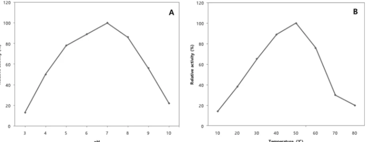
Effect of temperature and pH on mannanase activity The isolated B. amyloliquefaciens YJ17 produced an extracellular mannanase during grown with locust bean gum. The optimal pH for the mannanase activity was pH 7.0 (Fig. 3A). The optimal pH for the mannanase of B. amyloliquefaciens YJ17 was compared to pH 5.0 from B. subtilis NM-39 (Mendoza et al, 1994a), pH 6.0 from B. amyloliquefaciens CS47 (Cho, 2009) and pH 7.0 from B. subtilis KU-1 (Zakaria et al, 1998) and Bacillus sp. (Ooi and Kikuchi, 1995). The enzyme demonstrated broad pH stability within pH range of 5-8.

The mannanase from B. amyloliquefaciens YJ17 was optimally active at 50

o

C (Fig. 3B). The optimal temperature for the mannanase of B. amyloliquefaciens YJ17 was compared to 50-55

o

C from B. subtilis NM-39 (Mendoza et al, 1994a), KU-1 (Zakaria et al, 1998) and 5H (Khanongnuch et al, 1998), B. amyloliquefaciens CS47 (Cho, 2009) and 60

o

C from Bacillus licheniformis (Zhang et al, 2000). This thermal stability is important to use mannanase as an animal feed additive. For industrial application of mannanase from B.

amyloliquefaciens YJ17, it is necessary to carry out studies to determine the substrate specificity and stability of the purified mannanase under various conditions.

Fig. 2. Phylogenetic relationships of the isolate YJ17 and other closely related bacteria based on the partial 16S rRNA sequence. The branching pattern was generated by the neighbor-joing method. Bootstrap values (expressed as percentages of 10,000 replications) are shown at major branching points. Bar, 0.05 substitution per nucleotide position.

Fig. 3. Effects of pH (A) and temperature (B) on mannanase activities. (A) Enzyme activity was assayed at 50

o

C for 15 min in

sodium citrate buffers with the indicated pH values. (B) Enzyme activity was assayed at pH 6.0 for 30 min at the indicated

temperature.

(6)

Acknowledgements

This work was supported by Gyeongnam National University of Science and Technology Grant 2014.

References

Admark P, Varga A, Medve J, Harjunp V, Drakenberg T, Tjerneld F. 1998. Softwood hemicellulose-degrading enzymes from Aspergillus niger: purification and properties of a β- mannanase. J Biotechnol. 63:199-210.

Akino T, Nakamura N, Horikoshi K. 1987. Production of β- mannosidase and β-mannanase by an alkalophilic Bacillus sp.

Appl Microbiol Biotechnol. 26:323-327.

Arisan-Atac I, Hodits R, Kristufek D, Kubicek CP. 1993.

Purification and characterization of a β-mannanase of Trichoderma reesei C-30m. Appl Microbiol Biotechnol. 39:58- 62.

Claus D, Berkeley RCW. 1986. In Bergey's Manual of Systematic Bacteriology. pp. 1105-1139,Vol. 2, Williame & Wilkins.

Cho SJ. Isolation and characterization of mannanase producing Bacillus amyloliquefaciens CS47 from horse feces. Life Sci.

19:1724-1730.

Ethier N, Talbot G, Sygusch J. 1998. Gene cloning, DNA sequencing, and expression of thermostable β-mannanase from Bacillus stearothermophilus. Appl Environ Microbiol.

64:4428-4432.

Ferreira HM, Filho EXF. 2004. Purification and characterization of a β-mannanase from Trichoderma harzianum strain T4.

Carbohydr Polymer. 57: 23-29.

Khanongnuch C, Asada K, Tsuruga H, Ooi T, Kinoshita S, Lumyong S. 1998. β-Mannanase and xylanase of Bacillus subtilis 5H active for bleaching of crude pulp. J Ferment Bioeng. 86:461-466.

Kurakake M, Komaki T. 2001. Production of β-mannanase and β-mannosidase from Aspergillus awamori K4 and their properties. Curr Microbiol. 42: 377-380.

Logan NA, Berkeley RC. 1984. Identification of Bacillus strains using the API system. J Gen Microbiol. 130:1871-1882.

Mandel M, Marmur J. 1968. Use of ultraviolet absorbance- temperature profile for determining the guanine plus cytosine content of DNA. Methods Enzymol. 12:195-206.

McCleary BV. 1988. β-mannanase, pp. 596-610, In Wood WA, and Kellogg ST. (eds.), Methods in Enzymology, Vol. 160, Academic Press Inc., NewYork.

Mendoza NS, Arai M, Kawaguchi T, Yoshida T, Joson LM. 1994a.

Purification and properties of mannanase from Bacillus

subtilis. World J Microbiol Biotechnol. 10:551-555.

Mendoza NS, Arai M, Kawaguchi T, Cubol FS, Panerio EG, Yoshida T. 1994b. Isolation of mannan-utilizing bacteria and the culture conditions for mannanase production. World J Microbiol Biotechnol. 10:51-54.

Miller. 1959. Use of dinitrosalicylic acid reagent for determination of reducing sugars. Analytical Chemistry 31:426-428.

Ooi T, Kikuchi D. 1995. Purification and some properties of β- mannanase from Bacillus sp. World J Microbiol Biotechnol.

11:310-314.

Petty LA, Carter SD, Senne BW, Shriver JA. 2002. Effects of β- mannanase addition to corn-soybean meal diets on growth performance, carcass traits, and nutrient digestibility of weaning and growing-finishing pig. J Anim Sci. 80:1012-1019.

Sachslehner A, Haltrich D. 1999. Purification and some properties of a thermostable acidic endo-β-1,4-d-mannanase from Sclerotium (Athelia) rolfsii. FEMS Microbiol Lett. 177:47- 55.

Schaeffer P, Millet J, Aubert JP. 1965. Catabolic repression of bacterial sporulation. Proc Natl Acad Sci. USA 54:704-711.

Slominski BA, Meng X, Campbell LD, Guenter W, Jones O. 2006.

The use of enzyme technology for improved energy utilization from full-fat oilseeds. Part II: Flaxseed. Poult Sci.

85:1031-1037.

Takahashi R, Kusakabe I, Kobayashi H, Murakami K, Maekawa A, Suzuki T. 1984. Purification and some properties of mannanase from Streptomyces sp. Agri Biol Chem. 48:2189- 2195.

Wozniewski T, Blaschek W, Franz G. 1992. Isolation and characterization of an endo-•‚-mannanase of Lilium testaceum bulbs. Phytochemistry 31:3365-3370.

Yamaura I, Matsumoto T. 1993. Purification and some properties of endo-1,4-•‚-D-mannanase from a mud snail, Pomacea insularus (de Ordigny). Biosci Biotechnol Biochem. 57:1316- 1319.

Yoon KH, Lim BL. 2007. Cloning and strong expression of a Bacillus subtilis WL-3 mannanase gene in B. subtilis. J Microbiol Biotechnol. 17:1688-1694.

Zakaria MM, Yamamoto S, Yagi T. 1998. Purification and characterization of an endo-1,4- β-mannanase from Bacillus subtilis KU-1. FEMS Microbiol Lett. 158:25-31.

Zhang J, He ZM, Hu K. 2000. Purification and characterization of β-mannanase from Bacillus licheniformis for industrial use.

Biotechnol Lett. 22:1375-1378.

Zou XT, Qiao XJ, Xu ZR. 2006. Effect of β-mannanase (Hemicell) on growth performance and immunity of broilers. Poult Sci.

85:2176-2179.

수치

Fig. 1. Transmission electron micrograph of the isolate YJ17.
Table 2. Continued
Fig. 3. Effects of pH (A) and temperature (B) on mannanase activities. (A) Enzyme activity was assayed at 50 o C for 15 min in sodium citrate buffers with the indicated pH values

참조

관련 문서

When the car is heading up the hill the weight or gravitational force points opposite to that of the displacement, producing and angle greater than 90W. degrees, producing

Modern Physics for Scientists and Engineers International Edition,

 To further localize the wave within x by producing a wave packet, we need to combine more than two waves over a range of wave vectors ∆k.  Define ∆x to be ∆λ/2

In 1883, Renoir completed two paintings that look very similar, The Dance in the City and The Dance in the Country.. Both paintings show a couple who

The proposed approach is to compare Hindi contexts from the largest available Hindi Corpus with the Human judgments of degree of similarity existing between the benchmark word

• Since time is required in building up or depleting a charge distribution, the stored charge must inevitably lag behind the current in a time-dependent problem.. This is

16S rRNA 유전자 염기서열 분석을 통한 녹조 미생물 균총분석. 16S rRNA의 변이부위의 종 특이적 부위를 유전자증폭을 통한 DNA 단편 서열분석 Gene bank에 2천만 개 염기서열 등록,

1 John Owen, Justification by Faith Alone, in The Works of John Owen, ed. John Bolt, trans. Scott Clark, "Do This and Live: Christ's Active Obedience as the