Warty Squamous Cell Carcinoma of the Vulva in Older Women: Association with Human Papillomavirus
4
0
0
전체 글
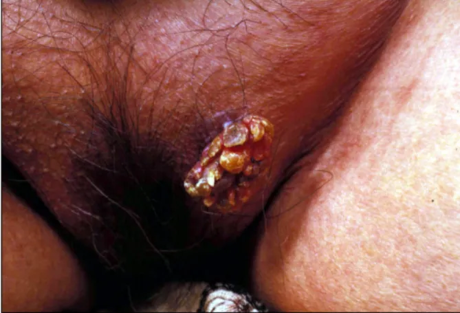
(2) 156. Yong-Hyun Jang, et al.. Fig. 1. A verrucous and pedunculated mass on the left vulva.. Fig. 3. HPV DNA chip microarray format (Left). DNA chip microarray for HPV types show the presence of other type HPV DNA in this case, but not the high and low risk HPV types. (Left: DNA chip microarray format; right: results in this case, other HPV DNA types positive.). Fig. 2. (a) A papillomatous pattern with hyperkeratosis, parakeratosis and acanthosis (haematoxylin and eosin; original magnification: × 40). Round and tapering dermal papillae with a fibrovascular core (×100). (b) Invasion of the dermis by irregularly shaped, jagged nests of epithelium, the cells which are predominantly mature squamous cells showing atypicality (haematoxylin and eosin; original magnification: × 50). The invading tumor masses are composed of atypical squamous cells (× 200). (c) Cells within the tumor nest demonstrate koilocytosis with large, wrinkled, hyperchromatic nuclei, and horn pearl is present (haematoxylin and eosin; original magnification: ×400).. oligonucleotide chip that enables the rapid and easy detection and genotyping of 18 high risk HPV types (i.e., 16/ 18/ 26/ 31/ 33/ 35/ 39/ 45/ 51/ 52/ 56/ 58/ 59/ 66/ 68/ 69/ 70/ 71), 14 low risk HPV types (i.e, 6/ 11/ 32/ 34/ 40/ 42/ 43/ 44/ 53/ 54/ 55/ 57/ 61/ 62) and other types HPV DNA (i.e., positive result in any HPV types, but type is not specified). For HPV genotyping, a commercially available HPV DNA chip was purchased from Biomedlab Co. (Seoul, Korea). The HPV DNA chip contains 32 type-specific probes. The manufacturer's protocol describes the preparation and testing of specimens, and the genotyping experiment was performed using a proYonsei Med J Vol. 46, No. 1, 2005. cedure provided by Biomedlab. Briefly, target HPV DNA was amplified by the polymerase chain reaction (PCR) using the primers (HPV and βglobin) and conditions provided by Biomedlab and labeled using Cy5-dUTP (NEN Life Science Products, Inc., Boston, MA, USA). The PCR product was hybridized onto the chip at 40 for 2 hours and washed with 3 × SSPE and with 1 × SSPE for 2 minutes each. Hybridized signals were visualized with a DNA Chip Scanner (GSI Lumonics, Scanarray lite, Ottawa, Canada). In our case, the DNA microarray showed the existence of other types of HPV DNA, but not the 18 high risk and 14 low risk HPV types (Fig. 3)..
(3) Warty Squamous Cell Carcinoma of the Vulva in Older Women: Association with Human Papillomavirus. DISCUSSION WSCC is a rare specific variant of invasive squamous cell carcinoma, usually described as a hybrid feature of condyloma and invasive squamous cell carcinoma. It has been described in the vulva, uterine cervix, penis, anus, oral mucosa and urinary bladder.2,3,6-11 WSCC typically occurs 1 in younger women. However, our case developed in old age. The gross appearance of WSCC resembles verrucous carcinoma, being large and exophytic with a papillary appearance, but the surface has a characteristic feathery appearance.7,8 Histologically, the tumors are mainly papillomatous with acanthosis and hyperkeratosis, and they have characteristic histologic findings that include prominent fibrovascular cores of dermal papilla, striking nuclear koilocytotic atypia and jagged, irregular interface between tumor and stroma.8,10 WSCC is a slow growing tumor. However, unlike giant condyloma or verrucous carcinoma, warty carcinomas have a risk of regional metastasis. As such, it must be differentiated from other verruciform neoplasms, such as giant condyloma acuminatum and verrucous carcinoma. Microscopic finding is helpful in differentiating warty carcinoma from giant condyloma. The latter exhibit a pale pink hyperplastic epidermis with uniform-appearing nuclei and much less atypia.12 On the other hand, the former has more cytologic variability, with clear nuclear pleomorphism. In addition, the interface between the base of the process and the underlying tissue is variable and irregular, a feature that is much less evident in condylomatous lesions. WSCC is distinguished from verrucous carcinoma on the basis of long and undulating, condylomatous papilla, with prominent fibrovascular cores and a base that is rounded or irregular and jagged. Furthermore, koilocytotic atypia is prominent and diffuse, while it is absent in the “pure” verrucous carcinoma.7,8,13 HPV infection is strongly associated with warty and basaloid squamous cell carcinoma.14 Several HPV types (e.g., 6, 11, 16, 18, 33) have been detected in the vast majority of WSCC cases.15-17 In the study by Hording et al.,18 84% of basaloid and warty carcinomas were found to harbor HPV type 16 or 33, high risk types, by PCR, while in. 157. the same study only 4% of the cases of keratinizing ISCC, which typically develops in older women, had evidence of HPV. In our case, the existence of HPV DNA was proven by DNA chip microarray, but we could not detect the specific HPV DNA type that is often associated with WSCC. Whang et al.19 evaluated the clinical efficacy of the DNA microarray for the detection of HPVs in various cervical lesions. They detected HPV DNAs in 158 and 174 of the 234 cervical samples by PCR-RFLP (restriction fragment length polymorphism) and HPV microarray, respectively, and suggested that HPV oligonucleotide microarray is a highly comparable method to the previously used PCR-RFLP method for the detection of HPV. In conclusion, we describe here vulvar WSCC occurring in a woman of old age that is associated with an unknown HPV type. This suggests that WSCC may occur regardless of age and that various types of HPV can be associated with the pathogenesis of WSCC.. REFERENCES 1. Al-Ghamdi A, Freedman D, Miller D, Poh C, Rosin M, Zhang L, et al. Vulvar squamous cell carcinoma in young women: a clinicopathologic study of 21 cases. Gynecol Oncol 2002;84:94-101. 2. Rastkar G, Okagaki T, Twiggs LB, Clark BA. Early invasive and in situ warty carcinoma of the vulva: clinical, histologic, and electron microscopic study with particular reference to viral association. Am J Obstet Gynecol 1982;143:814-20. 3. Kurman RJ, Toki T, Schiffman MH. Basaloid and warty carcinomas of the vulva. Distinctive types of squamous cell carcinoma frequently associated with human papillomaviruses. Am J Surg Pathol 1993;17:133-45. 4. Jones RW, Baranyai J, Stables S. Trends in squamous cell carcinoma of the vulva: the influence of vulvar intraepithelial neoplasia. Obstet Gynecol 1997;90:448-52. 5. Trimble CL, Hildesheim A, Brinton LA, Shah KV, Kurman RJ. Heterogeneous etiology of squamous carcinoma of the vulva. Obstet Gynecol 1996;87:59-64. 6. Ng WK, Cheung LK, Li AS. Warty (condylomatous) carcinoma of the cervix. A review of 3 cases with emphasis on thin-layer cytology and molecular analysis for HPV. Acta Cytol 2003;47:159-66. 7. Bezerra AL, Lopes A, Landman G, Alencar GN, Torloni H, Villa LL. Clinicopathologic features and human papillomavirus and prevalence of warty and squamous cell carcinoma of the penis. Am J Surg Pathol 2001;25: Yonsei Med J Vol. 46, No. 1, 2005.
(4) 158. Yong-Hyun Jang, et al.. 673-8. 8. Cubilla AL, Velazques EF, Reuter VE, Oliva E, Mihm MC Jr, Young RH. Warty (condylomatous) squamous cell carcinoma of the penis: a report of 11 cases and proposed classification of ‘verruciform’ penile tumors. Am J Surg Pathol 2000;24:505-12. 9. Noel JC, Sornin de Leysat C, Peny MO, van de Stadt J, Fayt I, De Dobbeleer G. Warty carcinoma of the anus: a variant of squamous cell carcinoma associated with anal intraepithelial neoplasia and human papillomavirus infection. Dermatology 2001;203:262-4. 10. Piattelli A, Rubini C, Fioroni M, Iezzi T. Warty carcinoma of the oral mucosa in an HIV+ patient. Oral Oncol 2001;37:665-7. 11. Koss LG. Warty carcinoma of bladder containing HPV type 11. Int J Surg Pathol 2000;8:367. 12. Brown TJ, Yen-Moore A, Tyring SK. An overview of sexually transmitted diseases. J Am Acad Dermatol 1999;41:661-77. 13. Schwartz RA. Verrucous carcinoma of the skin and mucosa. J Am Acad Dermatol 1995;32:1-21. 14. Kim KH, Kim YS. Role of human papillomavirus and p53 tumor suppressor gene in cervical carcinogenesis.. Yonsei Med J Vol. 46, No. 1, 2005. Yonsei Med J 1995;36:412-25. 15. Ostrow RS, Shaver MK, Turnquist S, Viksnins A, Bender M, Vance C, et al. Human papillomavirus-16 DNA in a cutaneous invasive cancer. Arch Dermatol 1989;125:666-9. 16. Petry KU, Kochel H, Bode U, Schedel I, Niesert S, Glaubitz M, et al. Human papillomavirus is associated with the frequent detection of warty and basaloid highgrade neoplasia of the vulva and cervical neoplasia among immunocompromised women. Gynecol Oncol 1996;60:30-4. 17. Cho NH, Joo HJ, Ahn HJ, Jung WH, Lee KG.. Detection of human papillomavirus in warty carcinoma of the uterine cervix: comparison of immunohistochemistry, in situ hybridization and in situ polymerase chain reaction methods. Pathol Res Pract 1998;194:713-20. 18. Hording U, Daugaard S, Junge J, Lundvall F. Human papillomaviruses and multifocal genital neoplasia. Int J Gynecol Pathol 1996;15:230-4. 19. Hwang TS, Jeong JK, Park M, Han HS, Choi HK, Park TS. Detection and typing of HPV genotypes in various cervical lesions by HPV oligonucleotide microarray. Gynecol Oncol 2003;90:51-6..
(5)
수치
관련 문서
The expression levels of apoptotic related proteins and caspase dependent proteins by the ostreolysin purified from P.ostreatus on cell viability in FaDu human
The proliferations of MC3T3-E1 cell seeded on pristine PCL scaffold and surface modified scaffolds for 1, 3, and 5 days... ALP activity of the MC3T3-E1 cell seeded
Concentration-Dependent inhibition of cell viability by adenosine in human oral fibroblast and FaDu human head and neck squamous cell carcinoma · · ·
It also suggest that cell proliferation inhibits by a novel signal transduction for adenosine effect in human FaDu hypopharynx squamous cell carcinoma cells....
Differential Expression of Desmoglein1, Desmoglein3, Epithelial Membrane Antigen, Ber-EP4 and CD10 in Basal Cell Carcinoma and Squamous Cell Carcinoma
Objective: The purpose of this study was to analyze recent trend in incidence of basal cell carcinoma and squamous cell carcinoma in patients from the Gwangju City
These results suggest that the bilobalide inhibits the cell proliferation and induces the apoptotic cell death in FaDu human pharyngeal squamous cell carcinoma via both
In the present study we investigated the proliferation effect of human oral cancer KB cell treated with pulsatilla koreana extract.. We analyzed the effects of this
