INTRODUCTION
Hepatocellular carcinoma (HCC) is a devastating disease with poor prognosis and is one of the most common causes of can- cer-related deaths worldwide.1,2 The prognosis for HCC pa- tients is determined by tumor status, liver function reserve,
general performance, and treatment efficacy.3 To improve pa- tient survival, several staging systems have been described for HCC including the Barcelona Clinic Liver Cancer (BCLC) clas- sification, Okuda et al., Cancer of the Liver Italian Program, and French scores.4-7 Among them, only the BCLC classification has been approved by the European Association for Study of the Liver and the American Association for the Study of Liver Disease for the prognostic classification and treatment selec- tion of HCC.4,8,9 This staging system, which has been validated in regards to its clinical usefulness in several studies, is associ- ated with treatment indications and aspects of estimated life expectancy.10,11
BCLC stage B HCC includes Child-Pugh scores A and B, as well as preserved performance status, with asymptomatic mul- tifocal tumors without vascular invasion or extrahepatic spread.
For this group of patients, the BCLC staging system indicates
Predictors of Micrometastases in Patients
with Barcelona Clinic Liver Cancer Classification B Hepatocellular Carcinoma
Suk-Won Suh and Yoo Shin Choi
Department of Surgery, College of Medicine, Chung-Ang University, Seoul, Korea.
Purpose: Transarterial chemoembolization (TACE) is indicated for Barcelona Clinic Liver Cancer (BCLC) B hepatocellular carci- noma (HCC). Whether TACE provides any long-term survival benefits remains unclear. We aimed to investigate micrometastases predictors with which to identify patients who would benefit from surgical resection (SR).
Materials and Methods: First, we analyzed risk factors of micrometastases, microvascular invasion, and poor histologic grade in 38 patients with newly diagnosed resectable BCLC stage B HCC limited to one or two segments with well-preserved liver function and who underwent SR between January 2006 and December 2013. Second, we validated identified risk factors in 54 newly diag- nosed resectable BCLC B HCC patients with well-preserved liver function who underwent TACE during the same period to deter- mine their influence on survival.
Results: Risk factors of micrometastases in SR patients were α-fetoprotein (AFP) ≥110 [hazard ratio (HR)=5.166; 95% confidence interval (CI), 1.031–25.897; p=0.046] and prothrombin induced by vitamin K absence-II (PIVKA-II) ≥800 (HR=5.166; 95% CI, 1.031–25.897; p=0.046). The cumulative probability of tumor recurrence (p=0.009) after SR differed according to levels of AFP and PIVKA-II. After validation of these risk factors in the TACE group, patients with SR and AFP <110 and PIVKA-II <800 had superior survival outcomes than other patients (HR=0.116; 95% CI, 0.027–0.497; p=0.004).
Conclusion: AFP and PIVKA-II levels predict micrometastases and survival. Therefore, they should be considered when selecting SR for BCLC B HCC.
Key Words: Predictors, micrometastases, Barcelona Clinic Liver Cancer classification B hepatocellular carcinoma, surgical resection, transarterial chemoembolization
pISSN: 0513-5796 · eISSN: 1976-2437
Received: November 22, 2016 Revised: February 1, 2017 Accepted: February 15, 2017
Corresponding author: Dr. Yoo Shin Choi, Department of Surgery, Chung-Ang University Hospital, 102 Heukseok-ro, Dongjak-gu, Seoul 06973, Korea.
Tel: 82-2-6299-1545, Fax: 82-2-824-7869, E-mail: [email protected]
•The authors have no financial conflicts of interest.
© Copyright: Yonsei University College of Medicine 2017
This is an Open Access article distributed under the terms of the Creative Com- mons Attribution Non-Commercial License (http://creativecommons.org/licenses/
by-nc/4.0) which permits unrestricted non-commercial use, distribution, and repro- duction in any medium, provided the original work is properly cited.
Yonsei Med J 2017 Jul;58(4):737-742 https://doi.org/10.3349/ymj.2017.58.4.737
transarterial chemoembolization (TACE) as the main treatment option.4 A previous study showed that TACE provided a better 2-year survival rate than supportive treatment;12 however, it is unclear whether TACE provides any long-term survival benefit.
BCLC stage B is a heterogeneous category that includes pa- tients who vary widely in tumor stage, liver function, and dis- ease etiology. Owing to this heterogeneity, TACE may not be the optimal therapy for all patients with BCLC stage B HCC. Recent studies have verified that surgical resection (SR) can be safely performed on a subset of patients with BCLC stage B HCC who exhibit good liver function, resulting in a long-term surviv- al rate comparable to that of patients treated with TACE.1,3,13,14 However, the indication of SR for BCLC stage B HCC remains controversial and needs further assessment.
If HCC progresses, venous outflow might be obstructed and portal backflow could develop. This can be a route of microme- tastases, one of the major causes of multinodular HCC.15 Mi- crometastases, which cannot often be detected by various im- aging modalities, can remain in remnant liver, holding a strong possibility of causing local recurrence after SR. Therefore, we hypothesized that if a patient has few risk factors for the pres- ence of micrometastases, such as microvascular invasion or poor histologic differentiation,16 the intrahepatic spread of the tumor might be limited. In the event of limited intrahepatic tu- mor spread, SR may confer a greater chance of curative treat- ment and may represent a better treatment option. Additional- ly, determining preoperative risk factors for intrahepatic tumor spread in patients with multinodular HCC would allow for a more accurate prediction of the possibility of curative SR. SR could, therefore, be recommended in patients with no risk fac- tors who are predicted to have a good prognosis.
The aims of this study were to identify preoperative risk fac- tors for the micrometastases of tumors in patients who under- went SR for BCLC stage B HCC and to validate these identified risk factors in terms of overall survival in a group of patients who underwent TACE.
MATERIALS AND METHODS
Patients
This retrospective cohort study was approved by the Institu- tional Review Board of Chung-Ang University Hospital (Seoul, Korea) and was exempted from the requirement to obtain in- formed consent. The first step of this study was to identify pre- operative risk factors in 38 patients with well-preserved liver function, indicated as Child-Turcotte-Pugh (CTP) A liver cir- rhosis, and newly diagnosed resectable BCLC stage B HCC limited to one or two segments, and who underwent SR be- tween January 2001 and December 2013 at Chung-Ang Uni- versity Hospital. Patients with a previous treatment history of HCC or insufficient clinical data were excluded from analysis.
The risk for micrometastases, which represents the possibility
of intrahepatic spread of the tumor, was defined as the pres- ence of Edmonson and Steiner histologic grade 3 or 4, and/or microvascular invasion. The following data were collected for all patients to determine risk factors for tumor aggressiveness:
age, sex, body mass index, presence of hepatitis B virus and hepatitis C virus, preoperative laboratory results (levels of total bilirubin, albumin, and the international normalized ratio), tu- mor marker levels [α-fetoprotein (AFP) and prothrombin in- duced by vitamin K absence-II (PIVKA-II)], and tumor charac- teristics (tumor size, histologic grade, and presence of micro- vascular invasion).
The second step of the study was to validate identified risk factors in 52 patients who met the same criteria as the SR group, including well-preserved liver function and resectable BCLC stage B HCC limited to one or two segments initially treated with TACE during the same period as the first step of the study.
We validated the identified risk factors after adjusting for sev- eral parameters that might have influenced the overall surviv- al of patients in the TACE group, such as demographics, labo- ratory results, and radiologic findings. Clinical variables were the same as those outlined for the first step of the study, with the following exceptions: pathologic results were collected from patients who underwent TACE, and risk factors for over- all survival after TACE were identified. The probability of over- all survival was analyzed according to the type of treatment and the presence of identified risk factors.
All patients were followed-up at 1, 3, and 6 months post- treatment and every 3 to 6 months thereafter, as necessary. At each visit, imagining studies, such as computed tomography, magnetic resonance imaging, and ultrasonography, were per- formed. Serologic tests, such as tumor marker analyses and biochemical liver function tests, were performed at each visit. If tumor recurrence was found after SR, multiple modalities, such as re-SR, radiofrequency ablation, or TACE, in consideration of tumor characteristics were performed as early as possible before progression of the recurrent tumor. Also, TACE was re- peatedly preformed until the disappearance of viable tumor was noted, and if the tumor progressed, TACE was continued.
Procedure
The SR was performed under low central venous pressure at less than 5 mm Hg. Anatomic partial hepatectomy was per- formed in a standardized manner; however, if the patients had poor liver function, non-anatomic partial hepatectomy was also performed. All SRs were performed based on a curative aim by ligating the feeding vessels, and securing at least 2 cm of resection margin using ultrasonographic guidance and af- ter resection, the frozen section was performed to confirm the minimal length of resection margin.
The TACE was performed under local anesthesia: a 2.4F highly flexible microcatheter (Progreat, Terumo Corporation, Tokyo, Japan) was introduced into the abdominal aorta via the superficial femoral artery using the Seldinger technique.
Hepatic arterial angiography was performed using fluoroscopy to guide the catheter into the celiac artery. Then, the feeding arteries, tumor stain, and vascular anatomy surrounding the tumor were identified. A microcatheter was introduced through the catheter and directed to the feeding arteries. A mixture of doxorubicin hydrochloride (Adriamycin; Ildong Pharm., Seoul, Korea) and iodized oil (Lipiodol; Laboratoire Guerbet, Aul- nay-Sous-Bois, France) was injected under fluoroscopic con- trol, followed by embolization of the feeding arteries using 1-mm diameter absorbable gelatin sponge particles (Gelform;
Upjohn, Kalamazoo, MI, USA). The volume of ionized oil ranged from 2 to 10 mL, and the amount of Adriamycin ranged from 10 to 70 mg.
Statistical analysis
For intergroup comparisons, the distribution of the data was first evaluated for normality using the Shapiro-Wilk test. Nor- mally distributed data are presented herein as mean±standard deviation. Groups were compared using Student’s t-test. De- scriptive variables were subjected to χ2 analysis or Fisher’s ex- act test, as appropriate. The optimal cut-off values for the levels of AFP and PIVKA-II were determined by the area under the receiver operating characteristic analysis. Multivariate analysis using an ordinary logistic regression model was performed to investigate risk factors for intrahepatic spread of the tumor in SR patients, as well as the influence of these factors on overall survival in TACE patients. Survival and recurrence after each type of procedure, as well as the presence of identified risk fac- tors, were plotted using the Kaplan-Meier method and com- pared using the log-rank test. A multivariate analysis using the Cox proportional hazard regression method was performed to investigate the risk factors for recurrence-free survival and
overall survival. A p-value of <0.05 was considered statistically significant. Statistical analyses were conducted using SPSS ver. 19.0 (SPSS Inc., Chicago, IL, USA).
RESULTS
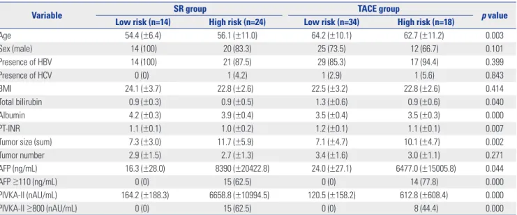
A comparison of patient demographics in each group (low risk/SR, high risk/SR, low risk/TACE, high risk/TACE) is shown in Table 1. Mean age was significant highest in the TACE/low risk group (p=0.003). Laboratory results indicative of liver function showed slight differences in each group, al- though all laboratory results were within normal limits, repre- senting well-preserved liver function as CTP A liver cirrhosis.
Expression of the tumor markers, such as AFP and PIVKA-II, showed significant differences in each group, and high risk groups had elevated serum levels, compared with low risk groups (Table 1).
In the first step of the study, 60.5% (23/38) of patients who underwent SR were designated as being at high risk for micro- metastases, indicating the possibility of intrahepatic spread.
These patients displayed the presence of either poor histologic grade (34.2%) or microvascular invasion (50.0%). AFP ≥110 [hazard ratio (HR)=5.166; 95% confidence interval (CI), 1.031–
25.897; p=0.046] and PIVKA-II ≥800 (HR=5.166; 95% CI, 1.031–
25.897; p=0.046) were the only significant risk factors identified using multivariate analysis (Table 2).
Patients with AFP <110 and PIVKA-II <800 had lower tumor recurrence (57.1%) and long disease-free survival (36.7±28.5 months), compared with the patients who had AFP ≥110 or PIVKA-II ≥800 (87.5% and 14.5±21.8 months). Multiple (33.3%) or extrahepatic tumor recurrence (19.0%) were observed more
Table 1. Demographics of Patients in the SR and TACE Groups
Variable SR group TACE group
p value Low risk (n=14) High risk (n=24) Low risk (n=34) High risk (n=18)
Age 54.4 (±6.4) 56.1 (±11.0) 64.2 (±10.1) 62.7 (±11.2) 0.003
Sex (male) 14 (100) 20 (83.3) 25 (73.5) 12 (66.7) 0.101
Presence of HBV 14 (100) 21 (87.5) 29 (85.3) 17 (94.4) 0.399
Presence of HCV 0 (0) 1 (4.2) 1 (2.9) 1 (5.6) 0.843
BMI 24.1 (±3.7) 22.8 (±2.6) 22.5 (±3.2) 22.8 (±2.6) 0.414
Total bilirubin 0.9 (±0.3) 0.9 (±0.5) 1.3 (±0.6) 0.9 (±0.6) 0.040
Albumin 4.2 (±0.3) 3.9 (±0.4) 3.5 (±0.4) 3.5 (±0.3) 0.000
PT-INR 1.1 (±0.1) 1.0 (±0.2) 1.2 (±0.1) 1.1 (±0.1) 0.007
Tumor size (sum) 7.3 (±3.0) 11.7 (±5.9) 7.1 (±4.7) 10.1 (±4.7) 0.002
Tumor number 2.9 (±1.5) 2.7 (±1.3) 3.4 (±1.6) 3.0 (±1.1) 0.271
AFP (ng/mL) 16.3 (±28.0) 8390 (±20422.8) 24.0 (±27.1) 6477.0 (±15005.8) 0.044
AFP ≥110 (ng/mL) 0 (0) 15 (62.5) 0 (0) 14 (77.8) 0.000
PIVKA-II (nAU/mL) 164.2 (±188.3) 6658.8 (±10994.5) 120.5 (±158.2) 612.8 (±608.4) 0.000
PIVKA-II ≥800 (nAU/mL) 0 (0) 15 (62.5) 0 (0) 8 (44.4) 0.000
HBV, hepatitis B virus; HCV, hepatitis C virus; BMI, body mass index; PT-INR, prothrombin time-international normalized ratio; AFP, α-fetoprotein; PIVKA-II, pro- thrombin induced by vitamin K absence-II; SR, surgical resection; TACE, transarterial chemoembolization.
Data are presented as the mean±standard deviations or numbers with percentages in parentheses unless otherwise indicated.
often in patients with AFP ≥110 or PIVKA-II ≥800; however, most patients who had AFP <110 and PIVKA-II <800 showed only single tumor recurrence of less than 2 cm (Table 3). The cumulative probability of tumor recurrence (p=0.009) after SR differed significantly according to AFP and PIVKA-II levels (Fig. 1).
In the second step of the study, we validated the identified risk factors stated above in the TACE group. Overall survival rates at 1, 3, and 5 years, respectively, were as follows: 78.6, 42.9, and 42.9% for Group 1 (SR, AFP <110 and PIVKA-II <800);
20.8, 12.5, and 12.5% for Group 2 (SR, AFP ≥110 or PIVKA-II
≥800); 73.5, 41.2, and 8.8% for Group 3 (TACE, AFP <110 and PIVKA-II <800); and 83.3, 50.0, and 16.7% for Group 4 (TACE, AFP ≥110 or PIVKA-II ≥800). The survival curve of Group 1 dif- fered significantly from those of the other groups (p=0.001) (Fig.
2). Multivariate analysis of overall survival showed a significant difference between Group 1 and the other groups (HR=0.116;
95% CI, 0.027–0.497; p=0.004).
DISCUSSION
The BCLC staging system is considered the standard of care for patients with HCC.16,17 TACE is the first line treatment op- tion of BCLC stage B HCC, because it confers a median sur- vival of 20 months, which represents an improvement when compared with supportive treatment.12,18 It remains unknown, however, whether TACE confers a survival advantage over other treatment options, such as SR. The indications for SR have expanded with advances in surgical techniques and pre- operative preparation in recent decades. Several studies have reported improved overall survival after SR for BCLC B HCC as compared with TACE.19-21 Our results were consistent with the results of these reports, demonstrating that patients treat- ed with SR had superior overall survival than those treated with TACE (57.4±6.4 months vs. 27.2±2.8 months; p=0.000).
Many studies have examined prognostic factors for survival of BCLC stage B HCC patients after SR, and patients with select risk factors are predicted to have lower overall survival. There- Table 2. Risk Factor Analysis for Tumor Aggressiveness in the SR Group
Variable Univariate analysis Multivariate analysis
Low risk group (n=15) High risk group (n=23) p value Relative risk (95% CI) p value
Age 54.1 (±10.1) 56.4 (±9.2) 0.491
Sex (male) 14 (93.3) 20 (87.0) 0.531
Presence of HBV 14 (93.3) 21 (91.3) 0.821
Presence of HCV 0 (0) 1 (4.3) 0.413
BMI 24.4 (±3.7) 22.5 (±2.5) 0.068
Total bilirubin 1.0 (±0.3) 0.9 (±0.4) 0.245
Albumin 4.1 (±0.3) 3.9 (±0.4) 0.192
PT-INR 1.1 (±0.1) 1.0 (±0.2) 0.497
Tumor size (sum) 8.1 (±3.5) 11.4 (±6.1) 0.064
Tumor number 2.9 (±1.4) 2.7 (±1.3) 0.703
AFP ≥110 (ng/mL) 3 (20.0) 12 (52.2) 0.047 5.166 (1.031–25.897) 0.046
PIVKA-II ≥800 (nAU/mL) 3 (20.0) 12 (52.2) 0.047 5.166 (1.031–25.897) 0.046
SR, surgical resection; HBV, hepatitis B virus; HCV, hepatitis C virus; BMI, body mass index; PT-INR, prothrombin time-international normalized ratio; AFP, α-fetoprotein;
PIVKA-II, prothrombin induced by vitamin K absence-II.
Data are presented as the mean±standard deviations or numbers with percentages in parentheses unless otherwise indicated.
Table 3. Recurrence Patterns of Hepatocellular Carcinoma in the SR Group According to AFP and PIVKA-II Levels
AFP <110 and PIVKA-II <800 (n=14) AFP ≥110 or PIVKA-II ≥800 (n=24) p value
Recurrence 8 (57.1) 21 (87.5) 0.034
Disease free survival (month) 36.7±28.5 14.5±21.8 0.011
Pattern of tumor recurrence Intrahepatic
Single tumor
<1 cm 1 (12.5) 2 (9.5)
≥1 and <2 cm 4 (50.0) 5 (23.8)
≥2 and <3 cm 0 (0) 3 (14.3)
Multiple tumor 2 (25.0) 7 (33.3)
Extrahepatic 1 (12.5) 4 (19.0)
SR, surgical resection; AFP, α-fetoprotein; PIVKA-II, prothrombin induced by vitamin K absence-II.
Data are presented as the mean±standard deviations or numbers with percentages in parentheses unless otherwise indicated.
fore, the identification of patients suitable for SR is of critical importance.7,22,23 Based on the clinical observation of a marked heterogeneity in the clinical benefit of TACE, one study has proposed a subclassification of the BCLC stage B score into four stages (B1–B4) to facilitate therapeutic decisions, and pa- tients who classified as the B1 subgroup showed acceptable survival rates after SR.24
Among the patients who underwent SR, patients who had
an identified risk factor had significant earlier tumor recur- rence, compared with other patients (14.5±21.8 months vs.
36.7±28.5 months; p=0.011). Early recurrence of HCC is the likely result of local tumor recurrence, which may have arisen either from micrometastases that were left untreated by SR or from the intrahepatic spread of the primary tumor via the por- tal venous system. Micrometastases are common in patients with HCC and are noted more often in HCC patients with either microvascular invasion or poor histologic differentiation.16,25 The possibility remains that some micrometastases will per- sist, increasing the possibility of intrahepatic tumor spread and thereby presenting difficulty in achieving curative SR. Our study showed that patients in the SR group with an identifi- able risk factor of AFP ≥110 or PIVKA-II ≥800 had higher rates of multiple tumor recurrence (33.3%) and extrahepatic metas- tasis (19.0%). These features represents increased tumor ag- gressiveness, as defined by the possibility of micrometastases or intrahepatic tumor spread, when compared with SR patients who had AFP <110 and PIVKA-II <800.
Several studies have demonstrated the clinical significance of serum AFP or PIVKA-II in patients with HCC. However, these two markers involve different synthetic pathways, such that the levels of these two markers show independent pat- terns.26-29 Therefore, it appears that AFP and PIVKA-II are com- plementary markers for clinical usefulness. Accordingly, we defined the high-risk group as AFP ≥110 or PIVKA-II ≥800 to reflect levels of both markers. Despite high recurrence rates of HCC, Group 1 had significantly superior survival outcomes than the other groups. Patients with AFP <110 and PIVKA-II
<800 usually had long disease-free survival (36.7±28.5 months) and single tumor recurrence (62.5%), the latter of which we suspect arose as a result of de novo tumorigenesis due to un- derlying cirrhosis.
There are some limitations to this study. It is a retrospective study, forcing us to rely on the integrity of completed medical records for our analysis. In addition, the background charac- teristics of the SR and TACE groups were significantly differ- ent; selection bias, therefore, cannot be eliminated. The study population was relatively small, and further studies with pro- spectively randomized large populations are warranted to con- firm these promising results.
In conclusion, serum levels of AFP and PIVKA-II are useful predictors of tumor aggressiveness and micrometastasis in BCLC B HCC. AFP and PIVKA-II levels can also predict early tumor recurrence after SR. The type of treatment and the com- bined levels of AFP and PIVKA-II carry significant prognostic value in relation to overall survival rates. The findings in the current study may be helpful to determining optimal treatment strategies in BCLC B HCC. We propose that if a patient has no risk of micrometastasis as defined by AFP and PIVKA-II levels, SR should be considered for first-line therapy.
Fig. 1. Recurrence-free survival and overall survival in surgical resection patients. The cumulative probability of tumor recurrence. The frequency of recurrence was significantly different according to the levels of AFP and PIVKA-II, especially during the first year (1-year recurrence-free sur- vival: AFP <110 and PIVKA-II <800, 78.6%; AFP ≥110 or PIVKA-II ≥800, 20.8%). The curves continued to diverge during 2 years of follow-up, but then continued parallel (p=0.009). AFP, α-fetoprotein; PIVKA-II, prothrom- bin induced by vitamin K absence-II.
1.0
0.8
0.6
0.4
0.2
0.0
1 2 3 4 5 6 7 8 9 Log rank test, p=0.009
Low risk group High risk group
Probability of recurrence-free survival
Fig. 2. The cumulative probability of overall survival in all patients. A sig- nificant difference was found in overall survival according to the type of procedure and combined levels of AFP and PIVKA-II (p=0.001). The curve of Group 1 significantly diverged from that of the other groups (HR=0.116;
95% CI, 0.027–0.497; p=0.004). SR, surgical resection; TACE, transarterial chemoembolization; AFP, α-fetoprotein; PIVKA-II, prothrombin induced by vitamin K absence-II; HR, hazard ratio; CI, confidence interval.
1.0
0.8
0.6
0.4
0.2
0.0
1 2 3 4 5 6 7 Log rank test, p=0.000
SR, low risk group SR, high risk group TACE, low risk group TACE, high risk group
Probability of overall survival
Duration of follow up (yrs)
Duration of follow up (yrs)
ACKNOWLEDGEMENTS
This work was supported by Samjin Pharm Co., Ltd., Seoul, Korea.
REFERENCES
1. Zhong JH, Xiang BD, Gong WF, Ke Y, Mo QG, Ma L, et al. Compari- son of long-term survival of patients with BCLC stage B hepato- cellular carcinoma after liver resection or transarterial chemoem- bolization. PLoS One 2013;8:e68193.
2. Bosetti C, Levi F, Boffetta P, Lucchini F, Negri E, La Vecchia C.
Trends in mortality from hepatocellular carcinoma in Europe, 1980-2004. Hepatology 2008;48:137-45.
3. Zhong JH, Ke Y, Gong WF, Xiang BD, Ma L, Ye XP, et al. Hepatic resection associated with good survival for selected patients with intermediate and advanced-stage hepatocellular carcinoma. Ann Surg 2014;260:329-40.
4. Bruix J, Sherman M; American Association for the Study of Liver Diseases. Management of hepatocellular carcinoma: an update.
Hepatology 2011;53:1020-2.
5. Okuda K, Ohtsuki T, Obata H, Tomimatsu M, Okazaki N, Hasega- wa H, et al. Natural history of hepatocellular carcinoma and prog- nosis in relation to treatment. Study of 850 patients. Cancer 1985;
56:918-28.
6. A new prognostic system for hepatocellular carcinoma: a retro- spective study of 435 patients: the Cancer of the Liver Italian Pro- gram (CLIP) investigators. Hepatology 1998;28:751-5.
7. Chevret S, Trinchet JC, Mathieu D, Rached AA, Beaugrand M, Chastang C. A new prognostic classification for predicting survival in patients with hepatocellular carcinoma. Groupe d’Etude et de Traitement du Carcinome Hépatocellulaire. J Hepatol 1999;31:
133-41.
8. European Association For The Study Of The Liver; European Or- ganisation For Research And Treatment Of Cancer. EASL-EORTC clinical practice guidelines: management of hepatocellular carci- noma. J Hepatol 2012;56:908-43.
9. Livraghi T, Brambilla G, Carnaghi C, Tommasini MA, Torzilli G. Is it time to reconsider the BCLC/AASLD therapeutic flow-chart? J Surg Oncol 2010;102:868-76.
10. Cabibbo G, Enea M, Attanasio M, Bruix J, Craxì A, Cammà C. A me- ta-analysis of survival rates of untreated patients in randomized clinical trials of hepatocellular carcinoma. Hepatology 2010;51:
1274-83.
11. Cillo U, Vitale A, Grigoletto F, Farinati F, Brolese A, Zanus G, et al.
Prospective validation of the Barcelona Clinic Liver Cancer staging system. J Hepatol 2006;44:723-31.
12. Llovet JM, Bruix J. Systematic review of randomized trials for un- resectable hepatocellular carcinoma: chemoembolization im- proves survival. Hepatology 2003;37:429-42.
13. Jianyong L, Lunan Y, Wentao W, Yong Z, Bo L, Tianfu W, et al.
Barcelona clinic liver cancer stage B hepatocellular carcinoma:
transarterial chemoembolization or hepatic resection? Medicine (Baltimore) 2014;93:e180.
14. Yu SJ. A concise review of updated guidelines regarding the man- agement of hepatocellular carcinoma around the world: 2010- 2016. Clin Mol Hepatol 2016;22:7-17.
15. Sasaki A, Kai S, Iwashita Y, Hirano S, Ohta M, Kitano S. Microsat- ellite distribution and indication for locoregional therapy in small hepatocellular carcinoma. Cancer 2005;103:299-306.
16. Okusaka T, Okada S, Ueno H, Ikeda M, Shimada K, Yamamoto J, et al. Satellite lesions in patients with small hepatocellular carci- noma with reference to clinicopathologic features. Cancer 2002;
95:1931-7.
17. Bruix J, Sherman M, Llovet JM, Beaugrand M, Lencioni R, Bur- roughs AK, et al. Clinical management of hepatocellular carcino- ma. Conclusions of the Barcelona-2000 EASL conference. Euro- pean Association for the Study of the Liver. J Hepatol 2001;35:421- 30.
18. Sala M, Forner A, Varela M, Bruix J. Prognostic prediction in pa- tients with hepatocellular carcinoma. Semin Liver Dis 2005;25:
171-80.
19. Delis SG, Bakoyiannis A, Tassopoulos N, Athanassiou K, Kelekis D, Madariaga J, et al. Hepatic resection for hepatocellular carcinoma exceeding Milan criteria. Surg Oncol 2010;19:200-7.
20. Yang T, Lin C, Zhai J, Shi S, Zhu M, Zhu N, et al. Surgical resection for advanced hepatocellular carcinoma according to Barcelona Clinic Liver Cancer (BCLC) staging. J Cancer Res Clin Oncol 2012;
138:1121-9.
21. Torzilli G, Belghiti J, Kokudo N, Takayama T, Capussotti L, Nuzzo G, et al. A snapshot of the effective indications and results of surgery for hepatocellular carcinoma in tertiary referral centers: is it adher- ent to the EASL/AASLD recommendations?: an observational study of the HCC East-West study group. Ann Surg 2013;257:929-37.
22. Chan SL, Mo FK, Johnson PJ, Hui EP, Ma BB, Ho WM, et al. New utility of an old marker: serial alpha-fetoprotein measurement in predicting radiologic response and survival of patients with hepa- tocellular carcinoma undergoing systemic chemotherapy. J Clin Oncol 2009;27:446-52.
23. Liu W, Zhou JG, Sun Y, Zhang L, Xing BC. Hepatic resection im- proved the long-term survival of patients with BCLC stage B hepa- tocellular carcinoma in Asia: a systematic review and meta-analy- sis. J Gastrointest Surg 2015;19:1271-80.
24. Weinmann A, Koch S, Sprinzl M, Kloeckner R, Schulze-Bergka- men H, Düber C, et al. Survival analysis of proposed BCLC-B sub- groups in hepatocellular carcinoma patients. Liver Int 2015;35:
591-600.
25. Plessier A, Codes L, Consigny Y, Sommacale D, Dondero F, Cortes A, et al. Underestimation of the influence of satellite nodules as a risk factor for post-transplantation recurrence in patients with small hepatocellular carcinoma. Liver Transpl 2004;10(2 Suppl 1):S86-90.
26. Ishii M, Gama H, Chida N, Ueno Y, Shinzawa H, Takagi T, et al. Si- multaneous measurements of serum alpha-fetoprotein and pro- tein induced by vitamin K absence for detecting hepatocellular carcinoma. South Tohoku District Study Group. Am J Gastroen- terol 2000;95:1036-40.
27. Kang SH, Kim DY, Jeon SM, Ahn SH, Park JY, Kim SU, et al. Clinical characteristics and prognosis of hepatocellular carcinoma with different sets of serum AFP and PIVKA-II levels. Eur J Gastroen- terol Hepatol 2012;24:849-56.
28. Fujiyama S, Tanaka M, Maeda S, Ashihara H, Hirata R, Tomita K.
Tumor markers in early diagnosis, follow-up and management of patients with hepatocellular carcinoma. Oncology 2002;62 Suppl 1:57-63.
29. Toyoda H, Kumada T, Kiriyama S, Sone Y, Tanikawa M, Hisanaga Y, et al. Prognostic significance of simultaneous measurement of three tumor markers in patients with hepatocellular carcinoma.
Clin Gastroenterol Hepatol 2006;4:111-7.