원 저 Korean Circulation J 2003;33(12):1110-1117
좌심실 이완기 장애 평가에서 Atrioventricular Plane Displacement의 임상적 중요성
한림대학교 의과대학 한강성심병원 내과학교실
정연손·조구영·이태유·김근숙·최창순 류제헌·최민호·박우정·임종윤·이 영
Clinical Significance of Atrioventricular Plane Displacement for Evaluating Left Ventricular Diastolic Dysfunction
Youn Son Chung, MD, Goo-Yeong Cho, MD, Tae-Yu Lee, MD, Keun Sook Kim, MD, Chang Soon Choi, MD, Je Hyun Ryu, MD, Min Ho Choi, MD,
Woo-Jung Park, MD, Chong Yun Rhim, MD and Young Lee, MD
Department of Internal Medicine, Hangang Sacred Heart Hospital, College of Medicine, Hallym University, Seoul, Korea
ABSTRACT
Background and Objectives:Atrioventricular plane displacement (AVPD) has been used for evaluating systolic function. However, its relations with other echocardiographic variables reflecting diastolic function are not well documented. This study was designed to assess the relations between AVPD and those echocardiographic variables known to reflect diastolic function, especially using mitral annulus velocity. Subjects and Methods: Eighty-seven patients with normal left ventricular (LV) systolic function (normal echocardiography group (Group I, n=44), concentric left ventricular hypertrophy (LVH) group (Group II, n=43)) and 51 patients with LV dysfunction (Group III) were studied. To evaluate the correlation with echocardiographic variables reflecting LV systolic and diastolic function, we measured mitral inflow velocity and mitral annulus Doppler tissue velocity.
Results:AVPD was correlated negatively with age, the ratio of early diastolic mitral inflow velocity and early diastolic mitral annulus velocity (E/E’), isovolumic relaxation time, and E/A ratio. AVPD was correlated posi- tively with deceleration time, ejection fraction, and systolic mitral annulus velocity (S’). By multivariate analysis, AVPD was independently correlated with S’ (β=0.4, p<0.001) and E’ (β=0.5, p<0.001) in the normal LV function group, and with S’ (β=0.6, p<0.001) and E/E’(β=-0.3, p=0.005) in the LV dysfunction group.
Conclusion:AVPD may be used as a diagnostic tool for evaluating LV diastolic function. (Korean Circulation J 2003;33 (12):1110-1117)
KEY WORDS:Diastolic function;Atrioventricular plane displacement.
논문접수일:2003년 6월 18일 심사완료일:2003년 8월 29일
교신저자:조구영, 150-719 서울 영등포구 영등포동 94-200 한림대학교 의과대학 한강성심병원 내과학교실
전화:(02) 2639-5613·전송:(02) 2639-5628·E-mail:cardioch@medimail.co.kr
서 론
심초음파를 이용한 비침습적 이완기능 평가 방법으로 는 승모판을 통한 혈류 유입 속도와 폐정맥을 통한 유 입 혈류 속도를 측정하는 방법, 도플러 조직 영상(Dop- pler tissue image, DTI)을 이용하여 승모판륜의 운동 을 측정하는 방법, 색체 M-mode로 측정한 초기 확장 기 좌심실 혈류 전파 속도를 이용한 방법 등이 현재 일 반적으로 사용되고 있다.1-3) 그 외에도 이면성 보조 M 형 심초음파를 이용하여 atrioventricular plane displa- cement(AVPD)를 측정하여 이완기 좌심실 기능을 평 가하는 데에 사용한 연구들이 보고되었다.4-7) 이러한 AVPD는 다른 심초음파적 인자에 비해 심초음파 영상 이 좋지 않는 환자에게도 쉽게 측정할 수 있는 장점이 있다. 수축기에 심장의 장축 직경의 감소 때문에 atrio- ventricular plane이 심첨부쪽으로 이동하게 되고, 이완 기에는 반대로 기저부로 이동하게 되는데, 이런 운동을 AVPD라고 하며,8-10) 건강한 성인에게는 1~1.5 cm 정 도로 보고되었다.11) AVPD는 좌심실의 수축 기능을 반 영하는 지표로 이용되었으며, 심부전 환자에서 예후와 관련이 있는 중요한 인자로 알려져 있다.9)12-15) 하지만, AVPD는 수축 기능이 정상인 관상 동맥 질환, 나이, 심 박동수 등에 의해서도 영향을 받는 것으로 알려져 있으
며,5)16) 또한 심방세동이 있는 환자에서 감소되어 있고
정상 동률동으로 돌아온 후에 증가한다는 사실이 보고되 었다.17) 이것은 아마도 AVPD가 좌심실 이완기 기능과 도 연관이 되어 있다는 것을 시사한다고 생각할 수 있다.
저자들은 AVPD가 심장의 이완기 기능 평가에 의미가 있는, 유용한 지표인지를 확인하기 위하여 다른 심초음파 변수들, 특히 최근 좌심실 이완기 기능 평가에 많이 사용 되는 DTI를 통해 측정하는 승모판륜 속도와 어떤 연관 이 있는지를 알아 보고자 하였으며, 이러한 연관성을 증 명 된다면 심초음파 영상이 좋지 못한 환자에서 유용하 게 시행할 수 있을 것으로 보여 본 연구를 시행하였다.
대상 및 방법
대 상
2000년 3월에서 2001년 6월까지 본원 순환기 내과 에 내원한 환자 138명을 대상으로 하였다. 임상적으로 심근허혈이나 심부전 소견이 없으면서 좌심실 구혈률이
50%를 초과한 환자를 정상 심초음파 소견을 보인 군 (Group Ⅰ, n=44)과 동심성 좌심실 비대를 보인 군 (Group Ⅱ, n=43)으로 세분하였다. 그리고, 심실 구혈 률이 45%미만이면서 뉴욕심장협회 분류 운동 등급 Ⅲ 이상인 환자들을 좌심실 기능 부전군(Group Ⅲ, n=51) 으로 분류하였다. 심방세동이나 유의한 판막 질환이 있 는 환자는 제외하였다.
방 법
심초음파는 Vivid V(GE vingmed Sound, Norway) 를 사용하였고 2.5 MHz 탐촉자를 사용하였다. 좌심실 용적과 좌심실 구혈률은 미국 심초음파회에서 권고한 방 법에 따라 측정하였고,18) 심초음파 기록은 핵심 영상과 도플러 조직영상을 디지털로 저장하였으며, off-line으 로 Echopac(GE vingmed, V.6.3)을 이용해 분석하였 다. 동심성 좌심실 비대는 상대적 좌심실두께(relative wall thickness)가 0.45 이상이면서 좌심실 질량이 남 자에서는 150 g/m2 이상이고, 여자에서는 120 g/m2 이 상일 때로 정의하였다.19)
AVPD는 중격, 측벽, 전벽 및 하벽에서 측정하여 상 기 네 곳에서 얻은 값의 평균으로 하였으며, AVPD의 심방 분획률은 전체 AVPD에서 심방 기여분의 비율로 계산하였다(Fig. 1). 각 환자에서 이면성 심초음파와 도 플러 조직영상을 시행하여 심초음파 변수들을 구하였 다. 승모판 간헐파형 도플러 혈류는 심첨 4방도로 열린 승모첨판들의 끝 사이에 표본양을 위치시켜 초기(E), 후기(A) 최대 승모판 유입 속도와 비(E/A ratio), 감속 시간(deceleration time, DT)을 측정하였다. 도플러 조 직 영상은 심첨 4방도에서 승모판륜 5~10 mm 아래의 심실내 중격에서 4 mm의 표본양으로 측정하였다.
도플러 조직 영상을 통하여 수축기 동안의 승모판륜 의 최대 속도(S’), 수축기 동안의 승모판륜의 운동 시 간(systolic duration), 동적 이완기(IVRT:isovolumic relaxation time), 초기 이완기 최대 속도(E’) 및 후기 이완기 최대속도(A’) 등을 측정하였다.
통 계
통계처리는 SPSS package를 이용하였다. 일원 배치 분산 분석(ANOVA)과 student t-test를 이용하여 각 군에서 AVPD와 다른 심초음파 변수들의 평균을 비교 하였고, 선형 회귀 분석을 통하여 각각의 군에서 변수들
간의 상관 관계를 알아 보았다. 또, AVPD에 영향을 주 는 유의한 독립변수를 알아보기 위하여 다변량 회귀 분 석을 시행하였다. 측정된 모든 자료는 평균값±표준편 차로 표시하였고, 통계적 유의 수준은 p값이 0.05미만 인 경우로 정의하였다.
결 과
좌심실 기능 부전군(Group Ⅲ)을 정상 좌심실 기능
군(Group Ⅰ+Ⅱ)과 비교했을 때, 수축기 및 이완기말 좌심실 용적, 좌심실구혈률, E/A ratio, 그리고 DT 등 에서 의미있는 차이가 있었다. 도플러 조직영상의 변수 를 비교한 결과 S’, E’, systolic duration, IVRT, E/E’
등이 세 군 사이에 의미있는 차이를 보였다(Table 1).
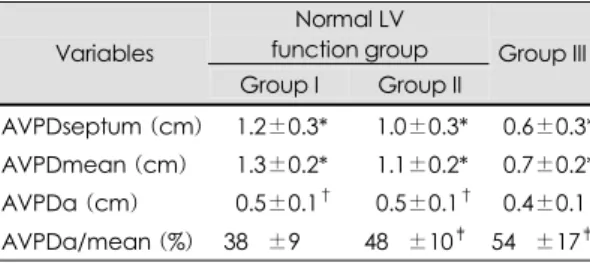
AVPD는 Group Ⅰ이 1.3±0.2 cm, Group Ⅱ가 1.1±
0.2 cm, Group Ⅲ가 0.7±0.2 cm로서 각군 모두 의미 있는 차이를 보였다(Table 2). AVPD에 대한 심방 분 획률(AVPDa/mean(%))은 Group Ⅰ이 Group Ⅱ 혹
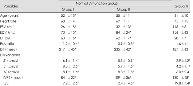
Table 1. Clinical and echocardiographic parameters
Normal LV function group Variables
Group I Group II Group III
Age (years) 052.0±15* 055.0±11 061.0±10
Heart rate 068.0±14 069.0±11 075.0±15
ESV (mL) 026.0±08* 032.0±13* 115.0±5
EDV (mL) 070.0±15* 084.0±24* 156.0±62
EF (%) 063.0±06* 062.0±07* 028.0±7
E/A ratio 001.2±00.4* 000.9±00.3* 001.6±1.1
DT (msec) 217.0±45* 235.0±42* 187.0±63 DTI variables
S’ (cm/s) 006.1±01.4† 005.1±00.9† 002.9±1.2†
E’ (cm/s) 008.8±02.6† 005.9±01.6† 004.2±1.1†
A’ (cm/s) 008.1±01.6* 008.3±01.8* 006.0±2.4
IVRT (msec) 084.0±22† 109.0±36† 130.0±48†
E/E’ 009.2±02.6† 012.4±04.5† 019.8±7.4†
ESV:end systolic volume, EDV:end diastolic volume, EF:ejection fraction, E/A ratio:ratio of early and late mitral inflow, DTI:doppler tissue image, DT:deceleration time, S’:systolic mitral annulus peak velocity, E’:early diastolic mitral annulus peak velocity, A’:late diastolic mitral annulus peak velocity, IVRT:isovolumic relaxation time, LV:left venricular, *:p<0.05 compared with Group III, †:p<0.05 compared with the other two groups
AVPD
AVPD a
B A
Fig. 1. The method of Atrioventricular plane displacement (AVPD) measurement. A:the AVPD toward and away from the cardiac apex was measured by M-mode echocardiogram from apical window. The septal and lateral AVPDs were measured in the four chamber view, anterior and inferior AVPD in the two chamber view. A mean value of the AVPD was calculated from the above four sites.
B:AVPDa/mean (%)= AVPDa
AVPDmean ×100.
AVPDa:atrial contribution of AVPD.
은 Group Ⅲ와 비교했을 때 의미있게 적었다(Table 2).
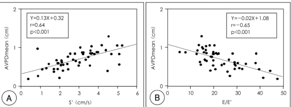
좌심실 기능이 정상인 군에서는 AVPD는 나이, E/E’, IVRT 와는 음의 상관관계, S’, E’, E/A ratio와는 양의 상관관계를 보였다. 좌심실 기능 부전군에서는 E/A ratio, E/E’이 음의 상관관계, 좌심실 구혈률, DT, S’, E’, A’,
systolic duration 이 양의 상관관계를 보였다. 단변량 분석에서 의미있는 인자들에 대해 다변량 분석을 시행 했을 때, 좌심실 기능이 정상인 군에서는 S’(β=0.4, p<
0.001), E’(β=0.5, p<0.001)이, 좌심실 기능 부전군에 서는 S’(β=0.6, p<0.001), E/E’(β=-0.3, p=0.005) 이 독립적 변수였다(Table 3)(Fig. 2, 3). 모든 군을 포함하였을 경우 S’(β=0.6, p<0.001), E/E’(β=0.3, p<0.001), 좌심실 구혈률(β=0.2, p=0.007)이 독립 적 변수였다(Table 4)(Fig. 4). AVPD의 심방 분획률은 AVPD가 증가할수록 감소하는 경향을 보였다(Fig. 5).
고 찰
본 연구는 AVPD를 좌심실 이완기 기능 평가에 사 용하는 것이 임상적으로 적합한지 알아보기 위해 현재 Table 3. Regression analysis in the normal left ventricular function group and left ventricular dysfunction group, predictive parameters for determining AVPD
Normal LV function group LV dysfunction group
Variables Univariate Multivariate Univariate Multivariate
β p β p β p β p
S’ -0.7 <0.001 0.4 <0.001 -0.6 <0.001 -0.6 <0.001
E’ -0.7 <0.001 0.5 <0.001 -0.5 <0.001
E/E’ -0.5 <0.001 -0.7 <0.001 -0.3 <0.005
IVRT -0.4 <0.001
Age -0.5 <0.001
EF -0.2 <0.05 -0.5 <0.001
E/A ratio -0.4 <0.001 -0.3 <0.017
DT -0.6 <0.001
A’ -0.5 <0.001
LV:left venricular, EF:ejection fraction, IVRT:isovolumic relaxation time, DT:deceleration time Table 2. AVPD of normal left ventricular function group
and left ventricular dysfunction group Normal LV function group Variables
Group I Group II
Group III
AVPDseptum (cm) 01.2±0.3* 01.0±0.3* 00.6±0.3*
AVPDmean (cm) 01.3±0.2* 01.1±0.2* 00.7±0.2*
AVPDa (cm) 00.5±0.1† 00.5±0.1† 00.4±0.1 AVPDa/mean (%) 38.0±9 48.0±10‡ 54.0±17‡ AVPDa:atrial contribution of AVPD, *:p<0.05 com- pared with the other two groups, †:p<0.05 compa- red with group III, ‡:p<0.05 compared with group I, AVPD:atrioventricular plane displacement
AVPDmean (cm)
2 4 6 8 10 12 S’ (cm/s)
3
2
1
0
Y=0.12X+0.55 r=0.68 p<0.001
AVPDmean (cm)
0 2 4 6 8 10 12 14 16 E’ (cm/s)
3
2
1
0
Y=0.06X+0.8 r=0.71 p<0.001
A B
Fig. 2. Correlations between AVPDmean and systolic mitral annulus peak velocity (S’), early diastolic mitral annulus peak velocity (E’) in normal left ventricular function group. AVPD:atrioventricular plane displacement.
이완기 기능 평가에 사용되는 변수들과 AVPD와의 관계 에 대해 알아 보았으며, 그 결과 AVPD는 좌심실의 이완 기 기능을 반영하는 심초음파적 인자 중 하나인 E/E’과 밀접한 연관이 있는 것으로 나타나 AVPD가 이완기 기 능을 평가하는 데 어느 정도 기여할 수 있을 것으로 여겨 진다.
AVPD는 심내막연을 잘 조영할 수 없는 경우나 비대칭 적 좌심실 수축 때문에 다른 방법으로는 심장 기능을 정확 히 측정하기 어려운 경우에 이용되어지고 있으나 많은 연 구에서 특히 허혈성 심질환이나 심부전증에서 좌심실 Table 4. Regression analysis in all patients, predictive
parameters for determining AVPD
Univariate Multivariate Variables
β p β p
S’ -0.8 <0.001 -0.6 <0.001 E/E’ -0.8 <0.001 -0.3 <0.001
EF -0.8 <0.001 -0.2 0.007
Age -0.5 <0.001
DT -0.4 <0.001
E/A ratio -0.2 <0.005
IVRT -0.4 <0.001
AVPD:atrioventricular plane displacement, EF:ejec- tion fraction, IVRT:isovolumic relaxation time, DT:de- celeration time
2
1
0
B
AVPDmean (cm)
0 10 20 30 40 60 E/E’
Y=-0.03X+1.5 r=-0.75 p<0.001
A
AVPDmean (cm)
0 2 4 6 8 10 12 S’ (cm/s)
3
2
1
0
Y=0.16X+0.29 r=0.84 p<0.001
Fig. 4. Correlations between AVPDmean and systolic diastolic mitral annulus velocity (S’), ratio of early diastolic mitral inflow and early diastolic mitral annulus velocity (E/E’) in the all patients. AVPD:atrioventricular plane dis- placement.
B
AVPDmean (cm)
0 10 20 30 40 50 E/E’
2
1
0
Y=-0.02X+1.08 r=-0.65 p<0.001
A
AVPDmean (cm)
0 1 2 3 4 5 6 S’ (cm/s)
2
1
0
Y=0.13X+0.32 r=0.64 p<0.001
Fig. 3. Correlations between AVPDmean and systolic mitral annulus peak velocity (S’), ratio of early mitral inflow and early diastolic mitral annulus peak velocity (E/E’) in the left ventricular dysfunction group.
0 1 2 AVPDmean (cm)
AVPDa/mean (%)
100
080 060 040
020
000
Y=-21.9X+69.9 r=-0.53, p<0.001
Fig. 5. Correlation between AVPDmean and AVPDa/
mean (%) in the all patients. AVPD:atrioventricular plane displacement.
구혈률 및 예후와 밀접한 관련이 있어 일반적으로 좌심 실 수축기능 평가에 많이 이용되어지고 있다.12-15)20)
본 연구에서도 AVPD는 좌심실 수축기능을 반영하 는 좌심실 구혈률이나 DTI로 측정된 최대 수축기 판륜 속도와 밀접한 연관이 있는 것으로 나타났다. 하지만 좌심실 수축기 기능이 정상인 환자에서도 나이가 증가 할수록, 심방세동이 있는 경우, 관동맥 질환이 있는 경 우 AVPD가 감소하는 경향을 보여 AVPD가 좌심실 수축 기능 이외에 이완기 기능에도 영향을 받을 것이라는 주 장이 제기되었다.5)16)17) 좌심실 구혈률은 일차적으로 심외막하, 원형근의 수축과 관련이 있는 반면에 AVPD 는 주로 심내막하, 세로근의 수축과 관련이 있다.21)22) 해 부학적인 혈류 분포로 인하여 심내막하 근섬유가 심외 막하 근섬유보다 심근 허혈에 더 취약하기 때문에 좌심 실 구혈률이 유지되는 경도의 관상 동맥 질환에서 이미 AVPD는 감소되어 있다.16) 즉 수축기 기능은 아직 손상 받지 않고 이완기 기능만이 감소한 경우에 AVPD를 측정 함으로써 이완기 기능 이상을 평가할 수 있다. Carha¨ll 등10)은 좌심실 기능이 정상인 군에서 연구한 결과 AVPD 는 좌심실 구혈률보다는 이완기말 용적등과 더 밀접한 상관 관계가 있다고 주장하였으며, Rydberg 등7)은 관 동맥 질환에서 AVPD가 좌심실 충만압의 지표로 이용 될 수 있음을 주장하였다. 또한 Alam 등23)은 심근 경 색 환자에서 이완기시 AVPD가 좌심실의 전반적인 이완 기 기능을 반영하는 인자로 이용될 수 있다고 보고하였 다. 본 연구에서도 좌심실 기능이 정상인 군에서 AVPD 는 DTI로 측정된 이완기시 초기 승모판륜 속도(E’)와 밀접한 연관(r=0.7, p<0.001)이 있었으며, 좌심실기능 부전군에서도 E ’(r=0.5, p<0.001), E/E ’(r=-0.7, p<0.001)과 밀접한 연관이 있어 AVPD가 이완기 기능 평가에 이용되어 질 수 있음을 알 수 있었다. Kranidis 등6)은 AVPD의 심방 분획률이 좌심실의 충만을 반영 하는 지표로 사용할 수 있다고 하였으며, Blomstrand 등24)도 A/E ratio와 AVPD의 심방분획률이 의미있는 상관관계를 보인다고 주장하였다. 본 연구에서도 AVPD 의 심방 분획률은 AVPD와 r=-0.53(p<0.001) 정도 로 음의 상관관계를 보였으며, DTI로 측정된 초기 이 완기 속도(E’) 혹은 E/E’과도 의미있는 상관관계(r=
-0.63, p<0.001;r=0.35, p<0.001, 자료:제시 안함) 를 보여 어느 정도는 좌심실 이완기 기능 평가에 이용되 어질 수 있음을 알 수 있었다.
결론적으로 본 연구에서 AVPD는 좌심실 기능이 정상 이거나 좌심실 기능 부전이 있는 경우 모두에서 좌심실 의 이완기 기능을 반영하는 인자로 사용이 가능하다는 것을 알게 되었다. 특히 심초음파 영상이 좋지 못해 좌심 실기능을 평가하기 힘든 환자에서 이완기 기능을 평가 하는 데 도움을 줄 수 있을 것으로 여겨진다.
본 연구의 제한점으로는 첫째, 좌심실의 이완기 기능 평가에 심도자를 통하여 측정하지 않아 AVPD가 이완 기 기능을 정확하게 반영할 수 있는지, 또한 다른 심초음 파적 인자와 비교하여 AVPD가 이완기 기능을 더 정 확하게 반영하는지를 알 수 없었다. 하지만 Nagueh 등
25)에 의하면 심초음파로 측정한 E/E’이 심도자로 측정 한 폐모세혈관압과 밀접한 연관이 있다고 알려져 있고, 또한 본 연구에서도 AVPD와 E/E’이 의미 있는 연관 이 있어 AVPD가 좌심실 이완기 기능 평가에 어느 정도 기여할 것으로 여겨진다. 둘째, 좌심실 기능 부전군에 서는 허혈성 심근병증이 포함되어 있어 국소벽운동장애 가 AVPD측정에 영향을 주었을 것으로 보인다. 이러한 문제점을 극복하기 위해 중격, 측벽, 하벽, 전벽 4군데 서 측정한 값의 평균을 취하였기 때문에 이 점을 최소화 하였을 것으로 여겨진다.
요 약
배경 및 목적:
Atrioventricular plane displacement(AVPD)는 심 장의 수축 기능을 평가하는 지표로 사용되고 있으나, 좌 심실 수축 기능이 정상인 경우에도 일부 질환에서는 감 소하는 경향이 있다. 본 연구는 AVPD와 기존의 이완기 심기능의 지표로 사용되는 심초음파 변수들, 특히 승모 판륜 속도를 이용한 도플러 조직 영상의 변수들과의 상 관 관계를 알아봄으로써 좌심실 이완기 기능을 평가함에 있어 AVPD의 임상적인 중요성에 대해 알아보기 위해 시행하였다.
방 법:
심근허혈이나 심부전 소견이 없으면서 좌심실 구혈 률이 50%를 초과한 환자를 정상 심기능 군(n=87)으로 분류하였고, 이들을 정상 심초음파군(Group Ⅰ, n=44) 과 동심성 좌심실 비대를 보인 군(Group Ⅱ, n=43)으 로 세분하였다. 심실 구혈률이 45%미만이면서 뉴욕심장 협회 분류 운동 등급 Ⅲ 이상인 환자들을 좌심실 기능
부전군(Group Ⅲ, n=51)으로 분류하였다.
AVPD는 중격, 측벽, 전벽, 하벽에서 측정하여 상기 네 곳에서 얻은 값의 평균으로 하였으며 AVPD심방 분 획은 전체 AVPD에서 심방 기여분의 비율로 계산하였다.
이면성 심초음파와 도플러 조직 영상을 통해 좌심실의 수축기 및 이완기 기능을 평가하였다.
결 과:
좌심실 기능이 정상인 군에서는 AVPD는 나이, E/E’, IVRT 와는 음의 상관관계, S’, E’, E/A ratio와는 양의 상관관계를 보였다. 좌심실 기능 부전군에서는 E/A ratio, E/E’이 음의 상관관계, 좌심실 구혈률, DT, S’, E’, A’, systolic duration이 양의 상관관계를 보였다. 다변량 분 석을 시행했을 때, 좌심실 기능이 정상인 군에서는 S’
(β=0.4, p<0.001), E’ (β=0.5, p<0.001)이, 좌심실 기능 부전군에서는 S’ (β=0.6, p<0.001), E/E’ (β=
-0.3, p=0.005)이 독립적 변수였다. 모든 군을 포함하 였을 경우 S’(β=0.6, p<0.001), E/E’(β=0.3, p<
0.001), 좌심실 구혈률(β=0.2, p=0.007)이 독립적 변 수였다.
결 론:
AVPD는 좌심실 이완 기능을 평가할 수 있는 심초음 파적 인자 중 하나로 사용 가능할 것으로 여겨진다.
중심 단어:좌심실 이완기 기능;Atrioventricular plane displacement.
REFERENCES
1) Oh JK, Appleton CP, Hatle LK, Nishimura RA, Seward JB, Tajik AJ. The noninvasive assessment of left ventricular diastolic function with two-dimensional and Doppler echo- cardiography. J Am Soc Echocardiogr 1997;10:246-70.
2) Zamorano J, Wallbridge DR, Ge J, Drozd J, Nesser J, Erbel R. Non-invasive assessment of cardiac physiology by tis- sue Doppler echocardiography: a comparison with inva- sive hemodynamics. Eur Heart J 1997;18:330-9.
3) Barbier P, Grimaldi A, Alimento M, Berna G, Guazzi MD.
Echocardiographic determinants of mitral early flow pro- pagation velocity. Am J Cardiol 2002;90:613-9.
4) Alam M, Hőglund C. Assessment by echocardiogram of left ventricular diastolic function in healthy subjects using the atrioventricular plane displacement. Am J Cardiol 1992;69:565-8.
5) Willenheimer R, Israelsson B, Cline C, Rydberg E, Broms K, Erhardt L. Left atrioventricular plane displacement is related to both systolic and diastolic left ventricular per- formance in patients with chronic heart failure. Eur Heart J 1999;20:612-8.
6) Kranidis A, Kostopoulos K, Anthopoulos L. Evaluation of
left ventricular filling by echocardiographic atrioventri- cular plane displacement in patients with coronary artery disease. Int J Cardiol 1995;48:183-6.
7) Rydberg E, Willenheimer R, Brand B, Erhardt LR. Left ven- tricular diastolic filling is related to the atrioventricular plane displacement in patients with coronary artery dis- ease. Scand Cardiovasc J 2001;35:30-4.
8) Slager CJ, Hooghoudt TE, Serruys PW, Schuurbiers JC, Reiber JH, Meester GT, Verdouw PD, Hugenholtz PG.
Quantitative assessment of regional left ventricular motion using endocardial landmarks. J Am Coll Cardiol 1986;7:
317-26.
9) Willenheimer R, Cline C, Israelsson B. Left ventricular atriovetricular plane displacement: an echocardiographic technique for rapid assessment of prognosis in heart failure. Heart 1997;78:230-6.
10) Carhäll CJ, Lindstrom L, Wranne B, Nylander E. Atrioven- tricular plane displacement correlates closely to circula- tory dimensions but not to ejection fraction in normal young subjects. Clin Physiol 2001;21:621-8.
11) Hőglund C, Alam M, Thorstrand C. Atrioventricular valve plane displacement in healthy persons. Acta Med Scand 1988;224:557-62.
12) Alam M, Hőglund C, Thorstrand C. Longitudinal systolic shortening of the left ventricle: an echocardiographic study in subjects with and without preserved global function.
Clin Physiol 1992;12:443-52.
13) Alam M, Hőglund C, Thorstrand C, Philip A. Atrioventri- cular plane displacement in severe congestive heart failure following dilated cardiomyopathy or myocardial infarction.
J Intern Med 1990;228:569-75.
14) Jarnert C, Mejhert M, Ring M, Persson H, Edner M. Do- ppler tissue imaging in congestive heart failure patients due to diastolic or systolic dysfunction: a comparison with Doppler echocardiography and the atrio-ventricular plane displacement technique. Eur J Heart Fail 2000;2:151-60.
15) Willenheimer RB, Erhardt LR, Cline CM, Rydberg ER, Israelsson BA. Prognostic significance of changes in left ventricular systolic function in elderly patients with con- gestive heart failure. Coron Artery Dis 1997;8:711-7.
16) Rydberg E, Willenheimer R, Erhardt L. Left atrioventricular plane displacement at rest is reduced in patients with cor- onary artery disease irrespective of prior myocardial in- farction. Int J Cardiol 1999;69:201-7.
17) Alam M, Thorstand C. Left ventricular function in patients with atrial fibrillation before and after cardioversion. Am J Cardiol 1992;69:694-6.
18) Levy D, Savage DD, Garrison RJ, Anderson KM, Kannel WB, Castelli WP. Echocardiographic criteria for left ventricular hypertrophy. Am J Cardiol 1987;59:956-60.
19) Sahn DJ, de Maria A, Kisslo J, Weyman A. Recommenda- tions regarding quantitation in M-mode echocardiography:
results of a survey of echocardiographic measurements.
Circulation 1978;58:1072-83.
20) Cosme O, Grodman RS. Estimation of left ventricular sys- tolic function by nonvolumetric echocardiographic analysis in subjects with poor left ventricular visualization. Clin Cardiol 1997;20:247-51.
21) Lundbäck S. Cardiac pumping and the function of the ven- tricular septum. Acta Physiol Scand 1986;(Suppl 550): 1-101.
22) Sokolow M, McIlroy MB. Clinical cardiology. 4th ed. Los Altos: Lange Medical publications; 1986. p.21.
23) Alam M, Wardell J, Andersson E, Samad BA, Nordlander R.
Left ventricular regional diastolic dysfunction in patients with first myocardial infarction determined by diastolic motion of the atrioventricular plane. Echocardiography 1999;16:635-41.
24) Blomstrand P, Kongstad O, Broqvist M, Dahlström U, Wranne B. Assessment of left ventricular diastolic dys-
function from mitral annulus motion, a comparison with pulsed Doppler measurements in patients with heart failure.
Clin Physiol 1996;16:483-93.
25) Nagueh SF, Middleton KJ, Kopelen HA, Zoghbi WA, Qui- ñones MA. Doppler tissue imaging: a noninvasive tech- nique for evaluation of left ventricular relaxation and estimation of filling pressures. J Am Coll Cardiol 1997;
30:1527-33.