DOI: http://doi.org/10.15205/kschs.2018.03.31.1089
Analysis on the corneal thickness and anterior chamber depth of the Keratoconus using Pentacam
6)
Douk-Hoon Kim, MD, FIACLE
1*; Kishor Sapkota, PhD, MSc, FAAO, FIACLE
21
Department of Optometry, Masan University, Korea
2
University of Minho, Portugal
(Received January 30, 2018; Received February 10, 2018; Accepted February 20, 2018) Abstract
Purpose: To investigate the corneal thickness and anterior chamber depth (ACD) of eyes with Keratoconus in the Korean population with the Pentacam .
Methods: The subjects consisted of 84 eyes from Keratoconic adults aged 7-59 years during 2010. The thinnest area, apex zone, and pupil centre of the corneal thickness were measured using the Pentacam pachymetry. ACD value was also measured with Pentacam.
Results: There was a statistically significant relationship between thickness of the cornea at the apex area and the pupil centre (p=0.0001). However, there was no statistical difference (p>0.5) in the mean thick- ness of thinnest area, apex zone, pupil centre of cornea between right eye and left eye. Also, correlation between ACD and corneal thickness in all subjects had no statistical differences (p>0.05) in all subjects.
Conclusion: These results suggested that the regional thickness of cornea and ACD with Pentacam can provide correct and useful diagnostic information of the morphology of Keratoconus for the RGP contact lens and diagnosis of abnormal corneal refraction surgery.
Key words: ACD, apex area, Pentacam system, pupil center zone, thinnest area
*Corresponding author : [email protected]
1. Introduction
Keratoconus has a congenital deformation of the anterior corneal morphology leading to myopia, irregular astigmatism, and eventually to a marked decrease in visual acuity
1-2).
For the clinical keratoconus research and diagnosis, the topographic system has provided excellent information about the fine structure of the cornea. The data acquired by topography tools has suggested significant important information in clinical diagnosis and management of keratoconus suspects
3), axial myopia
4), contactlenswear
5), and keratoplasty
6). Ccorneal thickness of each region is an indicator of corneal hydration and metabolism
7).
On the other hand, the topographic results of the central corneal thickness have been used to analyze the diagnosis of the glaucoma
8)and intraocular pressure
9). Distribution of the central and paracentral corneal thickness and curvature has been studied in the European population
10). Especially, Age related changes in the central and peripheral corneal thickness seem to give the useful information to keratoplasty and refractive surgery
11). Recently, the relationship between the central corneal thickness and the anterior corneal curvature has been studied through the pachymetry system
10).
Furthermore, ethnic differences in the central corneal thickness have been described throughout the European, Hong Kong Chinese, Chinese, Japanese and Americans
8). Also, data on corneal pachymetry maps have been reported in Korean keratoconus population
12-13). However, thethinnest, apex, and pupil center corneal thickness and anterior chamber depth values of Korean keratoconus have not been reported yet. In this study, we research characteristics of the central, apex, thinnest corneal thickness and anterior chamber depth in Korean keratoconus by using
pachymetry data obtained from the Pentcam system. The purpose of our study was therefore to analysis in the thinnest, apex and pupil center corneal thickness and anterior chamber depth in Korean keratoconus subjects by the Pentacam system.
2. Materials and Methods
2.1. Subjects
This study included 84 keratoconus eyes of forty-nine Korean subjects, 35 had bilateral keratoconus and 14 had unilateral keratoconus.
Keratoconus was diagnosed with both subjective criteria like stromal thinning, Vogt striae, iron ring, munson sign, scissoring on retinoscopy as well as with objective criteria like corneal curvature 47.0D or more on SimK. They have no ocular disease, medication, systemic disease, contact lens wear, and refractive surgery. The subject's age ranged from 7 to 59 years. To exclude ocular disease, we tested the visual acuity and the IOP, and examined the slit-lamp and the funduscopy.
2.2. Examination
The corneal thickness and ACD values at a
distance of 5mm from central cornea were
obtained by measurements of the Pentacam
topography system (Bausch & Lomb, USA). The
measurements were obtained for each eye. The
Pachymetry was used for corneal topography
measurements while the subjects were silently
seated in a test room. The subjects were asked to
keep both eyes open and focus on a light source
in the center of the scan field. The measurements
automatically operated when the correct alignment
and focus of eye were achieved. The thicknesses
at the thinnest area, apex zone, and pupil center
of cornea were recorded automatically and analyzed by a SPSS 14 and a micro-soft excel program. All measurements were made from 10:00 A.M. to 12:00 A.M. During the test, the temperature ranged from 18
oto 21
oC, and the humidity ranged from 40% to 50%. Only the scans with the quality factor of 95% or more were selected for the analysis.
2.3. Data analysis
Analyses were performed by using SPSS software. Descriptive data were expressed as mean±standard deviation. The Pearson correlation and t-test were used to compare measurements in three zones of corneal thickness and ACD. P
values of less than 0.05 was considered as statistically significant.
3. Results
We acquired the corneal topographic data from 84 eyes (42 OD, 42 OS) with the mean age of 24.8±9.9 years (range 7-59 years). The mean age for the eyes was 24.3±9.2 years (range 7-54 years) for OD and 25.2±10.7 years (range 7-59 years) for OD. There was no significant difference in the mean age between OD and OS (p>0.523).
The OD and OS distributions of the thinnest area, apex zone and pupil center of the corneal thickness for these subjects were summarized in Table 1, and figure 1.
Item OD OS P-values
Ages 24.34±9.246 25.2±10.743 p=0.54
TA 484.90±72.644μm 482.075±64.391 μm p=0.836
AT 495.80±72.374μm 494.375±55.666 μm p=0.914
PZ 506.02±65.645μm 495.325±53.633μm p=0.571
ACD 3.33±0.376 mm 3.308±0.359mm p=0.778
thinnest corneal thickness, PA: apex zone thickness, PZ: pupil zone thickness, ACD: anterior chamber depth.
Table1. The distribution of the corneal thicknesses and ACD values in Korean subjects.
Fig.1. Typical keratoconus thickness with
Pentacam topography system.
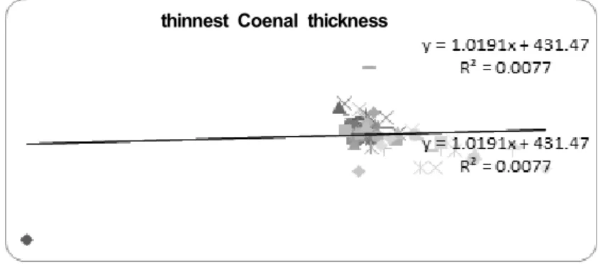
The average thinnest corneal thickness was 484.900±72.644μm (range, 549-406μm) and 482.075±64.391μm(range, 428-450μm) for OD and OS, respectively. The mean thinnest corneal thickness of all subjects was 483.489 μm (Figure 2).
There was no significant difference in the mean thinnest corneal thickness between OD and OS (P=0.836, T-test). Also the mean apex corneal thickness was 495.80±72.374μm and 494.375±
55.666μm for OD and OS, respectively. There was no significant difference in mean apex corneal thickness between OD and OS (p=0.914).
Also the average pupil center thickness was 506.02±65.645μm and 495.325±53.633μm for OD and OS, respectively. There was no significant difference in mean pupil center thickness between OD and OS (p=0.571). On the other hand, mean of anterior corneal depth was 3.33±0.376 mm and 3.308±0.359mm for OD and OD. There was no significant difference in mean anterior chamber depth between OD and OS (p=0.778). However, correlation between ACD and corneal thickness in all subjects was had strong statistical differences (p>0.05) in all subjects.
thinnest Coenal thickness
Fig. 2. The thinnest corneal thickness of keratoconus subjects
Table 2 showed that the correlation between thinnest cornea and anterior chamber depth had a statistical significance differences in OD (p=0.026) and OS (p=0.004, t-test). On the other hand, Table 2 showed that the correlation between apex region and anterior chamber depth had a statistical significance differences in OD (p=0.035, t-test) and OS (p=0.010). At the same
time, Table 2 showed that the correlation between pupil zone and anterior chamber depth had a statistical significance differences in OD (p=0.009, t-test) and OS (p=0.004). As a results, in all subjects, Table 2 showed that the correlation between corneal thickness and ACD had strong significant differences (p>0.1).
Item TA & ACD AT & ACD PCT & ACD
OD 0.026* 0.035* 0.009
OS 0.004* 0.010* 0.004*
*p<0.5
TA: thinnest corneal thickness, PA: apex thickness, PZ: pupil center thickness