Korean J Gastroenterol Vol. 69 No. 1, 83-86 https://doi.org/10.4166/kjg.2017.69.1.83 pISSN 1598-9992 eISSN 2233-6869
CASE REPORT
Korean J Gastroenterol, Vol. 69 No. 1, January 2017 www.kjg.or.kr
만성 췌장염에 동반된 췌장성지방층염
김의중, 주민수, 손기창, 조동호, 나가혜
1, 김학철, 조은영
원광대학교 의과대학 내과학교실, 피부과학교실1
Pancreatic Panniculitis in Patients with Chronic Pancreatitis: Case Report and Review of Literature
Eui Joong Kim, Min Su Chu, Ki Chang Sohn, Dong Ho Cho, Ga Hye Na1, Haak Cheoul Kim, and Eun Young Cho Departments of Internal Medicine and Dermatology1, Wonkwang University College of Medicine, Iksan, Korea
Pancreatic panniculitis is a rare complication characterized by subcutaneous fat necrosis associated with pancreatic disease. It has been postulated that pancreatic panniculitis is caused by the systemic activity of pancreatic enzymes that lead to microcirculatory disturbances. We report a 41-year-old heavy alcoholic woman with pancreatic panniculitis that coexisted with acute and chronic pancreatitis. She was diagnosed with chronic pancreatitis and alcoholic liver cirrhosis 5 years ago. She presented with multiple, ten- der, erythematous, subcutaneous nodules with heat sensation on both lower legs. Laboratory evaluation revealed an increase in the serum blood amylase and lipase. Histopathologic findings showed fat necrosis with inflammation around the necrotic subcutaneous fat tissue. The lesions subsided gradually with an improvement of acute pancreatitis. (Korean J Gastroenterol 2017;69:83-86) Key Words: Panniculitis; Chronic pancreatitis; Subcutaneous fat necrosis
Received August 28, 2016. Revised November 8, 2016. Accepted December 5, 2016.
CC This is an open access article distributed under the terms of the Creative Commons Attribution Non-Commercial License (http://creativecommons.org/licenses/
by-nc/4.0) which permits unrestricted non-commercial use, distribution, and reproduction in any medium, provided the original work is properly cited.
Copyright © 2017. Korean Society of Gastroenterology.
교신저자: 조은영, 54538, 익산시 무왕로 895, 원광대학교병원 내과
Correspondence to: Eun Young Cho, Department of Internal Medicine, Wonkwang University Hospital, 895 Muwang-ro, Iksan 54538, Korea. Tel: +82-63-859-2566, Fax: +82-63-855-2025, E-mail: [email protected]
Financial support: None. Conflict of interest: None.
서 론
지방층염(panniculitis)은 지방 조직 내의 염증으로 주로 피하지방층에 생기며 다양한 질환과 연관되어 발생한다.1 췌 장성지방층염(pancreatic panniculitis)은 췌장질환과 관련하 여 나타나는 드문 피부 병변으로 주요한 췌장질환 환자들의 0.3-3%에 걸쳐 발생하며, 주로 급성 또는 만성 췌장염 환자에 서 나타난다.2,3 혈액검사, 방사선학적 검사를 통한 췌장질환 의 확인과 함께 피부 병변의 병리조직학적 소견 즉, 피하 지방 의 염증 및 괴사 소견을 종합해 이 질환을 진단할 수 있으며,4 이 경우 관절염 등의 증상이 흔히 동반된다.5,6대다수의 췌장 성지방층염 증례에서는 췌장성지방층염이 췌장질환 발생 전 에 나타나 췌장성지방층염은 췌장염진단에 진단적 가치가 있 는 것으로 보고하고 있다. 저자들은 만성췌장염환자에서 발생
한 췌장성지방층염을 진단하고 치료한 예를 경험하였기에 췌 장성지방층염의 임상적 의의에 대하여 문헌고찰과 함께 보고 하는 바이다.
증 례
41세 여자가 수일 전부터 시작된 복부팽만과 상복부 불편 감으로 타 병원 입원 중 췌장 수치의 급격한 상승을 보여 본원 으로 전원되었다. 십수 년 전부터 거의 매일 소주 2병씩 마신 음주력이 있었으며, 알코올성 간경화, 만성 췌장염, 당뇨병 및 우울증으로 본원 외래를 통한 치료 중이었다. 내원 시 혈압 100/70 mmHg, 맥박 114회/분, 체온 36.8℃였고 만성 병색 소견을 보였으며 신체검사에서 상복부에 경미한 압통이 있었 으나 반발통은 없었고 복부 청진상 장음 감소가 관찰되었다.
84
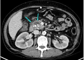
김의중 등. 만성 췌장염에 동반된 췌장성지방층염The Korean Journal of Gastroenterology Fig. 1. Low attenuating lesion with multiple calcifications &
peripancreatic fat infiltration (arrows) is seen at head portion of pancreas.
Fig. 2. Multiple, ill-defined, erythematous to brownish nodules are shown on both lower legs.
Fig. 3. The skin biopsy showing focal areas of adipocytes degeneration surrounded by inflammatory cells infiltration (H&E, ×100).
말초 혈액 검사에서 백혈구는 7,120/mm3 (중성구 68.9%), 혈 색소 9.9 g/dL, 혈소판 82,000/mm3였고 혈액 생화학 검사에 서 혈청 알부민 4.6 g/dL, AST 53 IU/L, ALT 30 IU/L, ALP 390 IU/L, Amylase 1,003 IU/L, Lipase 4,130 IU/L, CA19-9 96.1 U/mL, CRP 2.04 mg/L였다. 만성 췌장염의 급성악화를 확인하기 위해 실시한 복부컴퓨터촬영(computed tomog- raphy)에서 췌장 두부의 다발성 석회화와 췌장 가성 낭종 및 췌장주위의 지방침윤을 동반한 소견을 보여(Fig. 1) 만성 췌장 염에 동반된 급성 췌장염 진단 하에 금식과 함께 수액치료를 실시하였다. 입원 다음날 환자가 양쪽 pretibial area에 발적 과 부종 및 통증을 호소하고 그 부위에 열감과 압통을 호소해 봉와직염을 의심하고 경험적으로 1세대 cephalosporin을 투 여하였으나 입원 3일째 여전히 양쪽 pretibial area에 발적과
부종 및 통증을 호소하며 다수의 적갈색 결절성 병변이 관찰 되어 피부과에 의뢰하여 조직검사를 실시하였다(Fig. 2). 적갈 색의 결절성 병변의 조직검사 결과 피하지방의 결절상 지방 괴사를 보였고(Fig. 3) 이는 지방층염에 합당한 소견이었다.
이에 따라 지방층염 발생과 관련 있는 약물 복용력 및 감염 소견 등을 확인하였으나 특이 소견은 없었고, 췌장염 외에 동 반된 전신 질환이 없어서 췌장염에 의한 췌장성지방층염으로 진단할 수 있었다. 이에 따라 항생제 치료를 중단하고 췌장염 에 준한 일반적 수액치료를 실시하였다. 이후 상복부 통증 소 실과 췌장효소 수치의 감소를 보였고 하지의 피부병변도 호전 되어 퇴원하였으며, 퇴원 3주 후 외래 추적 관찰에서 피부병 변은 거의 호전되어 염증 후 과다색소침착만 보였으며 3개월 후 외래방문 시는 피부병변은 완전히 소실되었고 간경변과 만 성췌장염에 대해 외래 추적 중이다.
고 찰
췌장성지방층염은 급성 또는 만성췌장염, 췌장암, 췌장이 상, 가성낭종과 동반되어 나타나며, 내시경 역행 췌담관 조영 술 후에 생긴 경우도 보고되고 있다.7대개 중년에서 남자:여 자가 2:1 비율로 발생하지만, 소아에서도 발생할 수 있다.8췌 장염이 동반된 경우는 더 젊은 나이에서 발생하였으며 알코올 중독과 연관된 경우가 많았다.9
췌장성지방층염의 발생기전은 명확히 밝혀지지 않았지만, 아밀라아제, 리파제, 포스포릴라아제키나아제, 트립신 같은 췌장효소가 관련된 것으로 보인다.3혈액 내로 유리된 췌장효 소들은, 림프관 같은 미세순환의 투과성증가로 인해 피하지방 으로 들어가게 된다.5리파제 또는 아밀라아제는 중성지방을 가수분해하여 글리세롤과 유리지방산을 만들고 결국 지방괴 사와 염증을 유발한다. 피하지방 괴사조직에서 리파제와 항-
Kim EJ, et al. Pancreatic Panniculitis and Acute Exacerbation of chronic pancreatitis
85
Vol. 69 No. 1, January 2017 Table 1. Cases of Pancreatic Panniculitis Published in the Korean Literatures
Patient Age Sex Underlying disease Location
of skin lesion
Onset of skin lesion (Interval between skin lesion
and underlying disease)
Resolution of skin lesion
116 67 Female Pancreatic cancer Legs, hip, chest 3 months ago After a month
26 47 Male Chronic pancreatitis Legs 4 days ago After a week
317 50 Male Acute pancreatitis Legs 3 weeks ago Death after17days
42 72 Male Necrotizing pancreatitis Right leg Within a week After treamtment of
underlying disease
52 48 Male Pancreatic pseudocyst Legs With 5 days After treamtment of
underlying disease
63 63 Female Acute pancreatitis Legs 10 days ago After 3 weeks
79 45 Male Chronic pancreatitis Legs A week ago After treamtment of
underlying disease
812 54 Male Pancreatic cancer Legs, arms 9 months ago After operation of underlying
disease 919 32 Male Pancreatic pseudocyst,
hemorrhagic
Abdomen 15 days ago Death after 19 days
리파제 단클론 항체의 존재 확인을 통해서도 뒷받침된다.10 하 지만 췌장질환의 빈도에 비해 췌장성 지방층염의 빈도가 매우 낮은 점으로 미루어 볼 때 다른 여러 요인들이 작용할 것으로 생각된다. 더욱이 실험적으로, 정상인의 피하지방을 환자의 혈청과 배양했을 때, 지방괴사는 관찰되지 않았고, 정상 췌장 효소 수치를 보인 환자에게서 췌장성지방층염이 나타난 예도 있어 이러한 면을 뒷받침한다.9
췌장성지방층염의 피부병변은 다발성의 통증을 동반한 피 하결절로 나타나며 지방의 액화괴사로 인해 기름기 있는 황갈 색 물질이 배출되기도 한다.9호발 부위는 다리의 원위부, 특 히 발목과 무릎에 발생하며 그 외에 허벅지, 종아리, 팔, 복부 등에도 발생할 수 있다.11 급성 혹은 만성 췌장염에 동반된 지방층염은 췌장염의 호전상태에 따라 피부병변도 서서히 소 실되지만, 췌장암에 동반되는 경우는 만성적이고 지속적이며 잦은 재발을 보이고 궤양의 형성이나 하지 이외의 다른 부위 의 침범도 더 흔하게 나타난다.10
췌장성지방층염에서 급성 관절통과 부기는 또 다른 흔한 증상이다.12 이는 일차적인 윤활막염이 아닌, 관절 주위 지방 괴사로 발생한 것이며,13 관절 천자검사에서 아밀라아제, 리파 제 및 유리지방산을 포함한 무균, 크림색의 물질로 뒷받침된 다.14 침범 관절 부위는 피부병변 분포와 유사하며,12 가장 흔 한 침범부위는 발목관절인데 이는 진단적으로 중요한 의미가 있으며, 췌장염과 동반된 경우의 25%, 췌장암과 동반된 경우 35%에서 관절통이 동반된 것으로 보고되었고, 이런 경우에 일반적인 관절염 치료로 호전되지 않기 때문에 기저 췌장 질 환의 치료가 필수적이며,9 일반적으로 치료는 기저 췌장질환 을 교정해주고 지지적 치료를 병행해야 한다.3,14
췌장성지방층염의 진단은 췌장질환의 존재와 특징적인 피 부 조직 병리학적 소견으로 내릴 수 있다.5주된 소견으로 일
반적으로 혈관염을 동반하지 않는 소엽 지방층염과 응고 괴사 로 인해 특징적인 두꺼운 벽과 무핵으로 이루어진 유령 모양 세포들이 나타난다. 이 유령 모양 세포는 리파제에 상대적으 로 저항성을 나타내어 형성되는 모양이며, 비정상 조직 석회 화로 세포질 내부에는 호염기성 미세과립들이 침착되어 있다.
다시 말하면 이러한 미세과립들은 활성화된 췌장효소들에 의 해 지방의 비누화가 발생한 후 지방산에 칼슘이 침착되어 형 성되는데, 이들은 모두 췌장성지방층염의 특이소견이다. 그러 나 매우 초기단계에는 중격의 혈관에 있는 혈관내피세포의 손 상으로 인해 중격 지방층염을 보일 수 있으며 이후 손상된 혈관벽으로 췌장효소들이 혈관에서 지방으로 유출되어 지방 세포의 응고 괴사 소견을 보이며, 초기단계에는 호중구 침윤 을 볼 수도 있다. 오래된 병변에서는 유령 모양 세포와 지방괴 사가 뚜렷하지 않고 육아종성염증으로 대체되어 포말세포와 다핵거대세포가 주로 나타날 수 있으며, 섬유화와 지방위축을 관찰할 수 있다.9,11,14
지방층염은 췌장질환뿐 아니라 다양한 질환, 약물, 감염증 에 의해 생길 수 있으므로, 루푸스지방층염, 알파항트립신결 핍증, 결절성다발성동맥염, 웨버크리스찬병, 알레르기성혈관 염, 결절홍반, 경결홍반, 경구 피임약 등의 약물 복용력, 연쇄 구균 감염 및 상기도 감염 등과의 연관성을 확인하여야 한
다.9,15-17 본 증례는 기존 만성 췌장염 환자에서 급성악화로
입원치료 중 통증과 부기를 동반한 결절성 피하병변을 호소하 여 조직검사를 실시하였고 그 결과 지방층염의 특징적인 소견 이 확인되어, 이후 지방층염과 연관될 수 있는 타 질환들과 병력들을 배제한 후 만성 췌장염의 급성 악화와 연관된 췌장 성지방층염으로 진단하였다.
췌장성지방층염의 약 40%는 췌장질환에 선행하여 나타나 기 때문에 췌장질환을 진단하는데 의의가 있을 수 있다.6,18
86
김의중 등. 만성 췌장염에 동반된 췌장성지방층염The Korean Journal of Gastroenterology
현재까지 보고된 대부분의 국내 증례들에서 췌장염에 선행하 여 하지에 피부병변이 발생하였고(Table 1),2,3,6,9,12,16,17,19
국외 보고에서도 췌장성지방층염 증례의 48.9%가 췌장질환보다 피부병변이 선행한다고 하였다.20 그러나 최근 내시경적 역행 성 담도체관 조영술 및 내시경적 유두괄약근 절개술 후 발생 한 췌장성지방층염 증례들이 보고되고 있는데, 이 경우 피부 증상은 췌장염 발생과 동시에 나타나거나 일부는 췌장염 발생 이후에 나타나기도 하였으며 모두 췌장염에 대한 보전적 치료 로 호전되었다.7,19
본 증례는 앞서 발표된 증례들과는 다르게 만성 췌장염의 급성 악화로 진단되어 입원치료 중 피부병변이 발생하였다.
췌장성지방층염을 췌장염의 전구 징후로 보는 경향이 있으나, 본 증례와 같이 중복 이환 또는 질병경과 중에도 발생할 수 있다. 따라서 췌장질환이 있고 본 증례와 같은 피부병변을 호 소하는 경우 췌장성지방층염을 적극적으로 의심해야 하고, 췌 장성지방층염은 피부병변에 대한 직접적인 치료 없이 원인이 되는 췌장질환에 대한 치료로 호전되므로 원인질환에 대한 적 절한 치료와 피부병변 소실여부에 대한 관찰이 필요하겠다.
REFERENCES
1. Kim YJ, Uhm WS, Kim TH, Lee CW, Park CK, Jun JB. Lobular pan- niculitis in a patient with dermatomyositis. J Korean Rheum Assoc 2008;15:76-80.
2. Rongioletti F, Caputo V. Pancreatic panniculitis. G Ital Dermatol Venereol 2013;148:419-425.
3. Laureano A, Mestre T, Ricardo L, Rodrigues AM, Cardoso J.
Pancreatic panniculitis - a cutaneous manifestation of acute pancreatitis. J Dermatol Case Rep 2014;8:35-37.
4. Park HY, Ahn SY, Goo JW, Choi EH. Two cases of pancreatic panniculitis. Korean J Dermatol 2008;46:126-129.
5. Hong JS, Lee SY, Park J, et al. A case of pancreatic panniculitis.
Korean J Dermatol 2008;46:674-677.
6. Kim CW, Moon SK, Shin DH, Choi JS, Kim KH. A case of pancreatic panniculitis. Korean J Dermatol 2007;45:599-602.
7. Fernández-Jorge B, García-Silva J, Almagro M, Ruzo JS, Rey EC,
Fonseca E. Pancreatic panniculitis after endoscopic retrograde pancreatography with sphincterotomy. Am J Gastroenterol 2007;
102:463-464.
8. Kitagami H, Kondo S, Hirano S, Kawakami H, Egawa S, Tanaka M. Acinar cell carcinoma of the pancreas: clinical analysis of 115 patients from the Pancreatic Cancer Registry of Japan Pancreas Society. Pancreas 2007;35:42-46.
9. Kim SH, Chang SH, Park HJ. A case of pancreatitis presenting with pancreatic panniculitis. Korean J Pathol 2009;43:566-569.
10. Requena L, Sánchez Yus E. Panniculitis. Part II. Mostly lobular panniculitis. J Am Acad Dermatol 2001;45:325-361.
11. Moro M, Moletta L, Blandamura S, Sperti C. Acinar cell carcinoma of the pancreas associated with subcutaneous panniculitis. JOP 2011;12:292-296.
12. Zheng ZJ, Gong J, Xiang GM, Mai G, Liu XB. Pancreatic panniculitis associated with acinar cell carcinoma of the pancreas: a case report. Ann Dermatol 2011;23:225-228.
13. Narváez J, Bianchi MM, Santo P, et al. Pancreatitis, panniculitis, and polyarthritis. Semin Arthritis Rheum 2010;39:417-423.
14. Garcia-Romero D, Vanaclocha F. Pancreatic panniculitis. Dermatol Clin 2008;26:465-470.
15. Lyon MJ. Metabolic panniculitis: alpha-1 antitrypsin deficiency panniculitis and pancreatic panniculitis. Dermatol Ther 2010;
23:368-374.
16. Kim HJ, Lee KG. Subcutaneous fat necrosis associated with pan- creatic adenocarcinoma: a case report. Korean J Pathol 1996;
30:155-160.
17. Lee WS, Kim MY, Kim SW, Paik CN, Kim HO, Park YM. Fatal pancre- atic panniculitis associated with acute pancreatitis: a case report. J Korean Med Sci 2007;22:914-917.
18. Zundler S, Erber R, Agaimy A, et al. Pancreatic panniculitis in a patient with pancreatic-type acinar cell carcinoma of the liv- er--case report and review of literature. BMC Cancer 2016;
16:130.
19. Jang YS, Kim MS, Park CS, Park JY, Park NH. Pancreatic pan- niculitis associated with acute pancreatitis and hemorrhagic pseudocysts: a case report. J Korean Soc Radiol 2012;67:249-251.
20. Sharma M, Reddy DN, Kiat TC. Endoscopic retrograde chol- angiopancreatography as a risk factor for pancreatic panniculitis in a post-liver transplant patient. ACG Case Rep J 2014;2:36-38.