Abstract (J Korean Assoc Oral Maxillofac Surg 2011;37:396-402)
Ⅰ.
서 론
암종의 발생 및 성장 과정에는 다양하고 복잡한 요인들 이 작용하며 아직까지도 많은 부분들이 명확히 밝혀지지 않았다. 하지만, 분자생물학 분야의 발전과 함께 암종과 관 련된 여러 인자들 중 특히 유전자 변이에 대한 연구가 활발
히 시행되었으며 현재 수백 개 이상의 유전자가 암의 발생 및 성장에 관여한다고 알려져 있다1. 그 중에서 상피성장인 자(epidermal growth factor, EGF)는 세포 표면의 수용체(epi- dermal growth factor receptor, EGFR)와 결합하여 다양한 형 태의 세포 증식을 유도할 수 있는 강력한 분열 유발성 능력 을 갖고 있는 단백질로써 Cohen 등이 생쥐의 악하선으로부 터 추출한 이후 사람의 urine, 타액, 세포외액에서도 추출하 게 되었다2,3.
상피성장인자 수용체(EGFR)는 다양한 세포의 세포막에 존재하는 분자량 170 kDa의 막투과성 당단백질이다4. 상피 성장인자 수용체는 상피의 증식이 빠르게 일어나는 여러 종류의 상피세포와 상피조직에서 관찰된다5. 상피성장인 자와 세포의 세포막에 존재하는 상피성장인자 수용체와의 김 경 욱
330-716 충남 천안시 신부동 산7-1번지
단국대학교 치과대학 부속치과병원 구강악안면외과 Kyung-Wook Kim
Department of Oral and Maxillofacial Surgery, Dental Hospital, Dankook University
San 7-1, Sinbu-dong, Choenan, 330-716, Korea TEL: +82-41-550-1994 FAX: +82-41-551-8988 E-mail: kkwoms@dku.edu
구강편평상피암종에서 상피성장인자 수용체의 과발현과 K-ras 유전자 변이
문병출1∙한세진1∙정동준2∙김경욱1
1단국대학교 치과대학 구강악안면외과학교실, 2순천향대학교 의과대학 병리학교실
Epidermal growth factor receptor overexpression and K-ras mutation detection in the oral squamous cell carcinoma
Byeong-Chool Moon1, Se-Jin Han1, Dongjun Jeong2, Kyung-Wook Kim1
1Department of Oral and Maxillofacial Surgery, School of Dentistry, Dankook University
2Department of Pathology, School of Medicine, Soonchunhyang University, Cheonan, Korea
Introduction: Epidermal growth factor is a single-chain polypeptide consisting of 53 amino acids with potent mitogenic activity that stimulates the proliferation of a range of normal and neoplastic cells through an interaction with its specific receptor (epidermal growth factor receptor, EGFR). This interaction plays a key role in tumor progression including the induction of tumor cell proliferation. An increased EGFR copy number have been asso- ciated with a favorable response to EGFR tyrosine kinase inhibitors therapy. In contrast, K-ras mutations tend to predict a poor response to such thera- py. The aim of this study was to determine the correlation between the clinicopathological factors and the up-regulation of EGFR expression and K- ras mutations in oral squamous cell carcinoma.
Materials and Methods: This study examined the immunohistochemical staining of EGFR, K-ras mutation detection with peptide nucleic acid (PNA)-based real-time polymerase chain reaction (PCR) clamping in 20 specimens from 20 patients with oral squamous cell carcinoma.
Results: 1. In the immunohistochemical study of poorly differentiated and invasive oral squamous cell carcinoma, a high level of EGFR staining was observed. The correlation between immunohistochemical EGFR expression and histological differentiation, as well as the tumor size of the specimens was significant (Pearson correlation analysis, significance [r] >0.5, P<0.05). 2. In PNA-based real-time PCR clamping analysis, a K-ras mutation was not detected in all specimens.
Conclusion: These findings suggest that the up-regulation of the EGFR may play a role in the progression and invasion of oral squamous cell carci- noma that is, independent of a K-ras mutation.
Key words: Squamous cell carcinoma, Epidermal growth factor receptor, ras Proteins
[paper submitted 2011. 5. 6 / revised 2011. 9. 30 / accepted 2011. 9. 29]
결합은 궁극적으로 DNA의 복제와 세포 분열을 야기시키 며, 이런 특성이 종양 세포에서 증식 및 발암 기전에 중요 한 역할을 하는 것으로 생각되고 있다.
한편, 구강암은 사람에게 발생되는 모든 암종 중 약 2.65% 정도를 차지하며, 구강암 중 구강편평세포암종이 가 장 빈발하고 전이 및 재발률이 높아 장기 생존율이 50% 미 만일 정도로 예후가 불량한 양태를 보인다6-8. 이에 대해 외 과적 수술 및 방사선 치료가 주로 사용되고 있으며, 치료 기술의 많은 발전에도 불구하고 국소적 재발과 전이로 인 하여 그 예후가 크게 개선되지 않았다9-11.
예후 개선을 위한 노력으로 최근에는 수술적인 치료 방 법과 병행하여 다양한 항암치료 요법이 사용되고 있다. 그 중 EGFR tyrosine kinase inhibitors therapy는 암세포의 EGFR tyrosine kinase를 억제하는 small molecules를 투여하 여 kinase의 활성을 방해하고 암세포의 성장을 일으키는 일 련의 세포내 신호전달체계를 차단시켜 결국, 암세포의 증 식과 침습을 억제하는 항암요법으로 임상적으로도 그 효 과가 입증된 치료방법이다. 하지만, 이러한 항암요법의 적 용 시 암세포 내 K-ras gene의 mutation이 있을 경우, 표적치 료에 저항성이 생긴다는 문제가 있다12.
따라서 본 연구는 구강편평상피암종에서 면역조직화학 적 염색을 이용한 EGFR의 발현 양상을 검사하여 환자의 임상병리학적 정보와 EGFR 발현과의 상관관계를 알아보 는 한편, K-ras mutation을 검사하여 EGFR tyrosine kinase inhibitors therapy를 위한 표지자로서 그 가능성을 알아보고 자 하였다.
Ⅱ.
연구 대상 및 방법
1. 연구 대상실험에 사용된 조직편은 단국대학교 치과대학 부속 치과 병원 구강악안면외과에서 구강편평상피세포암으로 최종 진단 받은 환자 20명의 수술 후 절제된 조직 20편을 사용하 였다.
절제된 조직은 10% neutral buffered formalin으로 8-12시간 고정 후 통상적인 방법으로 paraffin block으로 만들어졌다.
2. 연구 방법
1) EGFR 면역조직화학적 염색
절취한 조직을 고정한 후 Poly-L-Lysine으로 처리된 슬라 이드에 4 μm 파라핀 절편을 제작하였다. 통상적인 방법으 로 탈 파라핀 후 antigen retrieval을 위하여 0.01 M Citrate buffer (pH 6.0)로 pressure cooker에서 15분 처리한 후 endogenous peroxidase와 nonspecific binding을 막기 위하여 20% 과산화수소용액/methanol에 15분 처리 후 normal goat serum에 20분 처리하였다.
EGFR에 대한 monoclonal antibody (CD34, Abcam Co., Cambridge, MA, USA)를 1:25로 희석한 후 조직에 얹어 4℃
에서 8시간 이상 incubation 하였다. 그 후 PBS (phosphate buffered saline, pH 7.0)로 3회 수세 후 lab Vision Kit 에 있는 일차항체 enhancer에 20분간 incubation 하였고 PBS로 3회 수세한 후, Polymer로 40분간 실온에서 incubation 하였다.
역시 3회 PBS로 수세 후 DAB (Diaminobenzidine)로 발색하 여 hematoxylin으로 대조염색 후 광학현미경으로 관찰하였 으며, 병리의사에 의해 염색이 mild 또는 negative이며 염색 된 세포가 20% 이내일 경우에는 low-level staining으로, 염 색된 세포가 20% 이상 골고루 강한 염색을 나타내는 mod- erate, severe한 경우는 high-level staining으로 구분하였다.
2) K-ras mutation 검출 (1) DNA 추출
Paraffin block에 microtome을 사용하여 15 um 절편 3개를 얻어 탈파라핀, 함수 과정을 거친 후 종양 부위를 24 게이 지 바늘로 긁어 eppendorf tube에 넣어 DNA를 추출하였다.
DNA 추출은 aiamp DNA FFPE Tissue Kit (Qiagen, Hilden, Germany)으로 추출하였다. 추출한 DNA는 nanodrop으로 DNA 양과 질을 측정하였다. 추출된 DNA 양은 10 ng-500 ng이였고 260 nm/280 nm는 1.7-2.2였다.
(2) PNA-based real-time polymerase chain reaction (PCR) clamping
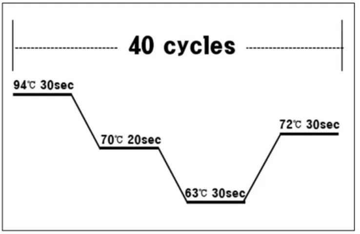
K-ras mutation은 PNA Clamp K-ras Mutation Detection Kit (Panagene Ltd., Daejeon, Korea)에 기술된 방법으로 수행하 였다. 즉, 총 PCR 반응액 양을 20 uL로 하였고 이에 50 ng의 추출 DNA, 13 uL의 real-time SYBR Green PCR master mix (Applied Biosystems, Foster City, CA, USA), primer 및 PNA probe를 codon 12와 codon 13에 각각 첨가하였다. PCR의 positive control에는 PNA probe를 첨가하지 않았으며, PNA control에는 mutation이 없는 wild type template DNA를 첨가 하였다. PCR 조건은 94℃에서 5분간 DNA 변성을 유도한 후 94℃에서 30 sec, 70℃에서 20 sec, 63℃에서 30 sec 그리 고 72℃에서 30 sec 등의 4단계 과정을 40회 반복한 후 72℃
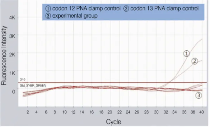
에서 5분 동안 거치하였다.(Fig. 1) PNA probe는 wild type의 K-ras gene에 100% 결합되기 때문에 PCR을 방해하여 PCR 이 이루어지지 않는 반면 codon 12와 13의 mutant K-ras 유 전자는 PNA probe가 100% 결합되지 않기 때문에 PCR이 이루어진다. 따라서 실시간으로 형광염료의 농도가 기계 적으로 검출 된다.(Fig. 2)
3) 통계학적 분석
면역조직화학적 염색 결과에 따른 EGFR expression level 과 암종의 임상적, 조직학적 양상과의 관계를 알아보기 위 해 pearson correlation analysis를 사용하였으며, 유의성은 연관계수 [r]>0.5, P<0.05로 하였다.
Ⅲ.
결 과
1. 면역조직화학적 염색 소견정상적인 구강편평상피조직의 경우, 상피세포의 기저부 에서만 EGFR의 발현이 관찰되며, 고등도 분화 구강편평상 피세포 암종에서 EGFR의 발현이 관찰되지 않거나 미약하
였다.(Figs. 3, 4)
반면, 중등도 분화 구강편평상피세포 암종의 경우, 종양 세포의 세포막에서 증가한 EGFR 발현이 관찰되었으며, 저 등도 분화 구강편평상피세포 암종의 경우, 정상조직에 비 해 결체조직내로 침습한 종양세포 내에서 상당히 증가된 EGFR 발현을 관찰할 수 있었다.(Figs. 5, 6)
Fig. 1.Real-time PCR cyclining condition.
Byeong-Chool Moon et al: Epidermal growth factor receptor overexpression and K-ras mutation detection in the oral squamous cell carcinoma. J Korean Assoc Oral Maxillofac Surg 2011
Fig. 3. Negative immunohistochemical staining for EGFR of well differentiated oral squamous cell carcinoma (anti- EGFR Immunoperoxidase, ×100). EGFR, epidermal growth factor receptor.
Byeong-Chool Moon et al: Epidermal growth factor receptor overexpression and K-ras mutation detection in the oral squamous cell carcinoma. J Korean Assoc Oral Maxillofac Surg 2011
Fig. 4.Mild immunohistochemical staining for EGFR of mod- erate differentiated oral squamous cell carcinoma (anti- EGFR Immunoperoxidase. ×100). EGFR, epidermal growth factor receptor.
Byeong-Chool Moon et al: Epidermal growth factor receptor overexpression and K-ras mutation detection in the oral squamous cell carcinoma. J Korean Assoc Oral Maxillofac Surg 2011
Fig. 2. The PNA probe which is complementary to the wild type K-ras binds to the K-ras gene completely and the PCR is inhibited, while on mutant K-ras gene, the PNA probe binds to the mutan K-ras gene imcompletely and PCR is not inhibited resulting in amplicon.
Byeong-Chool Moon et al: Epidermal growth factor receptor overexpression and K-ras mutation detection in the oral squamous cell carcinoma. J Korean Assoc Oral Maxillofac Surg 2011
2. 면역조직화학적 염색 결과에 따른 EGFR expression과 암종의 임상적, 조직학적 양상과의 관계
EGFR low-level staining은 총 20예 중에 15예(75%)였으며 high-level staining은 5예(25%)였다. 암종의 조직학적 분화
도 양상과 EGFR 발현과의 연관성이 유의하게 나타났고 TNM 분류 중 종양크기에 따른 분류 항목과도 EGFR 발현 의 차이와 유의성이 있게 나타났다. 그 외 다른 요인들은 EGFR 발현의 차이와 통계학적으로 유의성이 없었다.
(Table 1)
Fig. 5. Moderate immunohistochemical staining for EGFR of poor differentiated oral squamous cell carcinoma (anti- EGFR Immunoperoxidase, ×100). EGFR, epidermal growth factor receptor.
Byeong-Chool Moon et al: Epidermal growth factor receptor overexpression and K-ras mutation detection in the oral squamous cell carcinoma. J Korean Assoc Oral Maxillofac Surg 2011
Fig. 6. Severe immunohistochemical staining for EGFR of poor differentiated and invasive oral squamous cell carci- noma (anti-EGFR Immunoperoxidase, ×100). EGFR, epi- dermal growth factor receptor.
Byeong-Chool Moon et al: Epidermal growth factor receptor overexpression and K-ras mutation detection in the oral squamous cell carcinoma. J Korean Assoc Oral Maxillofac Surg 2011
Table 1.The correlation between immunohistochemical EGFR expression and clinical and pathological factors
Variable Case (n) EGFR staining pattern r
Low-staining (n=) High-staining (n=) P
Sex Male 11 8 3 -0.071
Female 9 7 2 0.396
Age 60≤ 5 4 1 0.188
60> 15 11 4 0.351
Histological differentiation Well 13 13 0 0.697*
Moderate 3 2 1 0.021*
Poor 4 0 4
Tumor size T1 0 0 0
T2 11 10 1 0.599*
T3 7 5 2 0.032*
T4 2 0 2
Nodal status N(-) 11 8 3 0.441
N(+) 9 7 2 0.335
Metastasis M(-) 17 13 4 0.394
M(+) 3 2 1 0.239
TNM stage I 0 0 0
II 8 6 2 0.419
III 6 5 1 0.072
IV 6 4 2
n, number of patients; r, correlation coefficient; EGFR, epidermal growth factor receptor; TNM, tumor-node-metastasis (*Pearson correlation analysis, significance [r]>0.5, P<0.05)
Byeong-Chool Moon et al: Epidermal growth factor receptor overexpression and K-ras mutation detection in the oral squamous cell carcinoma. J Korean Assoc Oral Maxillofac Surg 2011
3. K-ras mutation 검출
실험에 사용된 총 20개의 모든 구강편평상피암종 조직에 서 K-ras mutation이 검출되지 않았다.(Fig. 7)
Ⅳ.
고 찰
정상 세포의 성장과 분화는 특이 인자들에 의해 조절 되 며, 이를 성장인자라고 한다5. 성장인자는 세포막에 존재하 는 특정 수용기와 상호작용하는 단백질로서, 표현형의 조 절, 세포의 운동성과 세포골격구조의 변화, 세포 증식 속도 의 변화 등을 포함하는 다양한 생물학적 반응들을 야기한 다. 성장인자는 특정 세포에서만 생산되는 것이 아니라 여 러 종류의 세포에서 생산되며, 다양한 범위의 서로 중복되 는 생물학적 기능을 가지고 있고 일반적으로 비교적 짧은 거리에서 작용한다. 대부분의 정상 세포들은 여러 가지 성 장인자를 분비하고 반응하는 것으로 알려져 있다13,14.
여러 성장인자들 중 상피성장인자는 53 amino-acids로 구 성된 single-chain polypeptide로서, 세포 표면의 수용체와 결 합하여 다양한 형태의 세포들의 증식을 유도할 수 있는 강 력한 분열 유발성 능력을 갖고 있는 단백질이다15,16. Cohen 등이 생쥐의 악하선으로부터 처음으로 추출하였으며 이후 사람의 urine, 타액, 세포외액에서도 추출되게 되었고2,3많 은 관련 연구가 시행되었다17,18.
상피성장인자 수용체는 다양한 세포의 세포막에 존재하 며, 상피의 증식이 빠르게 일어나는 여러 종류의 상피세포 와 상피조직에서 관찰된다19. 상피성장인자와 세포의 세포 막에 존재하는 상피성장인자 수용체와의 결합은 수용체의
세포 내 domain의 tyrosine kinase를 활성화시킨다. 이와 같이 활성화된 tyrosine kinase는 downstream 기질을 인산화시킴 으로써 궁극적으로는 DNA의 복제와 세포 분열을 야기 시 킨다. 따라서 상피성장인자는 상피성장인자 수용체와의 결 합으로 세포의 증식을 촉진시키는데 중요한 역할을 한다.
상피성장인자 수용체는 종양세포의 증식 및 발암기전에 서도 중요한 역할을 하며, 인체 내 여러 부위에서 발생하는 상피성 암종에서 그 발현과 임상적 소견과의 연관성이 연 구되었고, 치료결과에 대한 예후 예측이나 재발 가능성 등 에 대한 중요한 지표로서 그 가능성이 연구된 바 있다20. 대 부분의 편평상피세포암종에서 상피성장인자 수용체는 과 발현되는 경향을 나타내는데, Ishitoya 등4은 두경부에 발생 한 편평상피암종 15예 중 8예(53%)에서 상피성장인자 수 용체의 과발현이 관찰되었다고 보고하였으며, Kim 등21은 37예의 두경부 편평상피암종 중 20예(54%)에서 상피성장 인자 수용체의 발현이 관찰되었다고 보고하였다. 본 연구 에서도 마찬가지로 20예의 구강편평상피암종 중 9예에서 는 발현이 관찰되지 않았으나 6예에서 미약한 발현을 나타 냈으며, 5예(25%)에서 과발현을 관찰할 수 있었다.
본 연구의 EGFR 면역조직화학적 검사 결과, 암종 주위의 정상적인 구강편평상피조직의 경우, 그 발현이 증식하는 기저세포들에서 주로 관찰되었으며 점막 표층으로 세포 분화도가 증가함에 따라 상피성장인자 수용체의 발현은 현저히 감소되었다. 이러한 양상은 EGFR이 정상적인 경 우, 상피세포에서는 그 발현이 제한된다는 다른 연구들의 결과들과도 일치 한다5,22. 또한 조직병리학적 조직 소견 상 분화가 잘 되어 있고 침습성이 덜한 암종 조직이나 고등도 분화 구강편평상피암종에서는 EGFR의 발현이 미미하게 관찰되었다. 반면, 저분화도의 침습적인 구강편평상피세 포암종에서는 EGFR의 강한 발현이 관찰되었는데 이는 EGFR의 발현 정도가 암종의 분화도 또는 침습성과 상관관 계가 있다고 생각해 볼 수 있었으며, 면역조직화학적 염색 결과에 따른 EGFR expression level과 암종의 임상적, 조직 학적 양상과의 관계를 알아보기 위한 통계학적 분석에서 증명되었다. 이를 위해 사용된 통계 방법인 Pearson correla- tion analysis의 상관 계수 r은 그 값이 0.5 이상일 경우, 연관 성이 매우 높다고 해석할 수 있으며 0.4 이상인 경우는 중 등도의 연관성을 나타내며 그 이하인 경우는 아주 낮은 연 관성을 나타낸다고 해석할 수 있다. 그 결과 본 연구에서는 조직학적 분화도에 따른 EGFR 발현과의 연관성이 유의하 였으며, 또한 종양 크기에 따른 분류와의 연관성이 유의하 게 나타났다. 이러한 결과는 Christensen 등5및 Kim 등21의 연구와도 유사한 결과이다. 반면에 Kunikata 등19은 편평상 피암종의 조직학적 분화도와 상피성장인자 수용체의 발현 과의 상관관계가 유의하지 않았다고 하였으며, Ishitoya 등4 은 고등도 분화 편평상피암종에서 상피성장인자 수용체의 과발현이 더 많이 관찰되었다고 보고하였는데 이러한 상 이한 결과들로 보았을 때 앞으로 더 많은 검체들에 대한 연
Fig. 7. K-ras mutation detection with PNA-based real-time PCR clamping.
Byeong-Chool Moon et al: Epidermal growth factor receptor overexpression and K-ras mutation detection in the oral squamous cell carcinoma. J Korean Assoc Oral Maxillofac Surg 2011
① codon 12 PNA clamp control ② codon 13 PNA clamp control
③ experimental group
Fluorescence Intensity
4K
3K
2K
1K
0
346 Std_SYBR_GREEN
Cycle
2 4 6 8 10 12 14 16 18 20 22 24 26 28 30 32 34 36 38 40
구가 시행되어져야 더욱 명확한 상관관계를 알 수 있을 것 으로 생각되었다.
한편, 현재까지 구강편평상피암종의 치료는 주로 수술적 인 절제 및 방사선 치료가 대표적인 치료방법들이었다. 하 지만, 림프절로의 전이 또는 술 후 재발 등은 그 예후를 불 량하게 만들며 결국 환자의 생존률을 감소시키게 된다. 따 라서 이러한 치료방법의 한계를 극복하고 경부 림프절 전 이 병소의 발생을 억제하거나 재발된 국소부위 악성 병소 를 조절할 수 있는 새로운 치료법을 연구하게 되었다. 그 중 편평상피암종의 상피성장인자 수용체의 과발현과 연계 하여 이를 저지하기 위한 EGFR tyrosine kinase inhibitors therapy가 소개되었으며, 현재 대장암이나 폐암 환자에서 임상적으로 적용되어 효과를 보고 있는 항암요법이다. 하 지만, 이 치료방법의 효과는 때때로 제한적일 수 있는데 이 는 암종 세포내 K-ras mutation 여부와 연관성이 깊다. K-ras 는 알려진 바와 같이 세포내 상피성장인자 수용체와 연결 되어 tyrosine kinase의 인산화 과정에 관여하게 되는데 많 은 암종에서 그 변이가 관찰되었으며, 이는 암종의 발생 및 성장에서 중요한 역할을 하게 된다23. 따라서 K-ras 변이를 가지는 암종에서는 EGFR tyrosine kinase inhibitors therapy 의 효과가 감소되며 치료결과도 미미하게 되므로 이러한 치료방법을 적용시키기 전에 암종의 K-ras 변이의 검출이 필수적이다. Erminia 등12은 암종의 K-ras 변이 연구에서 전 체 표본 중 22.8%에서 K-ras 변이가 검출되었다고 보고하 였다. 본 연구에서는 20예 전체 표본 모두에서 K-ras 변이 가 검출되지 않아 상이한 결과를 나타냈는데 이는 암종의 부위 및 검출방법의 차이로 인한 것으로 생각되었다. 하지 만, 검체수가 적었기 때문에 이러한 결과를 모든 구강편평 상피암종에 일반화하는 것은 무리이며, EGFR tyrosine kinase inhibitors therapy가 적용될 환자의 암종 조직에 대해 치료 전 개별적으로 K-ras 변이 검출을 시행하는 것이 알맞 을 것으로 생각되었다.
Ⅴ. 결 론
EGF는 53 amino-acids로 구성된 single-chain polypeptide 로서, EGFR과 결합하여 다양한 형태의 세포들의 증식을 유도할 수 있는 강력한 분열 유발성 능력을 갖고 있는 단백 질이다. 상피성장인자와 세포의 세포막에 존재하는 상피 성장인자 수용체와의 결합 후 일련의 인산화 과정을 통해 서 세포의 DNA복제와 세포 분열을 야기하며 이런 특성이 종양 세포에서 증식 및 발암 기전에 중요한 역할을 하는 것 으로 생각되고 있다. 이에 구강편평상피암종에서 면역조 직화학적 염색을 이용한 EGFR의 발현 양상을 검사하여 환 자의 임상병리학적 정보와 EGFR 발현과의 상관 관계를 알 아보고 K-ras mutation을 검출하여 EGFR tyrosine kinase inhibitors therapy를 위한 표지자로서 그 가능성을 알아보고 자 하였다.
실험에 사용된 조직편은 단국대학교 치과대학 부속 치과 병원 구강악안면외과에서 구강편평상피세포암으로 최종 진단 받은 환자 20명의 수술 후 절제된 조직 20편을 사용하 였다. 각각의 조직편에서 EGFR의 발현을 확인하기 위해 면역조직화학적 검사, K-ras mutation 검출을 시행하였으 며, 통계학적으로 검증하였다.
1. 면역조직화학적 검사: 정상 구강편평상피의 경우, EGFR의 발현은 상피조직의 기저부에서만 관찰되었 다. 반면에 저등도 분화 구강편평상피세포 암종의 경 우, 결체조직으로 침습된 암세포에서 매우 증가된 EGFR의 발현을 관찰할 수 있었다.
2. K-ras mutation 검출: 20개의 종양 조직편 모두에서 K- ras 20개의 종양 조직편 모두에서 KRAS 돌연변이가 관 찰되지 않았다.
3. 통계학적 검증: 조직학적 분화도에 따른 분류 및 tumor size에 따른 EGFR 발현의 차이가 유의성이 있게 나타 났다.
결론적으로 구강편평상피암종에서 정상조직 및 비침습 적 종양에 비해 EGFR의 과발현이 나타났으며 이는 암종의 증식과 침습에 기여하리라 여겨지며 이를 억제하기 위한 EGFR tyrosine kinase inhibitors therapy를 시행하기 전 그 효 과에 대한 예후를 판별하기 위해 구강편평상피암종에서 K-ras 변이 검출을 시행하는 것이 적절하리라 생각된다.
References
1. Kang JH, Kim KW, Lee JH. Study on mRNA expression of p21 and p73 in the cell lines of primary and metastatic squamous cell carcinoma. J Korean Assoc Oral Maxillofac Surg 2001;27:483- 490.
2. Cohen S. Isolation of submaxillary gland protein accelerating in- cisor eruption and eyelid opening in the new-born animal. J Biol Chem 1962;237:1544-62.
3. Cohen S, Carpenter G. Human epidermal growth factor: isolation and chemical and biological properties. Proc Natl Acad Sci U S A 1975;72:1317-21.
4. Ishitoya J, Toriyama M, Oguchi N, Kitamura K, Ohshima M, Asano K, et al. Gene amplification and overexpressi on of EGF receptor in squamous cell carcinomas of the head and neck. Br J Cancer 1989;59:559-62.
5. Christensen ME, Therkildsen MH, Hansen BL, Albeck H, Hansen GN, Bretlau P. Epidermal growth factor receptor expres- sion on oral mucosa dysplastic epithelia and squamous cell carci- nomas. Eur Arch Otorhinolaryngol 1992;249:243-7.
6. Sankaranarayanan R, Masuyer E, Swaminathan R, Ferlay J, Whelan S. Head and neck cancer: a global perspective on epi- demiology and prognosis. Anticancer Res 1998;18:4779-86.
7. Parkin D.M, Bray F, Ferlay J, Pisani P. Estimating the world can- cer burden: Globocan 2000, Int J Cancer 2001;94:153-6.
8. Carvalho AL, Ikeda MK, Magrin J,Kowalski LP. Trends of oral and oropharyngeal cancer survival over five decades in 3267 pa- tients treated in a single institution. Oral Oncol 2004;40:71-6.
9. Pisani P, Parkin DM, Bray F, Ferlay J. Estimates of the worldwide mortality from 25 cancers in 1990. Int J Cancer 1999;83:18-29.
10. Parkin DM, Pisani P, Ferlay J. Estimates of the worldwide inci- dence of 25 major cancers in 1990. Int J Cancer 1999;80:827-41.
11. Carvalho AL, Magrin J, Kowalski LP. Sites of recurrence in oral
and oropharyngeal cancers according to the treatment approach.
Oral Dis 2003;9:112-8.
12. Massarelli E, Varella-Garcia M, Tang X, Xavier AC, Ozburn NC, Liu DD, et al. KRAS mutation is an important predictor of resistance to therapy with epidermal growth factor receptor tyro- sine kinase inhibitors in non small cell lung cancer. Clin Cancer Res 2007;13:2890-6.
13. James R, Bradshaw RA. Polypeptide growth factors. Ann Rev Biochem 1984;53:259-92.
14. Modjtahedi H, Hickish T, Nicolson M, Moore J, Styles J, Eccles S, et al. Phase I trial and tumor localization of the anti-EGFR monoclonal antibody ICR62 in head and neck or lung cancer. Br J Cancer 1996;73:228-35.
15. Carpenter G, Zendegui JG. Epidermal growth factor, its receptor, and related proteins. Exp Cell Res 1986;164:1-10.
16. Ino M, Ushiro K, Ino C, Yamashita T, Kumazawa T. Kinetics of epidermal growth factor in saliva. Acta Otolaryngol Suppl 1993;
500:126-30.
17. Gardner DP, Shimizu N. Loss of cytotoxic effect of epidermal growth factor(EGF) on EGF receptor overexpressing cells is as- sociated with attenuation of EGF receptor tyrosine kinase activi-
ty. J Cellular Physiol 1994;158:245-55.
18. Ozanne B, Richards CS, Hendler F, Burns D, Gusterson B. Over- expression of the EGF receptor is a hallmark of squamous cell carcinomas. J Pathol 1986;149:9-14.
19. Kunikata M, Yamada K, Yamada T, More M, Tatemoto Y, Osaki T. Epidermal growth factor receptor in squamous cell carcinoma and other epidermal lesions of squamous origin - an immunohis- tochemical study. Acta Histochem Cytochem 1992;25:387-94.
20. Pusztai L, Lewis CE, Lorenzen J, Mcgee JO. Growth factors:
regulation of normal and neoplastic growth. J Pathol 1993;169:
191-201.
21. Kim KW, Kim MJ. Expression of the epidermal growth factor re- ceptor and cell cycle analysis in the head and neck squamous cell carcinomas. J Korean Assoc Oral Maxillofac Surg 2000;26:154- 163.
22. Shirasuna K, Hayashido Y, Sugiyama M, Yoshioka H, Matsuya T. Immunohistochemical localization of epidermal growth factor (EGF) and EGF receptor in human oral mucosa and its malignan- cy. Virchows Arch A Pathol Anat Histopathol 1991;418:349-53.
23. Kranenburg O. The KRAS oncogene: past, present, and future.
Biochim. Biophys. Acta 2005;1756:81-2.