J Lung Cancer 2010;9(2):103-105
103
IgG4-Related Lung Disease Presenting as a Consolidative Mass: A Case Report
Immunoglobulin G4 (IgG4)-related sclerosing disease involving the lung is a rare condition, and this is characterized by an elevated serum IgG4 level, fibrotic inflammation with numerous IgG4-positive plasma cells and a response to steroid therapy. We present here a case of pulmonary IgG4-related disease in a 75-year-old man who presented with cough and yellowish sputum for the previous 3 months. The chest images showed a consolidative mass in the right lower lobe that suggested mucinous bronchioloalveolar carcinoma.
The wedge resected specimen revealed an ill-defined gray-tan, firm lesion.
Microscopically, the lesion showed a diffuse lymphoplasmacytic infiltration with irregular fibrosis in the alveolar interstitium and bronchovascular bundles.
There were numerous IgG4-positve plasma cells and these cells were diffusely distributed. The serum IgG4 level was elevated on the postoperative check-up (249 mg/dL). After corticosteroid therapy for 7 months, the patient’s symptoms and radiologic abnormalities were improved. (J Lung Cancer 2010;9(2):103
105)
Key Words: Autoimmune disease, IgG4, Interstitial lung disease, Plasma cells, Lung neoplasms
Ha Young Park1 Joungho Han1 Guhyun Kang1 Chin A Yi2 and Man Pyo Chung3
Departments of 1Pathology, 2Radio- logy and Center for Imaging Science, and 3Division of Pulmonary and Critical Care Medicine, Samsung Medical Center, Sungkyunkwan Uni- versity School of Medicine, Seoul, Korea
Received: September 8, 2010 Revised: September 15, 2010 Accepted: September 16, 2010 Address for correspondence Joungho Han, M.D.
Department of Pathology, Samsung Medical Center, Sungkyunkwan Uni- versity School of Medicine, 50, Irwon- dong, Gangnam-gu, Seoul 135-710, Korea
Tel: 82-2-3410-2800 Fax: 82-2-3410-0025 E-mail: [email protected]
Immunoglobulin G4 (IgG4)-related sclerosing disease is characterized by an elevated serum IgG4 level, fibrotic inflammation with numerous IgG4-positive plasma cells and a response to steroid therapy (1-4). It commonly involves the pancreas, the common bile duct and the retroperitoneum (5).
Involvement of the lung is rare, and several cases have been described with variable patterns (2,4,6,7). We recently experi- enced a case of IgG4-related lung disease in a 75-year-old man.
He presented with consolidation on chest computed tomography (CT), and so wedge resection was performed. The microscopic findings showed lymphoplasmacytic sclerosing inflammation with numerous IgG4-positive plasma cells. The patient’s serum levels of total IgG and IgG4 were elevated. He was treated with high-dose prednisolone (30 mg/day) and he demonstrated a good responsive.
CASE REPORT
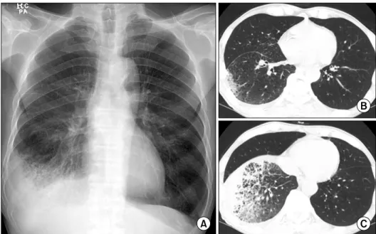
A 75-year-old man, who had smoked for 40-pack years, complained of cough, yellowish sputum and 10 kg weight loss over the previous 3 months. He was found to have air space consolidation and ground-glass attenuations in the periphery of the right lower lobe with a small amount of pleural effusion on the chest PA and computed tomography (CT) scan (Fig. 1).
He was admitted to our hospital for further evaluation. The physical examination was unremarkable. The results of the pulmonary function tests are as follows: a FEV1/FVC of 51%, a FVC of 3.39 L (95% of the predicted value) and a FEV1 of 1.71 L (73% of the predicted value), which was consistent with a moderate obstructive pattern. The peripheral blood test results were as follows: a hemoglobin level of 11.0 g/dL; a WBC of
104 J Lung Cancer 2010;9(2):103-105
Fig. 1. (A) The chest X-ray shows parenchymal consolidation at the right costophrenic angle with pleu- ral effusion. (B, C) The chest com- puted tomography scans reveal air space consolidation and ground- glass attenuations.
Fig. 2. (A, B) Scanning views of the VATS wedge resected specimen show a subpleural patchy mononuclear cells infiltration. (C) The low power view shows a dense lymphoid cell infiltration with fibrosis in the alveolar interstitium and bronchovascular bundles (H&E satin, ×40). (D) The lesion is composed of mature plasma cells and some lymphocytes (H&E stain, ×200). (E) Phlebitis is also present in the interlobular septal vein (H&E stain, ×100). (F) Many plasma cells are positive for IgG4 (×400).
IgG4-Related Lung Disease 105
7,290/μL (segmented neutrophils, 68.8%; lymphocytes, 19.6%); a C-reactive protein level of 4.99 mg/dL; and an ESR of 76 mm/hr. The serum electrolytes and the renal/liver function tests were normal. A test for fluorescence anti-nuclear antibody was weakly positive at a titer of 1:40. Bronchoscopy revealed an obstruction in the mediobasal segment of the right lower lobe by a fibrotic membrane. Transbronchial lung biopsy and bronchoalveolar lavage were performed, and the patho- logical report was chronic inflammation. For histologic con- firmation, the patient underwent video-assisted thoracoscopic wedge resection. Microscopically, the lesion showed a dense lymphoplasma cell infiltration in the peribronchiolar intersti- tium and subpleural connective tissue, and an interlobular septum (Fig. 2A-D). The features of organizing pneumonia with eosinophilic infiltration were also noted. There were lympho- cytic infiltrates in the small vessels that almost mimicked vasculitis (Fig. 2E). Immunohistochemically, the plasma cells in the interstitial spaces were positive for IgG4 (1:2,000, The Binding Site, Birmingham, UK) (Fig. 2F). The following serum tests revealed elevated levels of IgG4 (1,320 mg/dL) and IgG4 (249 mg/dL). He was treated with high-dose prednisolone, and the symptoms and radiologic abnormalities were resolved after 3 months.
DISCUSSION
IgG4-related lung disease shares the microscopic features of IgG4-related sclerosing disease of the other organs. The present case revealed a diffuse lymphoplasmacytic infiltration, irregular fibrosis and lymphocytic infiltration in the small vessels, and all this led to a differential diagnosis of nonspecific interstitial pneumonia or plasmacytoma. The majority of inflammatory cells in nonspecific interstitial pneumonia are lymphocytes, while the infiltrated cells in the present case consisted of lymphocytes, plasma cells and eosinophils. The presence of IgG4-positive plasma cells also suggested IgG4-releated lung disease rather than nonspecific interstitial pneumonia. The plasma cells lacked atypism and they were intermingled with lymphocytes and eosinophils, which are findings incompatible with plasmacytoma. In addition, irregular interstitial fibrosis was prominent in this case, but the spindle cell proliferation fell short
of making the diagnosis of inflammatory pseudotumor.
Inoue et al. (8) recently classified IgG4-related lung disease into 4 categories according to the radiologic features (i.e., the solid nodular, round-shaped ground glass opacity, alveolar inter- stitial and bronchovascular subtypes), and they reported that the CT findings corresponded to IgG4-related sclerosing inflammat- ion along the intrapulmonary connective tissue. A recent study described that IgG4-related disease revealed a great variety of pulmonary and pleural lesions, and it is important to know the morphologic variety and clinicopathologic characteristics of this disorder (7). Our case can be classified as the alveolar interstitial subtype and any obliterative vascular change was indefinite, which is generally more common in the solid areas.
IgG4-related lung disease is effectively treated with high- dose corticosteroid. If a biopsy specimen shows interstitial lymphoplasmacytic infiltration with irregular fibrosis, then immunostaining and serological tests for IgG and IgG4 should be considered to rule out the possibility of IgG4-related disease.
REFERENCES
1. Hamed G, Tsushima K, Yasuo M, et al. Inflammatory lesions of the lung, submandibular gland, bile duct and prostate in a patient with IgG4-associated multifocal systemic fibrosclerosis.
Respirology 2007;12:455-457.
2. Kobayashi H, Shimokawaji T, Kanoh S, et al. IgG4-positive pulmonary disease. J Thorac Imaging 2007;22:360-362.
3. Takato H, Yasui M, Ichikawa Y, et al. Nonspecific interstitial pneumonia with abundant IgG4-positive cells infiltration, which was thought as pulmonary involvement of IgG4-related autoimmune disease. Intern Med 2008;47:291-294.
4. Shigemitsu H, Koss MN. IgG4-related interstitial lung disease:
a new and evolving concept. Curr Opin Pulm Med 2009;15:
513-516.
5. Kamisawa T, Okamoto A. IgG4-related sclerosing disease.
World J Gastroenterol 2008;14:3948-3955.
6. Yamashita K, Haga H, Kobashi Y, et al. Lung involvement in IgG4-related lymphoplasmacytic vasculitis and interstitial fibrosis: report of 3 cases and review of the literature. Am J Surg Pathol 2008;32:1620-1626.
7. Zen Y, Inoue D, Kitao A, et al. IgG4-related lung and pleural disease: a clinicopathologic study of 21 cases. Am J Surg Pathol 2009;33:1886-1893.
8. Inoue D, Zen Y, Abo H, et al. Immunoglobulin G4-related lung disease: CT findings with pathologic correlations.
Radiology 2009;251:260-270.