154 Accepted for Publication: September 9, 2009
*Corresponding author: Deok-Hwan Yang, 519-809, Department of Hematology/
Oncology, Chonnam National University Hwasun Hospital, Phone: +82-61- 3797636, Fax: +82-61-3797628, Email: [email protected] Chonnam Medical Journal
Vol. 45, No. 3, pp. 154∼160 DOI: 10.4068/cmj.2009.45.3.154
Clinical Correlation of CD4+CD25+ Regulatory T Cells in Early Immune Reconstitution after Myeloablative Allogeneic Stem Cell Transplantation
Deok-Hwan Yang1*, Jung-Sun Park1, Jae-Sook Ahn, Yeo-Kyeoung Kim, Je-Jung Lee and Hyeoung-Joon Kim Hematology-Oncology, Chonnam National University Hwasun Hospital, Hwasun, Jeonnam,
1Research Institute of Medical Sciences, Chonnam National University, Gwangju, Korea
We investigated whether CD4+CD25+ regulatory T cells (Tregs) not only reduce the incidence of acute graft-versus-host disease (aGVHD) but also inhibit the expression of NK cells during early immune reconstitution after allogeneic myeloablative hematopoietic stem cell transplantation (HSCT). In addition, we evaluated whether Tregs were associated with disease relapse. Twenty-nine patients underwent non-T cell-depleted allogeneic HSCT. Peripheral blood mononuclear cells (PBMCs) were separated at 3 weeks after HSCT. Fourteen patients developed grade 2-4 aGVHD and 13 patients relapsed. Patients with grade 2-4 aGVHD (median, 5.36 ng/μl) had significantly lower levels of FOXP3 gene expression than did those with grade 0-1 aGVHD (median, 7.45 ng/μl). However, the level of FOXP3 gene expression in patients with relapse (median, 7.46 ng/μl) was significantly higher than in those (median, 5.52 ng/μl) without relapse (P=0.02). However, we did not find an inverse correlation between the expression of NK cells and Tregs. The level of FOXP3 expression in CD4+CD25+ Tregs was related with the incidence of aGVHD and was associated with the risk of disease relapse. However, NK cells showed no correlation with the expression of Tregs during early immune reconstitution after myeloablative HSCT.
Key Words: Stem cells; Graft-vs-host disease; Gene expressions
INTRODUCTION
The Clonal expansion of donor T cells, which respond to the recipient’s environment, causes acute graft-versus- host disease (aGVHD) and graft-versus-leukemic (GVL) effects during early immune reconstitution after trans- plantation. The pathophysiology of aGVHD is thought to be due to donor T cell recognition of anti-genic differences by antigen-presenting cells (APCs), activated cytokine storm and mediated cellular and inflammatory
responses on the target tissue.1,2 Several animal studies have suggested that down-regulation of aGVHD and establishment of immune tolerance are mediated by antigen-specific regulatory T cells (Tregs).3-7
Tregs play a key role in hematopoietic stem cell trans- plantation (HSCT); they prevent aGVHD and promote engraftment and chimerism by specific induction of tolerance of the alloantigens. The expression of FOXP3, which encodes a forkhead/winged helix transcription factor and is required for the development and the functional activity of CD4+CD25+ Tregs, is used as a reliable molecular marker for quantifying Tregs in the peripheral blood.8-10 CD4+CD25+ Tregs can block the amplification of the NK cell response to APCs, and
depletion of Tregs can result in the enhancement of NK cell-mediated anti-tumor immunity.11,12 Importantly, several in vitro studies have reported that the suppression of proliferation and function of cytotoxic T cells by Tregs results in an unfavorable influence on allogeneic transplantation interfering with the GVL activity of donor T cells.13-15 However, conflicting results have been reported where inhibition of GVHD by Tregs did not interfere with the GVL effects after allogeneic HSCT.16,17
In this study, we evaluated whether the expression of CD4+CD25+ Tregs and NK cells is associated with the incidence of aGVHD or whether Tregs have a differentiated potential between GVL effects and aGVHD without causing disease relapse during early immune reconstitution after myeloablative HSCT.
Materials and Methods
1. Patients and transplant approach
This study was conducted between September 2002 and January 2007 after obtaining informed consent and the approval from our institutional review board. Twenty- nine patients who underwent non-T cell-depleted my- eloablative allogeneic HSCT were analyzed to investigate the relationship between early reconstitution of Tregs, NK cells, the incidence of aGVHD, and disease relapse.
The clinical characteristics of the patients are shown in Table 1.
All patients received non-T cell-depleted grafts after myeloablative conditioning with fludarabine (180 mg/m2) and busulfan (12.8 mg/kg) (n=13), cyclophosphamide (120 mg/kg) and total body irradiation (1,200 cGy) (n=7), or busulfan (16 mg/kg) and cyclophosphamide (120 mg/kg) (n=9). Nine patients received stem cells from unrelated donors and five patients were infused from HLA-mismatched donors. Three of five patients with HLA-mismatched HSCT had alemtuzumab (30 mg) added to the conditioning to reduce aGVHD. Cyclosporin A (CSA) or FK-506 was used for prophylaxis against GVHD,
starting on day −3, continued for 180 days, and then tapered over a 90-day period. A short course of methotrexate was added to the protocol in patients without alemtuzumab conditioning.
2. Cell isolation
Peripheral blood samples were obtained at 21 days after HSCT until the absolute neutrophil count reached above 1,000×109/L. Peripheral blood mononuclear cells (PBMC) were separated from blood samples by Ficoll- Hypaque (lymphoprep; 1.077 density) gradient centrifu- gation and were frozen in RPMI 1,640 media with 20%
fetal bovine serum and 10% dimethyl sulfoxide. Before analysis, frozen cells were thawed, washed and re- suspended. The level of FOXP3 mRNA expression was assessed by quantitative real-time reverse transcription PCR.
3. Flow cytometry
Immunophenotyping was performed on collected peri- pheral blood samples. Briefly, mononuclear cells isolated by Ficoll-Hypaque density gradient centrifugation were incubated with the following mouse monoclonal anti- bodies: Rhodamine 1-conjugated anti-human CD16, CD56 and fluorescein isothiocyanate (FITC)-conjugated anti-CD3, CD4-phycoerythrin (PE), and CD25-FITC (Coulter Immnuotech, Miami, FL, USA). CD4+CD25+
T cells in PBMCs were sorted on a BD FACS Aria cell sorter (BD Biosciences, San Jose, CA 95131, USA) using PE-conjugated anti-CD4, FITC-conjugated anti- CD25, and its isotype control antibody. The level of FOXP3 mRNA expression was measured in sorted CD4+CD25+T cells. At least 5,000 cells were counted and analyzed by use of BD FACS Aria cell sorter software.
4. RNA extraction and real-time reverse transcription PCR for FOXP3
Total RNA from cells was isolated by using TRIzol (Invitrogen, Karlsruhe, Germany). The first-strand cDNA was synthesized from 100 ng of total RNA by use of a
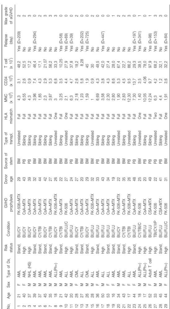
Table 1. Patient’s characteristics No.AgeSexType of Ds.Risk statusConditionGVHD prophylaxisDonor ageSource of stemType of transpl.HLA mismatchMNC (x 108 )CD34 (x 106 )T cell (x 107 )Relapse (day)Max grade of aGVHD 121FAMLStand.BU/CYFK-506+MTX29BMUnrelatedFull 4.3 3.148.2Yes (D+209)2 240FAMLHighBU/CYCsA+MTX43PBSiblingFull 6.55 2.652.5No2 337MNHL (HS)HighBU/CYCsA+MTX39PBSiblingFull 4.3 0.917.2No3 439MAMLStand.BU/CYCsA+MTX32BMSiblingFull 3.96 7.446.4Yes (D+294)0 537MAMLStand.CY/TBICsA+MTX44BMSiblingFull 1.85 4.317.1No2 645MAMLStand.BU/CYCsA+MTX42BMSiblingFull 2.3 2.921.07No3 735MAMLStand.CY/TBICsA+MTX27BMSiblingFull 3.87 5.356.2No3 818MALL(Ph+)HighBU/CYFK-506+MTX26BMUnrelatedOne 3 6.833.3No3 921MAMLStand.CY/TBICsA+MTX22BMSiblingFull 3.25 3.510.25Yes (D+58)0 1042MALLHighBU/FLU/CFK-50627BMUnrelatedOne 2.1 4.427.9Yes (D+69)0 1120MAMLHighBU/FLUCsA+MTX24PBSiblingFull 6.3 4.737.8Yes (D+39)0 1228FCMLStand.BU/CYCsA+MTX27BMSiblingFull 2.18 2.618.2No2 1327MALLStand.CY/TBICsA+MTX20BMSiblingFull 1.23 5.8 8.28Yes (D+202)0 1426MCMLStand.BU/CYCsA+MTX29BMSiblingFull 1.59 1.945Yes (D+725)0 1528MALLHighBU/FLUFK-506+MTX32BMUnrelatedFull 1 0.930No2 1636FAMLStand.BU/FLUCsA+MTX33PBSiblingFull 6.69 4.348.5No0 1750MALLHighBU/FLUCsA+MTX43BMSiblingFull 3.58 3.842.0Yes (D+447)0 1853FALLStand.BU/FLUCsA+MTX34BMSiblingFull 2.30 4.327.4No0 1916FAMLStand.BU/FLUCsA+MTX19BMSiblingFull 2.80 8.628.0No1 2034MAMLStand.BU/CYFK-506+MTX22BMUnrelatedFull 1.90 5.367.4No2 2143FAMLStand.CY/TBICsA+MTX35BMSiblingFull 2.83 4.522.7No2 2217FALLHighCY/TBIFK-506+MTX26PBUnrelatedFull12.30 9.586.2No3 2344FAMLStand.BU/FLUCsA+MTX48PBSiblingFull 7.3013.729.3Yes (D+197)0 2418MALL(Ph+)HighCY/TBI/CFK-50625BMUnrelatedOne 1.40 3.538.5Yes (D+341)1 2517MALL(Ph+)HighBU/FLUCsA+MTX20PBSiblingFull10.05 4.0678.0No0 2652FAdult T cellHighBU/FLUCSA+MTX49PBSiblingFull12.24 9.797.9Yes (D+98)1 2733MAMLHighTBI/CY/VPFK-50622PBUnrelatedTwo 6.3 4.268.2Yes (D+153)3 2845MAMLStand.BU/FLUCSA+MTX41PBSiblingFull 4.4 1.232.1No3 2944FALL(Ph+)HighBU/FLU/CFK-50631BMUnrelatedOne 1.91 3.632.2Yes (D+84)0
SuperScript III kit (Invitrogen) according to the manu- facturer's protocol. Human FOXP3 mRNA expression was quantified by using a SYBR green quantitative PCR kit (Takara, Japan) with the Rotor-gene 3,000 (CORBETT, Australia) and was corrected by amplification of a control, the human β-actin housekeeping gene. Amplification was carried out in a total volume of 20 μL for 40 cycles of 15 s at 95oC, 20 s at 60oC and 20 s at 72oC.
The samples were run in triplicate, and their relative expression was determined by normalizing the expression of each target gene to β-actin and then comparing this normalized value to the normalized expression in a reference sample from which the fold change was calculated. For FOXP3, the following primer combina- tions were used: forward, 5'-CGG ACA CTC AAT GAG ATC TA-3'; and reverse, 5'-ATC CTC CTT TCC TTG ATC TT-3'. The FOXP3 primers were synthesized by Integrated DNA Technologies. For β- actin, the primers were as follows: forward, 5'-GAT GAG ATT GGC ATG GCT TT-3'; reverse, 5'-CAC CTT CAC CGT TCC AGT TT -3'.
5. Statistical Analysis
To compare the level of FOXP3 expressions and the expression of NK cells between grade 0-1 aGVHD and grade 2-4 aGVHD or between relapse and no relapse, discrete or continuous variables were analyzed by use of the Fisher’s exact t test and Mann-Whitney U test, respectively. p values <0.05 were considered statistically significant.
Results
We studied all 29 patients who underwent non-T cell- depleted myeloablative allogeneic HSCT and analyzed the correlation between Tregs and immunophenotypic expression of NK cells at early engraftment. In addition, we analyzed whether the level of FOXP3 expressions differed in relapsed cases compared with cases who did not relapse. The population of Tregs in CD4+ CD25+
T cells was evaluated by real-time quantitative PCR for FOXP3 gene expression.
The median age of the patients was 34 years (range, 16∼53 years) and there were more males than females.
The median follow-up time was 17.2 months (range, 6.6∼57.8 months). Fourteen patients developed grade 2∼4 aGVHD and 13 patients experienced relapse. The median dose of infused CD34+ cells was 4.18×10⁶/kg, and T cell doses were 34.4×10⁷/kg. Only one patient, who had leukemic marrow before transplantation, failed to achieve an engraftment. Unrelated donor, HLA mismatching and alemtuzumab conditioning were signi- ficantly related to the incidence of aGVHD (p<0.05).
Alemtuzumab conditioning and aGVHD were strongly associated with relapse. Patients with grade 2∼4 aGVHD had a significantly lower relapse rate (n=1, 7.1%) than did those with grade 0∼1 aGVHD (n=12, 80%) (p=0.00). The overall median level of FOXP3 expression and the percentage of NK cells for all patients was a median 6.38 ng/μL (range, 0.36∼19.87 ng/μL) and 19.8% (range, 2.0∼60.0%), respectively.
1. The level of FOXP3 expression in CD4+CD25+ T cells correlates with aGVHD and a relapse in early engraftment
The expressions of the FOXP3 gene in CD4+CD25+
T cells showed that patients with grade 2∼4 aGVHD (median, 5.36 ng/μL; range, 0.36∼10.31 ng/μL) had significantly lower levels of FOXP3 gene expression than did those with grade 0∼1 aGVHD (median, 7.45 ng/μL; range, 1.40∼19.87 ng/μL; p=0.03) (Fig. 1A).
However, the level of FOXP3 gene expressions in patients with relapse (median, 7.46 ng/μL; range, 3.42
∼19.87 ng/μL) was significantly higher than in those without relapse (median, 5.52 ng/μL; range 0.36∼
10.31 ng/μL) (p=0.02) (Fig. 1B). The three patients treated with alemtuzumab conditioning had no aGVHD;
their level of FOXP3 expression was a median of 8.91 ng/μL (range, 4.76∼12.31 ng/μL). They all relapsed after a median of 6.6 months (range, 1.1∼31.4 months) from transplantation (p=0.04).
Fig. 1. The level of FOXP3 gene expression in CD4+CD25+ T cells in early engraftment after myeloablative HSCT. Patients who experienced grade 2∼4 aGVHD were compared with those who experienced grade 0∼1 aGVHD. Patients with relapse (A) were significantly higher than those without relapse (B).
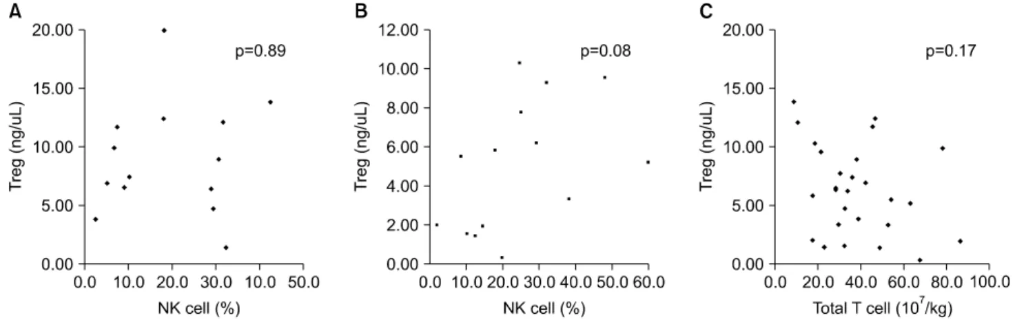
Fig. 2. No inversed correlation between Tregs and the expression of NK cells in early engraftment within grade 0∼1 aGVHD (A) or grade 2∼4 GVHD (B) and no direct relationship between donor grafted T cells and Tregs (C).
2. No inverse relationship between Tregs and NK cells and no correlation between NK cells and aGVHD or a relapse
When we investigated populations of NK cells during early engraftment, we found that the percentage of NK cells in patients with grade 2∼4 aGVHD was a median of 22.2% (range, 2.0∼60.0%) and that in patients with grade 0∼1 aGVHD was a median of 18.3% (range, 2.6
∼42.6%; p=0.55). There was no significant difference in the population of NK cells in the relapsed (median, 28.3%; range, 2.6∼60.0%) and non-relapsed patients (median, 18.0%; range, 2.0∼48.1%) nor between the two aGVHD groups (p=0.51). The analysis of the influence of Tregs on the activation of NK cells, during
the early post-transplantation period, showed that NK cell proliferation in response to FOXP3 expression, with or without aGVHD, was not influenced by early recon- stitution of CD4+FOXP3+ regulatory T cells (Fig. 2A, B). In addition, the grafted donor T-cell doses did not affect the level of FOXP3 gene expression during the early post-HSCT period (p=0.18) (Fig. 2C).
Discussion
After hematopoietic stem cell transplantation, the key donor immune cells controlling early transplant out- comes (eg, GVHD and relapse) are the transplanted nonthymic-dependent donor T cells and early recovering
NK cells because thymic function is defective or absent in adults, broad cell-mediated immune recovery occurs months to years after transplantation.18,19 The early post- transplant period is characterized by powerful immune reactions causing GVHD and GVL. To identify the GVHD-reacting T cells and to separate from those con- ferring GVL, in vitro and in vivo studies have suggested that distinct subsets of host-reacting and leukemia-re- acting T cells separate GVHD from GVL alloreactivity.20-22 This selective biology of GVHD and GVL activity has been investigated including the evaluation of regulatory T cells in allogeneic HSCT. Recent studies by Trenado et al16 and Edinger et al17 suggested that CD4+CD25+
regulatory T cells might preserve the GVL activity while suppressing GVHD in a mouse model of transplantation.
However, conflicting studies have reported that regulatory T cells may reduce anti-tumor immunity in murine models and in human subjects.23-25
In the present study, the early reconstitution of Tregs after HSCT was significantly related with the develop- ment of aGVHD. This finding is consistent with recent published studies.26,27 However, we did not confirm a role for Tregs in the suppression of the proliferation of NK cells or differential effects on aGVHD or GVL effects. This might be because of the different rate of relapse between the grade 0∼1 aGVHD group (80%) and the grade 2∼4 aGVHD group (7.1%). In addition, the three patients who were treated with the alemtu- zumab conditioning had relatively high levels of FOXP3 expression and experienced relapse without aGVHD.
Alemtuzumab is used to deplete recipient lymphocytes in vivo, thereby enhancing engraftment, and to deplete donor T cells to reduce the incidence of aGVHD. The affect of alemtuzumab on immunosuppression after HSCT is estimated to persist for over three months.28 However, alemtuzumab conditioning did not suppress the reconstitution of CD4+CD25+ Tregs during early engraftment and did not cause grade 2∼4 aGVHD in this study, although a limited number of patients were evaluated. We demonstrated that alemtuzumab con- ditioning was associated with CD4+CD25+ Tregs after
early HSCT and the severity of aGVHD as well as the risk of relapse.
Several studies29-31 reported on how donor T cells affect engraftment after HSCT and prevent the rejection of stem cells. Regulatory T-cell content, in the grafted donor T cells, may be part of an important mechanism underlying engraftment by inducing donor tolerance and preventing aGVHD. The demonstration that Tregs could separate aGVHD from GVL activity suggested that their immunosuppressive potential could be manipulated to reduce aGVHD without detrimental consequences on GVL effects. However, it remains unclear whether the early-engrafted Tregs have the same features at the time of relapse. Hence, just before relapse, patients have both donor-type and recipient- type Tregs in mixed chimerism. No studies have determined which type of Tregs play a dominant role in causing a relapse.
In summary, our findings suggest that CD4+CD25+
Tregs during early engraftment have an inverse corre- lation with the risk of aGVHD and a direct correlation with relapse. However, we failed to identify a corre- lation between the expression of CD4+CD25+ Tregs and NK cells in patients with non-T cell-depleted allogeneic HSCT.
Acknowledgement
This research was supported by Research Institute of Medial Sciences, Chonnam National University, Republic of Korea.
References
1. Shlomchik WD, Couzens MS, Tang CB, McNiff J, Robert ME, Liu J, et al. Prevention of graft versus host disease by inactivation of host antigen-presenting cells. Science 1999;285:412-5.
2. Matte CC, Liu J, Cormier J, Anderson BE, Athanasiadis I, Jain D, et al. Donor APCs are required for maximal GVHD but not for GVL.
Nat Med 2004;10:987-92.
3. Blazar BR, Taylor PA, Linsley PS, Vallera DA. In vivo blockade of CD28/CTLA4: B7/BB1 interaction with CTLA4-Ig reduces lethal murine graft-versus-host disease across the major histocompatibility complex barrier in mice. Blood 1994;83:3815-25.
4. Taylor PA, Lees CJ, Blazar BR. The infusion of ex vivo activated and expanded CD4(+)CD25(+) immune regulatory cells inhibits graft-versus- host disease lethality. Blood 2002;99:3493-99.
5. Hoffmann P, Ermann J, Edinger M, Fathman CG, Strober S. Donor- type CD4(+)CD25(+) regulatory T cells suppress lethal acute graft- versus-host disease after allogeneic bone marrow transplantation. J Exp Med 2002;196:389-99.
6. Hoffmann P, Edinger M. CD4+CD25+ regulatory T cells and graft- versus-host disease. Semin Hematol 2006;43:62-9.
7. Oluwole SF, Oluwole OO, DePaz HA, Adeyeri AO, Witkowski P, Hardy MA. CD4+CD25+ regulatory T cells mediate acquired transplant tolerance. Transpl Immunol 2003;11:287-93.
8. Hori S, Nomura T, Sakaguchi S. Control of regulatory T cell develop- ment by the transcription factor Foxp3. Science 2003;299:1057-61.
9. Khattri R, Cox T, Yasayko SA, Ramsdell F. An essential role for Scurfin in CD4+CD25+ T regulatory cells. Nat Immunol 2003;4:
337-42.
10. Yagi H, Nomura T, Nakamura K, Yamazaki S, Kitawaki T, Hori S, et al. Crucial role of FOXP3 in the development and function of human CD25+CD4+ regulatory T cells. Int Immunol 2004;16:1643-56.
11. Shimizu J, Yamazaki S, Sakaguchi S. Induction of tumor immunity by removing CD25+CD4+ T cells: a common basis between tumor immunity and autoimmunity. J Immunol 1999;163:5211-18.
12. Romagnani C, Della Chiesa M, Kohler S, Moewes B, Radbruch A, Moretta L, et al. Activation of human NK cells by plasmacytoid dendritic cells and its modulation by CD4+ T helper cells and CD4+
CD25hi T regulatory cells. Eur J Immunol 2005;35:2452-58.
13. Dieckmann D, Bruett CH, Ploettner H, Lutz MB, Schuler G. Human CD4+CD25+ regulatory, contact-dependent T cells induce interleukin 10-producing, contact-independent type 1-like regulatory T cells [corrected]. J Exp Med 2002;196:247-53.
14. Suri-Payer E, Amar AZ, Thornton AM, Shevach EM. CD4+CD25+ T cells inhibit both the induction and effector function of autoreactive T cells and represent a unique lineage of immunoregulatory cells. J Immunol 1998;160:1212-8.
15. Piccirillo CA, Shevach EM. Cutting edge: control of CD8+ T cell activation by CD4+CD25+ immunoregulatory cells. J Immunol 2001;
167:1137-40.
16. Trenado A, Charlotte F, Fisson S, Yagello M, Klatzmann D, Salomon BL, et al. Recipient-type specific CD4+CD25+ regulatory T cells favor immune reconstitution and control graft-versus-host disease while maintaining graft-versus-leukemia. J Clin Invest 2003;112:1688-96.
17. Edinger M, Hoffmann P, Ermann J, Drago K, Fathman CG, Strober S, et al. CD4+CD25+ regulatory T cells preserve graft-versus-tumor activity while inhibiting graft-versus-host disease after bone marrow transplantation. Nat Med 2003;9:1144-50.
18. Barrett J. Improving outcome of allogeneic stem cell transplantation by immunomodulation of the early post-transplant environment. Curr Opin Immunol 2006;18:592-98.
19. Wils EJ, Cornelissen JJ. Thymopoiesis following allogeneic stem cell transplantation: new possibilities for improvement. Blood Rev 2005;
19:89-98.
20. Rezvani K, Brenchley JM, Price DA, Kilical Y, Gostick E, Sewell AK, et al. T-cell responses directed against multiple HLA-A*0201-restricted epitopes derived from Wilms' tumor 1 protein in patients with leukemia and healthy donors: identification, quantification, and charac- terization. Clin Cancer Res 2005;11:8799-807.
21. Barrett J, Rezvani K. Neutrophil granule proteins as targets of leukemia-specific immune responses. Curr Opin Hematol 2006;13:
15-20.
22. Michalek J, Collins RH, Durrani HP, Vaclavkova P, Ruff LE, Douek DC, et al. Definitive separation of graft-versus-leukemia- and graft- versus-host-specific CD4+ T cells by virtue of their receptor beta loci sequences. Proc Natl Acad Sci USA 2003;100:1180-84.
23. Wei WZ, Morris GP, Kong YC. Anti-tumor immunity and auto- immunity: a balancing act of regulatory T cells. Cancer Immunol Immunother 2004;53:73-8.
24. Grauer OM, Nierkens S, Bennink E, Toonen LW, Boon L, Wesseling P, et al. CD4+FoxP3+ regulatory T cells gradually accumulate in gliomas during tumor growth and efficiently suppress antiglioma immune responses in vivo. Int J Cancer 2007;121:95-105.
25. Wolf AM, Wolf D, Steurer M, Gastl G, Gunsilius E, Grubeck- Loebenstein B. Increase of regulatory T cells in the peripheral blood of cancer patients. Clin Cancer Res 2003;9:606-12.
26. Rezvani K, Mielke S, Ahmadzadeh M, Kilical Y, Savani BN, Zeilah J, et al. High donor FOXP3-positive regulatory T-cell (Treg) content is associated with a low risk of GVHD following HLA-matched allogeneic SCT. Blood 2006;108:1291-7.
27. Schneider M, Munder M, Karakhanova S, Ho AD, Goerner M. The initial phase of graft-versus-host disease is associated with a decrease of CD4+CD25+ regulatory T cells in the peripheral blood of patients after allogeneic stem cell transplantation. Clin Lab Haematol 2006;28:
382-90.
28. Barge RM, Starrenburg CW, Falkenburg JH, Fibbe WE, Marijt EW, Willemze R. Long-term follow-up of myeloablative allogeneic stem cell transplantation using Campath "in the bag" as T-cell depletion: the Leiden experience. Bone Marrow Transplant 2006;37:1129-34.
29. Jiang Z, Adams GB, Hanash AM, Scadden DT, Levy RB. The contribution of cytotoxic and noncytotoxic function by donor T-cells that support engraftment after allogeneic bone marrow transplantation.
Biol Blood Marrow Transplant 2002;8:588-96.
30. Kim DH, Won DI, Lee NY, Sohn SK, Suh JS, Lee KB. Non-CD34+
cells, especially CD8+ cytotoxic T cells and CD56+ natural killer cells, rather than CD34 cells, predict early engraftment and better trans- plantation outcomes in patients with hematologic malignancies after allogeneic peripheral stem cell transplantation. Biol Blood Marrow Transplant 2006;12:719-28.
31. Urbano-Ispizua A, Rozman C, Pimentel P, Solano C, de la Rubia J, Brunet S, et al. The number of donor CD3(+) cells is the most important factor for graft failure after allogeneic transplantation of CD34(+) selected cells from peripheral blood from HLA-identical siblings. Blood 2001;97:383-7.