316
This is an Open Access article distributed under the terms of the Creative Commons Attribution Non-Commercial License (http://creativecommons.org/licenses/by-nc/4.0) which permits unrestricted non-commercial use, distribution, and reproduction in any medium, provided the original work is properly cited.
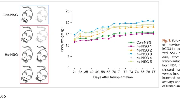
Fig. 1. Survival and weight changes of newborn NSG mice after hCD34+ cell injection. Humani- zed NSG mice were monitored daily from the 3rd week of transplantation. Most of the new- born NSG mice did well, but two showed features suggesting graft versus host disease (weight loss, hunched posture, and diminished activity) and died on the 26th day of transplantation.
BLOOD RESEARCH Volume 52ㆍNumber 4ㆍDecember 2017
Letters to the Editor
Transplantation of human umbilical cord blood CD34
+cells into the liver of newborn NOD/SCID/IL-2Rγ null (NSG) mice after busulfan
conditioning
TO THE EDITOR: Humanized mice carrying human hema- topoietic and immune systems are considered as ideal tools for studying hematopoiesis, infectious disease, and im- munology [1]. Various strains of immunodeficient mice have been developed to closely recapitulate human biological systems. Currently, NOD.Cg-PrkdcscidIl2rtm1Wjl/SzJ (NOD-scid IL2rnull, NSG) mice, which lack T-, B-, and NK cell activity, are considered as ideal candidates to establish humanized mice [1]. Humanized mouse models are still under develop- ment in order to improve human cell engraftment and func- tion [1]. Various humanization protocols exist, including those pertaining to the age of the recipient mice, condition- ing, cell source, and route of cell administration. As in human hematopoietic stem cell transplantation, systemic
(vein) administration is predominantly used to create humanized mice [1]. Here, we report that intrahepatic in- jection of human umbilical cord blood hematopoietic stem cells into the liver of newborn NSG mice after busulfan conditioning allowed significantly high human CD3+ T-cell engraftment.
NSG mice were purchased from The Jackson Laboratory (Bar Harbor, ME, USA) and maintained under specific patho- gen-free conditions at Laboratory of Animal Research Center in Korea Institute of Radiological Medical Sciences (Seoul, Korea). All experiments were performed according to guidelines of Institutional Animal Care and Use Committee (IACUC). Human umbilical cord blood CD34+ cells (StemPro CD34+ Cell Kit) were purchased from Life Technologies (Carlsbad, CA, USA). Newborn NSG mice (≤
48 hours after birth) were injected with busulfan (Korea Otsuka Pharmaceutical, Korea) in their retro-orbital sinus (25 mg/kg, 50–100 g per dose) 24 hours prior to transplantation. The next day, 3×10⁴ hCD34+ cells were injected into the liver. Twelve weeks later, mice were sacri- ficed and mononuclear cells were isolated from bone mar- row, liver, spleen, and peripheral blood. Single-cell suspen- sions were prepared by standard procedures and stained
bloodresearch.or.kr Blood Res 2017;52:316-39.
Letters to the Editor 317
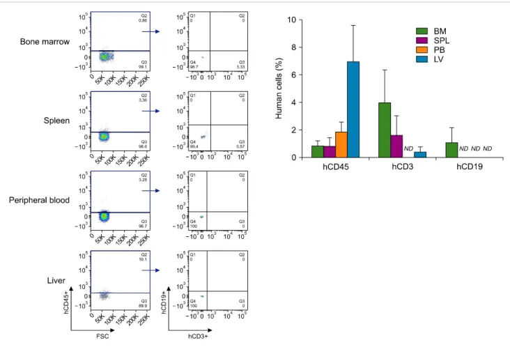
Fig. 2. Reconstitution of human cells in newborn NSG mice after intrahepatic transplantation of CD34+ cells. Mononuclear cells derived from bone marrow, spleen, peripheral blood and liver tissues of humanized NSG mice were isolated in the 12th week of transplantation and were stained with anti-hCD45, anti-hCD3, and anti-hCD19 antibodies. Data are means±SEM and representative of four mice per group, excluding lowest values (N=5).
Abbreviations: BM, bone marrow; LV, liver; ND, not detected; PB, peripheral blood; SPL, spleen.
with the following antibodies: hCD45-allophycocyanin (APC), hCD3-fluorescein isothiocyanate (FITC), and hCD19-phycoerythrin (PE) (BD Biosciences, San Jose, CA, USA). Flow cytometry was performed on FACSCanto II (BD Biosciences, San Jose, CA, USA).
Among the 8 NSG mice (including 2 control mice), 2 showed features suggesting graft versus host disease (weight loss, hunched posture, and diminished activity) and died on Day 26 of transplantation (Fig. 1). In the 12th week, the percentages of hCD45+ cells in the NSG mouse systems were 6.96% in the liver, 1.84% in the peripheral blood, 0.81% in the bone marrow, and 0.8% in the spleen.
Unexpectedly, CD19+ B-cell population was barely detected in mouse tissues, whereas significantly high human CD3+ T-cell population was observed. The population of hCD19+ B-cells was 1.09% in the bone marrow, and was not detected in any other tissue. The populations of hCD3+ T-cells were 3.98% in the bone marrow, 1.61% in the spleen, and 0.39%
in the liver (Fig. 2).
Even 12 weeks after the intrahepatic injection, human CD45+ cell reconstitution rate was high in the liver of NSG mice. The liver is the primary site of hematopoiesis during
the embryo and neonatal period [2]. As other liver functions increase several weeks after birth, the site of hematopoiesis gradually switches to the bone marrow [2]. It has been reported that the fetal liver provides a favorable micro- environment for hematopoiesis, and that macrophages were one of the major components comprising the early embry- onic hematopoietic microenvironment in mice [3]. The mi- croenvironment of the fetal liver enhances cell cycle pro- gression and proliferation of hematopoietic stem cells, with activation of Wnt signaling pathway [4]. On the contrary, microenvironment of the adult liver maintains hema- topoietic stem cells in a quiescent state, due to the prefer- ential role of Notch signaling pathway [4]. In adults, local damage of the liver stimulates liver regeneration and in- creases growth factors [5]. It was assumed that CD34+ cells from cord blood could be stimulated by stem cell factor or hepatocyte growth factor [6]. However, a significantly low engraftment of human CD34+ cord blood stem cells was found after intrahepatic transplantation in adult NOD/
SCID mice compared to newborn mice [6].
Limited data exist regarding humanized NSG mice gen- erated by intrahepatic injection of human hematopoietic
Blood Res2017;52:316-39. bloodresearch.or.kr
318 Letters to the Editor
stem cells. Organ-specific transplantation of hematopoietic stem cells is a useful method to study hematopoiesis and immune reconstitution [6]. It is reported that the differ- entiation pattern seems to differ between intrahepatic and intravenous transplanted CD34+ cells in NOD/SCID mice [6-8]. Intrahepatic transplantation of CD34+ cord blood stem cells into newborn NOD/SCID mice induced successful en- graftment of human cells. A high percentage of engrafted human cells was CD19+ B-cells, but lacked T-cell differ- entiation [6]. Others also reported that intrahepatic trans- plantation of cord blood CD34+ cells into newborn NSG allowed efficient multi-organ and multi-lineage hema- topoietic engraftment, predominantly of B-cells [7]. The two studies conditioned their newborn mice with irradiation and analyzed human cell engraftment earlier (≤10 wk).
Meanwhile, Choi et al. [8] reported that human T-cells developed in the liver of humanized NSG mice on intra- hepatic injection of human cord blood CD34+ cells. They used busulfan conditioning and analyzed human cell en- graftment for a prolonged period - until 20 weeks. Our study adopted busulfan conditioning and analyzed human cell engraftment during the 12th week after intrahepatic injection. Although this might explain the T-cell differ- entiation observed in our study, the reason we could not observe any B-cells in any of the mouse tissues is still elusive.
The peculiar finding of our study is that a significantly high human CD3+ T-cell population was detected in the bone marrow and spleen of the NSG mice, with barely detectable CD19+ B-cell population in all tissues. The extent to which the transplanted human stem cells would recon- stitute the hematopoietic system in NSG mice is still uncertain. The microenvironment and cytokines required for hematopoietic system development differs in human and mouse systems [1, 9]. NSG mice lack HLA molecules for human T-cell education, and have poorly organized lym- phoid architecture and deficiencies in development of lymph nodes [9]. Previous studies reported that most of the initially engrafted human cells in NSG mice were B-cells [10]. The engraftment level of human T-cells was lower than that of B-cells, and appeared 12–16 weeks after hema- topoietic stem cell transplantation [10]. It is presumed that T-cell development predominantly occurs in the thymus of NSG mice. However, only minute evidence exists support- ing this presumption. The transplanted human cord blood stem cells are detectable in mouse organs. However, in the thymus at different time intervals after long-term en- graftment, no CD3 expression was found [11]. Moreover, marginal enlargement of the thymus and minute increases in cellular number of the thymus were observed in human- ized NSG mice, compared to normal NSG mice [8]. Various studies tried to increase reconstitution of human hema- topoietic cells. The bone marrow, liver, thymus (BLT) model showed robust and stable engraftment of multiple human hematopoietic lineages, including T-cells [12]. Administrat- ion of recombinant human IL-7 improved T-cell develop- ment in humanized mice [13]. Currently, development of
new generation of immunodeficient mice strains, which express human hematopoietic growth factors, is underway [14, 15].
In this experiment, intrahepatic injection of human hema- topoietic stem cells into the liver of newborn NSG mice resulted in a significantly higher human CD3+ T-cell pop- ulation in the bone marrow and spleen, whereas CD19+
B-cell population was barely detectable in all tissues. We assume that intrahepatic injection of CD34+ cells in newborn NSG mice could facilitate T-cell reconstitution and un- identified factors in the fetal/newborn liver might contribute to T-cell development. Further studies are necessary to ex- plore the detailed cellular and molecular mechanisms re- garding the role of the liver in the reconstitution of human hematopoietic cells.
Yunmi Ko1, Yeon Ho Jeong2, Jun Ah Lee1,3
1Division of Clinical Translational Research, Korea Cancer Center Hospital, Seoul, 2Department of Medical Biotechnology, College of Biomedical Science, Kangwon National University, Chuncheon, 3Department of Pediatrics, Korea Cancer Center Hospital, Seoul, Korea
Correspondence to: Jun Ah Lee Department of Pediatrics, Korea Cancer Center Hospital,
75 Nowon-ro, Nowon-gu, Seoul 01812, Korea E-mail: [email protected]
Received on Feb. 3, 2017; Revised on Apr. 5, 2017; Accepted on Jun. 15, 2017 https://doi.org/10.5045/br.2017.52.4.316 Acknowledgments
This study was supported by a grant of the Korea Institute of Radiological and Medical Sciences (KIRAMS), funded by Ministry of Science, ICT and Future Planning, Republic of Korea (1711021931).
AuthorsÊ Disclosures of Potential Conflicts of Interest No potential conflicts of interest relevant to this article were reported.
REFERENCES
1. Shultz LD, Brehm MA, Garcia-Martinez JV, Greiner DL.
Humanized mice for immune system investigation: progress, promise and challenges. Nat Rev Immunol 2012;12:786-98.
2. Palis J, Robertson S, Kennedy M, Wall C, Keller G. Development of erythroid and myeloid progenitors in the yolk sac and embryo proper of the mouse. Development 1999;126:5073-84.
3. Sasaki K, Iwatsuki H. Origin and fate of the central macrophages of erythroblastic islands in the fetal and neonatal mouse liver.
Microsc Res Tech 1997;39:398-405.
4. Martin MA, Bhatia M. Analysis of the human fetal liver hema- topoietic microenvironment. Stem Cells Dev 2005;14:493-504.
5. Dalakas E, Newsome PN, Harrison DJ, Plevris JN. Hematopoietic stem cell trafficking in liver injury. FASEB J 2005;19:1225-31.
6. Wulf-Goldenberg A, Keil M, Fichtner I, Eckert K. Intrahepatic
bloodresearch.or.kr Blood Res 2017;52:316-39.
Letters to the Editor 319
transplantation of CD34+ cord blood stem cells into newborn and adult NOD/SCID mice induce differential organ engraftment.
Tissue Cell 2012;44:80-6.
7. Navarro-Montero O, Romero-Moya D, Montes R, et al.
Intrahepatic transplantation of cord blood CD34+ cells into new- born NOD/SCID-IL2Rγ(null) mice allows efficient multi-or- gan and multi-lineage hematopoietic engraftment without ac- cessory cells. Clin Immunol 2012;145:89-91.
8. Choi B, Chun E, Kim M, et al. Human T cell development in the liver of humanized NOD/SCID/IL-2Rγ(null)(NSG) mice gen- erated by intrahepatic injection of CD34(+) human (h) cord blood (CB) cells. Clin Immunol 2011;139:321-35.
9. Brehm MA, Shultz LD, Luban J, Greiner DL. Overcoming current limitations in humanized mouse research. J Infect Dis 2013;208(Suppl 2):S125-30.
10. Ishikawa F, Yasukawa M, Lyons B, et al. Development of func- tional human blood and immune systems in NOD/SCID/IL2 re- ceptor {gamma} chain(null) mice. Blood 2005;106:1565-73.
11. McKenzie JL, Gan OI, Doedens M, Dick JE. Human short-term repopulating stem cells are efficiently detected following intra- femoral transplantation into NOD/SCID recipients depleted of CD122+ cells. Blood 2005;106:1259-61.
12. Lavender KJ, Messer RJ, Race B, Hasenkrug KJ. Production of bone marrow, liver, thymus (BLT) humanized mice on the C57BL/6 Rag2(-/-)γc(-/-)CD47(-/-) background. J Immunol Methods 2014;407:127-34.
13. van Lent AU, Dontje W, Nagasawa M, et al. IL-7 enhances thymic human T cell development in "human immune system"
Rag2-/-IL-2Rgammac-/- mice without affecting peripheral T cell homeostasis. J Immunol 2009;183:7645-55.
14. Chen Q, Khoury M, Chen J. Expression of human cytokines dra- matically improves reconstitution of specific human-blood line- age cells in humanized mice. Proc Natl Acad Sci U S A 2009;106:21783-8.
15. Drake AC, Chen Q, Chen J. Engineering humanized mice for im- proved hematopoietic reconstitution. Cell Mol Immunol 2012;9:215-24.
Successful treatment of a patient with myelodysplastic syndrome accompanied by pyoderma
gangrenosum and BehçetÊs disease using allogeneic stem cell
transplantation
TO THE EDITOR: Myelodysplastic syndrome (MDS) is a het- erogeneous group of hematopoietic stem cell disorders with ineffective hematopoiesis and resulting cytopenias [1].
Approximately 10–20% of patients with MDS present with autoimmune diseases, such as vasculitis, rheumatoid arthri-
tis, and inflammatory bowel disease [2, 3]. Sometimes, pa- tients with MDS develop skin manifestations including pyo- derma gangrenosum (PG), Sweet syndrome, and skin in- filtration of malignant cells. The appearance of cutaneous lesions is associated with a poor prognosis [4-6]. Traditio- nally, hematopoietic stem cell transplantation (HSCT) has been used for refractory autoimmune diseases [7-9] and hematological disorders. Here, we report a case of MDS associated with PG and Behçet’s disease that was successfully treated with allogeneic stem cell transplantation.
A 53-year-old man was referred to our hospital in December 2011 for evaluation of pancytopenia. He had suf- fered from a chronic granulomatous skin lesion on the face for 2 years; there was no improvement despite treatment with methylprednisolone, methotrexate, and dapsone (Fig.
1A). Laboratory findings were as follows: white blood cell count 1,900/μL, neutrophils 1,010/μL, hemoglobin 8.5 g/dL, platelets 112,000/μL, and no blasts in the peripheral blood.
At this time, a minimal dose of methotrexate (5 mg/week) was being administered. A bone marrow biopsy showed 4.6% myeloblasts and increased megakaryocytes with dys- plastic features. We could not get cytogenetic information due to an inappropriate specimen for karyotyping.
He was diagnosed with MDS with refractory cytopenia with multilineage dysplasia (RCMD) and received a packed red blood cell transfusion every 2 months. In December 2013, he was hospitalized with fever, an aggravated skin lesion, and severe cytopenias (Fig. 1B). Lab tests revealed aggravated pancytopenia: white blood cell count 2,000/μL, neutrophils 240/μL, hemoglobin 4.9 g/dL, and platelets 32,000/μL.
A colonoscopy, performed to evaluate the marked anemia, showed an ulcerative lesion in the terminal ileum (Fig.
1B). A biopsy revealed a chronic ulcer with active in- flammation and polymerase chain reaction tests for Mycobacterium tuberculosis were negative. Serological tests revealed positivity for HLA B51 and a pathergy test was positive. Based on these results, he was diagnosed with Behçet’s disease (Fig. 1B). A skin biopsy of the lower lip showed ulceration with acute and chronic inflammation and focal suppurative inflammation, suggestive of pyoderma gangrenosum.
A subsequent examination of his bone marrow showed a persistent state of MDS with RCMD with a karyotype of 47, XY, dup(1)(q21q32), +8[20]. Based on the Interna- tional Prognostic Scoring System (IPSS), he was categorized into the intermediate-1 risk group; based on the revised IPSS, he was categorized into the high-risk group. He re- ceived three cycles of azacitidine (75 mg/m2 intravenously for 7 days, every 4 wk) bridge therapy before HSCT, which did not lead to a response. Unrelated peripheral blood HSCT with reduced-intensity conditioning (30 mg/m2/day fludar- abine intravenously for 6 days; 3.2 mg/kg/day busulfan intra- venously for 2 days; total 5 mg/kg anti-thymocyte im- munoglobulin for graft-versus-host-disease prophylaxis) was performed in March 2014, 27 months after the diagnosis