This is an Open Access article distributed under the terms of the Creative Commons At- tribution Non-Commercial License (http://creativecommons.org/licenses/by-nc/3.0/) which permits unrestricted non-commercial use, distribution, and reproduction in any medium, provided the original work is properly cited.
Adenine Nucleotide Translocator as a Regulator of Mitochondrial Function: Implication in the
Pathogenesis of Metabolic Syndrome
Eun Hee Kim, Eun Hee Koh, Joong-Yeol Park, Ki-Up Lee
Department of Internal Medicine, University of Ulsan College of Medicine, Seoul, Korea
Mitochondria play key roles in energy production and intracellular reactive oxygen species (ROS) generation. Lines of evidence have shown that mitochondrial dysfunction contributes to the development of metabolic syndrome. The causes of mitochondrial dysfunction are complex, but overnutrition and sedentary living are among the best known causes of mitochondrial dysfunction.
ATP synthesized in the mitochondria is exchanged for cytosolic ADP by adenine nucleotide translocator (ANT) to provide a continuous supply of ADP to mitochondria. We recently found that ANT function is essential for peroxisome proliferator-acti- vated receptor-γ coactivator 1-α (PGC-1α)’s action on endothelial cells. PGC-1α is a transcriptional coactivator of nuclear re- ceptors, playing an important role in fatty acid oxidation and mitochondrial biogenesis. Recent studies have shown that PGC-1α decreases intracellular ROS generation by increasing the expression of antioxidant genes. In our study, PGC-1α reduced cell apoptosis and ROS generation in endothelial cells by increasing ATP/ADP translocase activity of ANT and ANT1 expression.
Here we review the role of ANT in maintaining proper mitochondrial function, and possible role of ANT dysfunction in the pathogenesis of metabolic syndrome.
Keywords: Adenine nucleotide translocator; Metabolic syndrome; Mitochondrial dysfunction
Corresponding author: Ki-Up Lee
Department of Internal Medicine, University of Ulsan College of Medicine, 388-1 Pungnap2-dong, Songpa-gu, Seoul 138-736, Korea
E-mail: [email protected]
INTRODUCTION
Metabolic syndrome is a cluster of common metabolic risk fac- tors for atherosclerosis and type 2 diabetes occurring in obese subjects [1]. Metabolic syndrome is rapidly increasing in prev- alence worldwide as a consequence of the continued obesity
“epidemic”, and as a result will have a considerable impact on the global incidence of cardiovascular disease and type 2 dia- betes [2]. The major pathophysiologic mechanism of metabol- ic syndrome is insulin resistance [3]. Growing body of evidence has shown that mitochondrial dysfunction is an important pathogenic mechanism of diseases associated with insulin resis-
tance, i.e., diabetes, atherosclerosis, and fatty liver disease [4].
Mitochondrion is the main organ of energy production, mostly in the form of ATP via oxidative phosphorylation (OX- PHOS) and also a major site of intracellular reactive oxygen species (ROS) generation [5]. The ATP synthesized in the mi- tochondria is exchanged for cytosolic ADP by adenine nucle- otide translocator (ANT) to provide a continuous supply of ADP to mitochondria. ATP/ADP exchange by ANT is essential for the maintenance of ATP synthase activity [6]. On the other hand, in states of impaired function of ATP/ADP exchange, ANT plays a major role in generating ROS and inducing cell apoptosis [7,8]. In this article, we will review the role of ANT pISSN 1976-9180 · eISSN 2093-2650
in maintaining mitochondrial function, and possible role of ANT dysfunction in the pathogenesis of metabolic syndrome.
STRUCTURE AND FUNCTION OF MITOCHONDRIA
Structure
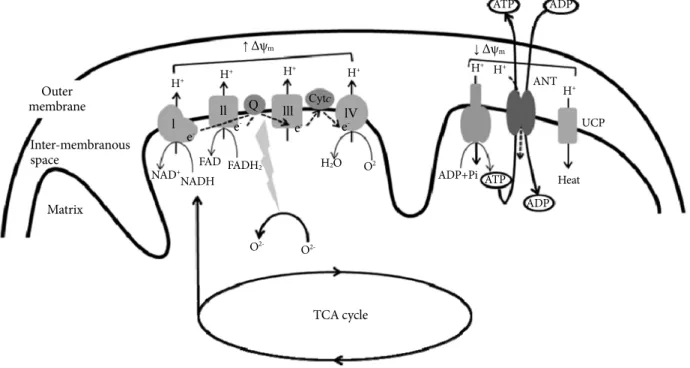
Mitochondrion is an intracellular double-membrane organelle present in most of eukaryotic cells [9]. Mitochondria form a reticulum that is in continuous communication through dy- namic fusion and fission events, moving actively to different regions of the cell through interactions with the cytoskeleton [10]. The mitochondrial reticulum is composed of an inner and outer membrane, between which lies the intermembranous space, and a matrix contained within the inner membrane. The surface of the inner membrane is folded into cristae, which gives mitochondrion its characteristic morphology (Fig. 1).
Mitochondrion has its own circular mitochondrial DNA (mtDNA) molecule, which encodes for 37 genes (13 of which are subunits of the electron transport chain, ETC) [11]. The majority of proteins regulating mitochondrial structure, func- tion and biogenesis are encoded by the nuclear DNA. However, mtDNA gene products are essential for normal functioning of the mitochondrial respiratory chain. The mtDNA has no in- trons and is poorly equipped with repair mechanism, render- ing it susceptible to oxidative damage and mutations. The mtD- NA mutations accumulate with age, and these mutations might have an important role in the process of senescence and diabe- tes [12].
Function
Inner membrane contains all the transmembrane proteins of the ETC as well as the mitochondrial ATPase. The matrix con- tains the enzymatic machinery for TCA cycle, which provides
Fig. 1. Mitochondrial electron-transport chain (ETC). Electrons derived from reducing equivalents (NADH and FADH2) are transported within ETC to molecular oxygen to produce water. As the electrons are transported, the free energy released is used to pump the protons into the intermembranous space. The proton gradient generated creates mitochondrial membrane potential (Δψm). The proton gradient produced is dissipated through the mitochondrial ATPase to produce ATP (OXPHOS or coupled res- piration). The ATP synthesized in the mitochondria is exchanged for cytosolic ADP by adenine nucleotide translocator (ANT).
Reactive oxygen species (ROS) is normally produced in the ETC during respiration, but delay of electron transport in the ETC re- sults in the overproduction of ROS. ROS generation is more likely to occur when the proton gradient is large (increase in Δψm). Ac- cumulation of ROS activates uncoupling protein (UCP), which dissipates the proton gradient without producing ATP (uncoupled respiration), decreases Δψm and ROS production. ANT also exhibits uncoupling activity or proton leak, and decreases ROS pro- duction and Δψm.
TCA cycle Matrix
Outer membrane
Inter-membranous space
NAD+NADHFAD FADH2
e-
↑ Δψm
l ll Q lll Cytc lV
↓ Δψm
e- e- e-
H2O O2
ADP+Pi ATP Heat
ADP
H+ H+ H+ H+ H+
H+ UCP ANT
ATP ADP
H+
O2- O2-
reducing equivalents, such as NADH and FADH2, to the ETC, and for β-oxidation, which generates acetyl-CoA from acyl chains. The ETC is composed of four large multisubunit com- plexes (complex I to IV) with more than 85 individual gene products. Electrons derived from reducing equivalents NADH and FADH2 are transported within the ETC to molecular oxy- gen to produce water. As the electrons are transported, the free energy released is used to pump the protons into the inter-mem- branous space. The proton gradient thus generated creates elec- trochemical gradient across the inner mitochondrial membrane (mitochondrial membrane potential, Δψm) (Fig. 1). The ener- gy contained in the proton gradient is then coupled to ATP production as protons flow back into the matrix through the mi- tochondrial ATPase. This process is known as OXPHOS, or cou- pled respiration [13]. The high proton gradient also drives the rapid entry of Ca2+ into the mitochondrial matrix, buffering its concentration in the cytoplasm [14].
Mitochondria are also a potent source of ROS [15]. ROS is normally produced in the ETC during respiration, but delay of electron transport in the ETC results in the overproduction of ROS. ROS generation is more likely to occur when the proton gradient is large: increase in Δψm is associated with delay of electron transport in ETC [16]. Consistent with this idea, we recently reported that overexpression of uncoupling protein 2 (UCP2), which dissipates the proton gradient without produc- ing ATP, decreased Δψm and ROS production in cultured en- dothelial cells [17].
Mitochondria are also the prime regulator of apoptosis [12].
When confronted with cellular stress, mitochondria open the mitochondrial permeability transition pore (mtPTP) [18]. Open- ing of the mtPTP allows the release of mitochondrial proteins, such as cytochrome c, caspases, and apoptosis initiating factor, to induce apoptosis [19].
MITOCHONDRIAL DYSFUNCTION IN METABOLIC SYNDROME
Skeletal muscle is the major organ that determines whole body insulin sensitivity. In 1963, Randle et al. [20] proposed that in- creased free fatty acids (FFA) availability and oxidation lead to insulin resistance in skeletal muscle by inhibiting glucose oxi- dation and glycogen synthesis. Subsequent studies have shown that both glucose oxidation and glycogen synthesis are impaired in state of high FFA availability [21-23]. However, recent studies have suggested that defective intracellular fatty acid metabolism
in skeletal muscle, rather than a simple oversupply of fatty acid fuel, is causally related to the development of insulin resistance [24]. In accordance with this concept, we previously showed that lipolysis in skeletal muscle was decreased in high fat-fed rats, suggesting that intracellular triglyceride accumulation in the insulin resistant state is the consequence of a diminished fatty acid oxidation capacity rather than the cause of insulin resistance [25].
Insulin resistance in the elderly or diabetic offspring is relat- ed to a reduction in the mitochondrial oxidative phosphoryla- tion capacity [26,27]. Petersen et al. [26] reported that insulin resistance in the skeletal muscle of insulin resistant offspring of patients with type 2 diabetes was associated with dysregula- tion of intramyocellular fatty acid metabolism, possibly be- cause of an inherited defect in mitochondrial oxidative phos- phorylation. It was suggested that insulin resistance in humans arises from defects in mitochondrial fatty acid oxidation, which leads to increases in intracellular fatty acid metabolites (fatty acyl CoA and diacylglycerol) that disrupt insulin signaling [28].
OVERNUTRITION AS A CAUSE OF MITOCHONDRIAL DYSFUNCTION IN METABOLIC SYNDROME
The cause of mitochondrial dysfunction in metabolic syndrome may be multifactorial [29]. Among them, overnutrition and underutilization of nutrition are shown to induce mitochon- drial dysfunction [30]. Chronic aerobic exercise increases mi- tochondrial content in muscle, thereby increasing ATP gener- ating capacity [31]. On the other hand, chronic disuse of mus- cle decrease mitochondrial content and oxidative capacity lead- ing to impaired glucose utilization [32].
It is well established that fasting prolongs lifespan. SIRT (mam- malian homologues of Sir2; silent information regulator 2), which was identified as a mediator of longevity, increases mi- tochondrial biogenesis and improves mitochondrial function [33]. Conversely, high-fat diet (HFD) has been shown to reduce mitochondrial function. Genes necessary for OXPHOS and mitochondrial biogenesis were downregulated in skeletal mus- cle of the mice given HFD [34]. It was also shown that HFD de- creases the expression of oxidative genes in healthy human indi- viduals [34]. As a consequence of mitochondrial dysfunction and impaired fatty acid oxidation, intracellular levels of lipid metabolites, i.e., long chain fatty acyl coenzyme A (LCAC), dia- cylglycerol, and ceramides, are increased in skeletal muscle, liv-
er, heart and pancreas β-cells of obese subjects [28]. Excess in- take of nutrients also increases ROS production in the mito- chondria. ROS-induced peroxidation may in turn leads to dam- age of mitochondria and further deterioration in oxidative ca- pacity [35].
ATP/ADP TRANSLOCASE (ANT)
Human ANT has four isoforms (ANT1, ANT2, ANT3, and ANT4), which display distinct tissue-specific expression pat- terns. ANT1 is predominantly expressed in the heart, skeletal muscle, and brain. ANT2 is predominantly expressed in the liver and in cells with increased proliferative activity. ANT3 is ubiquitously detected. ANT4 is expressed in the liver, testis, and undifferentiated embryonic stem cells [36]. Among them, we will primarily focus on ANT1.
FUNCTIONS OF ANT ATP/ADP TRANSLOCASE(ANT)
ATP/ADP translocase
ANT is a protein complex of two subunits that is located in the inner mitochondrial membrane and facilitates the exchange of mitochondrial ATP and cytosolic ADP [37]. ANT provides a continuous supply of ADP necessary to maintain the oxidative phosphorylation process. ATP/ADP exchange by ANT plays an essential role for the maintenance of ATP synthase activity and normal levels of Δψm. Impaired ATP/ADP translocase ac- tivity of ANT decreases intramitochondrial ADP and ATP synthesis, and increases Δψm [6].
Regulation of apoptosis
As described above, the prime function of ANT is to facilitate the ATP/ADP exchange across the inner mitochondrial mem- brane. However, in states where ATP/ADP translocase activity is impaired, ANT plays a major role in promoting apoptosis.
Mitochondrial membrane permeabilization (MMP) is a rate limiting step of apoptosis and is mediated by the mito- chondrial permeability transition pore (mtPTP) [38]. mtPTP is a nonspecific pore, permeable to all molecules of less than 1.5 kDa and is formed by the voltage-dependent anion channel (VDAC), members of the pro- and anti apoptotic Bax/Bcl2 protein family, cyclophilin D, and the ANT [39]. Additional proteins that were proposed to be part of the mtPTP complex are hexokinase, creatine kinase, and peripheral benzodiaz-
epine receptor [40]. mtPTP opening causes swelling of the mi- tochondrial matrix and outer membrane rupture. This is fol- lowed by release of cytochrome c and other proapoptotic pro- teins into cytosol [38].
ANT has been widely accepted as a component for the mtPTP complex, which was first proposed by Halestrap et al.
in 1990 [41]. ANT has been proposed to interact with VDAC, which is located in the outer mitochondrial membrane, to form a large protein-permeable conduit [42]. However, Wallace et al. have shown that mitochondria from livers of ANT-knock- out mice, in which the ANT has been genetically inactivated, still possess mtPTP activity [18]. This would imply that the ANT is not an essential component of the mtPTP. Despite these debates, ANT is still considered to play a major regulatory role in the genesis of mtPTP [42].
Mitochondrial uncoupler
Under physiological conditions, mitochondrial oxygen con- sumption is tightly coupled to ATP synthesis. The bulk of pro- ton re-enter the matrix via the F0F1 ATPase, which uses the energy to regenerate ATP from ADP (coupling of OXPHOS).
A small proportion of proton can bypass the F0F1 ATPase, so that mitochondrial oxygen consumption is not coupled to ATP synthesis (mitochondrial uncoupling) [43]. In the 1970s, a pro- tein responsible for non-shivering thermogenesis was identified in the inner mitochondrial membrane of brown adipose tissue mitochondria, and was named as uncoupling protein (UCP1) [44]. More recently, four more UCP homologues have been identified (UCP2, UCP3, UCP4 and UCP5/BCMP1 [brain mi- tochondrial carrier protein 1]) [45]. These proteins mediate pro- ton leak across the mitochondrial membrane and decrease Δψm [46]. Since ROS production increases with increasing Δψm, UCP-mediated uncoupling has been proposed to play a role in decreasing mitochondrial ROS production [17]. This may represent a mechanism by which mitochondria protect themselves from oxidative damage [47].
Several lines of evidence suggested that ANT is also a mito- chondrial uncoupler and is responsible for basal uncoupling or proton leak [43]. In rodents, ANT1 and ANT2 were shown to mediate uncoupling by fatty acids and to lower mitochon- drial membrane potential in heart and skeletal muscle [48]. It was also demonstrated that ANT1-deficient mice have a 50%
decrease in proton conductance in skeletal muscle [49]. In the heart, 4-hydroxy-2-nonenal-induced proton leak could be in- hibited by the ANT inhibitor carboxyatractyloside, but not by
the UCP inhibitor GDP [50]. These results suggest that ANT may decrease mitochondrial ROS production by functioning as an uncoupler.
INCREASED INTRACELLULAR LCAC IN METABOLIC SYNDROME IMPAIRS ATP/ADP TRANSLOCASE ACTIVITY
Central obesity is associated with increased cytosolic triglyc- eride stores in non-adipose tissue such as muscles, liver and pancreatic β-cells [51-53]. Cytosolic triglyceride is a source of LCAC, the metabolically active form of fatty acids. LCAC may accumulate under pathological conditions with excess lipid supply, such as obesity, and conditions with a mitochondrial fatty acid β-oxidation defect [54]. LCAC was shown to inhibit the ATP/ADP translocase activity of the ANT by competitive displacement of the nucleotide from its binding site on the pro- tein [55].
It has been thus hypothesized that increased concentrations of LCACs in the cell interfere with mitochondrial function through inhibition of the ANT. Inhibition of ATP/ADP trans- locase activity lowers cytosolic ATP and matrix ADP availabil- ity, and increases mitochondrial membrane potential (Δψm) [8]. These events promote the formation of ROS, resulting in impaired cellular functions and cell death. Inhibition of the mi- tochondrial ANT by LCACs has been thus proposed to contrib- ute to mitochondrial dysfunction in metabolic syndrome [8,56].
PGC-1α PREVENTS ENDOTHELIAL APOPTOSIS BY INCREASING ATP/ADP TRANSLOCASE ACTIVITY
Peroxisome proliferator-activated receptor-γ coactivator 1-α (PGC-1α) is a transcriptional coactivator of nuclear receptors,
playing an important role in energy metabolism [57]. PGC-1α increases mitochondrial biogenesis and fatty acid oxidation [58].
In addition, recent studies have reported that PGC-1α is a ma- jor regulator of intracellular ROS generation. It was suggested that PGC-1α increases the expression of antioxidant genes [59].
We recently found that PGC-1α prevents endothelial apop- tosis by increasing ATP/ADP translocase activity of ANT [7].
It is well known that fatty acids, such as linoleic acid (LA), in- crease ROS generation and cell apoptosis in endothelial cells [17]. LA treatment in human aortic endothelial cells increased intracellular and mitochondrial ROS generation and apopto-
Table 1. Effect of linoleic acid (LA) and PGC1-α on various functions of endothelial cells Δψm ROS Apoptosis Antioxidant
enzyme/
UCP2
ANT-1 expression
ATP/ADP translocase
activity FAO Intracellular lipid metabolites
Control + + + + + + + +
LA ++ ++ ++ ++ ++ - - ++
PGC-1 α + + + ++ ++ ++ +++ +
LA + PGC-1 α + + + ++ ++ ++ ++ +
PGC1-α, peroxisome proliferator-activated receptor-γ coactivator 1-α; ROS, reactive oxygen species; Δψm, mitochondrial membrane potential;
UCP2, uncoupling protein 2; FAO, fatty acid oxidation.
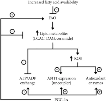
Fig. 2. Proposed model of PGC-1α actions on endothelial cells to prevent ROS generation and cell apoptosis. PGC1-α, peroxisome proliferator-activated receptor-γ coactivator 1-α;
ROS, reactive oxygen species; FAO, fatty acid oxidation;
LCAC, long chain fatty acyl coenzyme A; DAG, diacylglycerol;
ANT, adenine nucleotide translocator.
Increased fatty acid availability
Lipid metabolites (LCAC, DAG, ceramide)
ROS
ATP/ADP
exchange ANT1 expression (uncoupler)
PGC-1α +
+ +
+ + +
- -
-
Antioxidant enzymes FAO
sis. PGC-1α overexpression prevented LA-induced increases in ROS generation and apoptosis (Table 1, Fig. 2).
LA increased the mRNA and protein expression of antioxi- dant enzymes, including manganese superoxide dismutase, copper-zinc superoxide dismutase, catalase, glutathione per- oxidase, and uncoupling protein2 UCP2, consistent with the notion that oxidative stress can induce cellular antioxidant re- sponses [60]. As previously reported [59], adenoviral overex- pression of PGC-1α also increased the mRNA and protein ex- pression of the same antioxidant enzymes and UCP2. However, in the presence of LA, endogenous PGC-1α did not further increase the expression of antioxidant enzymes or UCP2. This result suggests that PGC-1α’s effect on antioxidant genes may not fully explain its effect to decrease intracellular ROS pro- duction and cell apoptosis.
Similar to other antioxidant genes, LA significantly increased ANT-1 expression. On the other hand, LA significantly de- creased ATP/ADP translocase activity, as measured by 14C- ADP import. This was associated with a significant increase in Δψm (hyperpolarization) and ROS generation. Interestingly, inhibitors of fatty acyl CoA synthase and ceramide synthase reduced LA-induced effects on ATP/ADP translocase activity, suggesting involvement of lipid metabolites, such as LCAC, diacylglycerol, and ceramide, in LA-induced impairment of ATP/ADP translocase activity. As expected, this was associat- ed with changes in intracellular ceramides levels. On the other hand, antioxidant N-aceylcysteine prevented LA-induced ANT- 1 expression, but did not affect ATP/ADP translocase activity regardless of LA treatment. These results suggest that increased ROS generation with LA may be responsible for the increase in ANT-1 expression. More importantly, changes in ANT-1 ex- pression may not account for the decrease in ATP/ADP trans- locase activity with LA.
PGC-1α overexpression completely reversed LA-dependent decreases in ATP/ADP translocase activity, and prevented LA- induced changes in Δψm. PGC-1α also increased ANT-1 ex- pression but did not increase ANT-1 expression above the lev- els induced by LA. In isolated aortic ring, LA treatment signif- icantly decreased endothelium-dependent vascular relaxation.
PGC-1α significantly inhibited LA-induced decreases in en- dothelium-dependent vasorelaxation, confirming that PGC- 1α has antiatherogenic effects in vascular endothelial cells.
Taken together, PGC-1α-dependent enhancement of ATP/
ADP translocase activity of ANT is critically required for the beneficial effects of PGC-1α on endothelial function.
CONCLUSION
From this brief review, we have shown that ANT function is important in the maintenance of mitochondrial function. The prime function of ANT is exchange of ATP and ADP across the inner mitochondrial membrane, which is important for both ATP production and maintenance of normal Δψm. ANT also plays a role as an uncoupler. These two functions are impor- tant to protect the mitochondria from increased ROS genera- tion associated with increased Δψm. However, in states where ATP/ADP translocase activity is impaired, ANT participates to play a role in the genesis of mtPTP and cell apoptosis. We have shown recently that PGC1-α regulates ROS generation and apoptosis in endothelial cells by enhancing ATP/ADP trans- locase activity of ANT. Understanding these mechanisms may help to find measures to prevent or treat metabolic syndrome.
REFERENCES
1. Isomaa B, Almgren P, Tuomi T, Forsen B, Lahti K, Nissen M, Taskinen MR, Groop L. Cardiovascular morbidity and mortali- ty associated with the metabolic syndrome. Diabetes Care 2001;
24:683-9.
2. Bestermann W, Houston MC, Basile J, Egan B, Ferrario CM, Lackland D, Hawkins RG, Reed J, Rogers P, Wise D, Moore MA. Addressing the global cardiovascular risk of hyperten- sion, dyslipidemia, diabetes mellitus, and the metabolic syn- drome in the southeastern United States, part II: treatment recommendations for management of the global cardiovascu- lar risk of hypertension, dyslipidemia, diabetes mellitus, and the metabolic syndrome. Am J Med Sci 2005;329:292-305.
3. Reaven GM. Banting lecture 1988: role of insulin resistance in human disease. Diabetes 1988;37:1595-607.
4. Kim JA, Wei Y, Sowers JR. Role of mitochondrial dysfunction in insulin resistance. Circ Res 2008;102:401-14.
5. Schonfeld P, Wojtczak L. Fatty acids as modulators of the cellu- lar production of reactive oxygen species. Free Radic Biol Med 2008;45:231-41.
6. Vander Heiden MG, Chandel NS, Schumacker PT, Thompson CB. Bcl-xL prevents cell death following growth factor with- drawal by facilitating mitochondrial ATP/ADP exchange. Mol Cell 1999;3:159-67.
7. Won JC, Park JY, Kim YM, Koh EH, Seol S, Jeon BH, Han J, Kim JR, Park TS, Choi CS, Lee WJ, Kim MS, Lee IK, Youn JH, Lee KU. Peroxisome proliferator-activated receptor-gamma
coactivator 1-alpha overexpression prevents endothelial apop- tosis by increasing ATP/ADP translocase activity. Arterioscler Thromb Vasc Biol 2010;30:290-7.
8. Bakker SJ, RG IJ, Teerlink T, Westerhoff HV, Gans RO, Heine RJ. Cytosolic triglycerides and oxidative stress in central obesi- ty: the missing link between excessive atherosclerosis, endothe- lial dysfunction, and beta-cell failure? Atherosclerosis 2000;148:
17-21.
9. Wallace DC. Mitochondrial diseases in man and mouse. Sci- ence 1999;283:1482-8.
10. Shaw JM, Nunnari J. Mitochondrial dynamics and division in budding yeast. Trends Cell Biol 2002;12:178-84.
11. Anderson S, Bankier AT, Barrell BG, de Bruijn MH, Coulson AR, Drouin J, Eperon IC, Nierlich DP, Roe BA, Sanger F, Schreier PH, Smith AJ, Staden R, Young IG. Sequence and organization of the human mitochondrial genome. Nature 1981;290:457-65.
12. Wallace DC. A mitochondrial paradigm of metabolic and de- generative diseases, aging, and cancer: a dawn for evolutionary medicine. Annu Rev Genet 2005;39:359-407.
13. Saraste M. Oxidative phosphorylation at the fin de siecle. Sci- ence 1999;283:1488-93.
14. Murgia M, Giorgi C, Pinton P, Rizzuto R. Controlling metabo- lism and cell death: at the heart of mitochondrial calcium sig- nalling. J Mol Cell Cardiol 2009;46:781-8.
15. Raha S, Robinson BH. Mitochondria, oxygen free radicals, dis- ease and ageing. Trends Biochem Sci 2000;25:502-8.
16. Korshunov SS, Skulachev VP, Starkov AA. High protonic po- tential actuates a mechanism of production of reactive oxygen species in mitochondria. FEBS Lett 1997;416:15-8.
17. Lee KU, Lee IK, Han J, Song DK, Kim YM, Song HS, Kim HS, Lee WJ, Koh EH, Song KH, Han SM, Kim MS, Park IS, Park JY.
Effects of recombinant adenovirus-mediated uncoupling pro- tein 2 overexpression on endothelial function and apoptosis.
Circ Res 2005;96:1200-7.
18. Kokoszka JE, Waymire KG, Levy SE, Sligh JE, Cai J, Jones DP, MacGregor GR, Wallace DC. The ADP/ATP translocator is not essential for the mitochondrial permeability transition pore.
Nature 2004;427:461-5.
19. Wang C, Youle RJ. The role of mitochondria in apoptosis. Annu Rev Genet 2009;43:95-118.
20. Randle PJ, Garland PB, Hales CN, Newsholme EA. The glu- cose fatty-acid cycle: its role in insulin sensitivity and the met- abolic disturbances of diabetes mellitus. Lancet 1963;1:785-9.
21. Lee KU, Lee HK, Koh CS, Min HK. Artificial induction of in- travascular lipolysis by lipid-heparin infusion leads to insulin
resistance in man. Diabetologia 1988;31:285-90.
22. Kim CH, Youn JH, Park JY, Hong SK, Park KS, Park SW, Suh KI, Lee KU. Effects of high-fat diet and exercise training on in- tracellular glucose metabolism in rats. Am J Physiol Endocrinol Metab 2000;278:E977-84.
23. Itani SI, Ruderman NB, Schmieder F, Boden G. Lipid-induced insulin resistance in human muscle is associated with changes in diacylglycerol, protein kinase C, and IkappaB-alpha. Diabe- tes 2002;51:2005-11.
24. Kelley DE, He J, Menshikova EV, Ritov VB. Dysfunction of mitochondria in human skeletal muscle in type 2 diabetes. Di- abetes 2002;51:2944-50.
25. Kim CH, Kim MS, Youn JY, Park HS, Song HS, Song KH, Park JY, Lee KU. Lipolysis in skeletal muscle is decreased in high-fat- fed rats. Metabolism 2003;52:1586-92.
26. Petersen KF, Dufour S, Befroy D, Garcia R, Shulman GI. Im- paired mitochondrial activity in the insulin-resistant offspring of patients with type 2 diabetes. N Engl J Med 2004;350:664-71.
27. Iossa S, Mollica MP, Lionetti L, Crescenzo R, Tasso R, Liverini G. A possible link between skeletal muscle mitochondrial effi- ciency and age-induced insulin resistance. Diabetes 2004;53:
2861-6.
28. Lowell BB, Shulman GI. Mitochondrial dysfunction and type 2 diabetes. Science 2005;307:384-7.
29. Lee HK, Cho YM, Kwak SH, Lim S, Park KS, Shim EB. Mito- chondrial dysfunction and metabolic syndrome-looking for environmental factors. Biochim Biophys Acta 2010;1800:282-9.
30. Frisard M, Ravussin E. Energy metabolism and oxidative stress:
impact on the metabolic syndrome and the aging process. En- docrine 2006;29:27-32.
31. Hood DA. Mechanisms of exercise-induced mitochondrial biogenesis in skeletal muscle. Appl Physiol Nutr Metab 2009;34:
465-72.
32. Wicks KL, Hood DA. Mitochondrial adaptations in denervat- ed muscle: relationship to muscle performance. Am J Physiol 1991;260:C841-50.
33. Rodgers JT, Lerin C, Haas W, Gygi SP, Spiegelman BM, Puig- server P. Nutrient control of glucose homeostasis through a complex of PGC-1alpha and SIRT1. Nature 2005;434:113-8.
34. Sparks LM, Xie H, Koza RA, Mynatt R, Hulver MW, Bray GA, Smith SR. A high-fat diet coordinately downregulates genes required for mitochondrial oxidative phosphorylation in skel- etal muscle. Diabetes 2005;54:1926-33.
35. Schrauwen P, Schrauwen-Hinderling V, Hoeks J, Hesselink MK.
Mitochondrial dysfunction and lipotoxicity. Biochim Biophys
Acta 2010;1801:266-71.
36. Dolce V, Scarcia P, Iacopetta D, Palmieri F. A fourth ADP/ATP carrier isoform in man: identification, bacterial expression, functional characterization and tissue distribution. FEBS Lett 2005;579:633-7.
37. Fiore C, Trezeguet V, Le Saux A, Roux P, Schwimmer C, Di- anoux AC, Noel F, Lauquin GJ, Brandolin G, Vignais PV. The mitochondrial ADP/ATP carrier: structural, physiological and pathological aspects. Biochimie 1998;80:137-50.
38. Halestrap AP. What is the mitochondrial permeability transi- tion pore? J Mol Cell Cardiol 2009;46:821-31.
39. Zoratti M, Szabo I. The mitochondrial permeability transition.
Biochim Biophys Acta 1995;1241:139-76.
40. Schwarz M, Andrade-Navarro MA, Gross A. Mitochondrial carriers and pores: key regulators of the mitochondrial apop- totic program? Apoptosis 2007;12:869-76.
41. Halestrap AP, Davidson AM. Inhibition of Ca2(+)-induced large-amplitude swelling of liver and heart mitochondria by cyclosporin is probably caused by the inhibitor binding to mi- tochondrial-matrix peptidyl-prolyl cis-trans isomerase and preventing it interacting with the adenine nucleotide translo- case. Biochem J 1990;268:153-60.
42. Leung AW, Halestrap AP. Recent progress in elucidating the molecular mechanism of the mitochondrial permeability tran- sition pore. Biochim Biophys Acta 2008;1777:946-52.
43. Boudina S, Abel ED. Mitochondrial uncoupling: a key con- tributor to reduced cardiac efficiency in diabetes. Physiology (Bethesda) 2006;21:250-8.
44. Nicholls DG, Locke RM. Thermogenic mechanisms in brown fat. Physiol Rev 1984;64:1-64.
45. Ledesma A, de Lacoba MG, Rial E. The mitochondrial uncou- pling proteins. Genome Biol 2002;3:REVIEWS3015.
46. Teshima Y, Akao M, Jones SP, Marban E. Uncoupling protein-2 overexpression inhibits mitochondrial death pathway in cardi- omyocytes. Circ Res 2003;93:192-200.
47. Brand MD, Esteves TC. Physiological functions of the mito- chondrial uncoupling proteins UCP2 and UCP3. Cell Metab 2005;2:85-93.
48. Roussel D, Chainier F, Rouanet J, Barre H. Increase in the ade- nine nucleotide translocase content of duckling subsarcolemmal mitochondria during cold acclimation. FEBS Lett 2000;477:
141-4.
49. Brand MD, Pakay JL, Ocloo A, Kokoszka J, Wallace DC, Brookes
PS, Cornwall EJ. The basal proton conductance of mitochon- dria depends on adenine nucleotide translocase content. Bio- chem J 2005;392:353-62.
50. Echtay KS, Esteves TC, Pakay JL, Jekabsons MB, Lambert AJ, Portero-Otin M, Pamplona R, Vidal-Puig AJ, Wang S, Roebuck SJ, Brand MD. A signalling role for 4-hydroxy-2-nonenal in regulation of mitochondrial uncoupling. EMBO J 2003;22:
4103-10.
51. Lee Y, Hirose H, Zhou YT, Esser V, McGarry JD, Unger RH.
Increased lipogenic capacity of the islets of obese rats: a role in the pathogenesis of NIDDM. Diabetes 1997;46:408-13.
52. Oakes ND, Cooney GJ, Camilleri S, Chisholm DJ, Kraegen EW.
Mechanisms of liver and muscle insulin resistance induced by chronic high-fat feeding. Diabetes 1997;46:1768-74.
53. Saloranta C, Groop L. Interactions between glucose and FFA metabolism in man. Diabetes Metab Rev 1996;12:15-36.
54. Franch J, Knudsen J, Ellis BA, Pedersen PK, Cooney GJ, Jensen J. Acyl-CoA binding protein expression is fiber type- specific and elevated in muscles from the obese insulin-resistant Zuck- er rat. Diabetes 2002;51:449-54.
55. Woldegiorgis G, Yousufzai SY, Shrago E. Studies on the inter- action of palmitoyl coenzyme A with the adenine nucleotide translocase. J Biol Chem 1982;257:14783-7.
56. Ciapaite J, Bakker SJ, Diamant M, van Eikenhorst G, Heine RJ, Westerhoff HV, Krab K. Metabolic control of mitochondrial properties by adenine nucleotide translocator determines palm- itoyl-CoA effects: implications for a mechanism linking obesi- ty and type 2 diabetes. FEBS J 2006;273:5288-302.
57. Puigserver P, Wu Z, Park CW, Graves R, Wright M, Spiegelman BM. A cold-inducible coactivator of nuclear receptors linked to adaptive thermogenesis. Cell 1998;92:829-39.
58. Lin J, Handschin C, Spiegelman BM. Metabolic control through the PGC-1 family of transcription coactivators. Cell Metab 2005;
1:361-70.
59. St-Pierre J, Drori S, Uldry M, Silvaggi JM, Rhee J, Jager S, Handschin C, Zheng K, Lin J, Yang W, Simon DK, Bachoo R, Spiegelman BM. Suppression of reactive oxygen species and neurodegeneration by the PGC-1 transcriptional coactivators.
Cell 2006;127:397-408.
60. Bianchi A, Becuwe P, Franck P, Dauca M. Induction of Mn- SOD gene by arachidonic acid is mediated by reactive oxygen species and p38 MAPK signaling pathway in human HepG2 hepatoma cells. Free Radic Biol Med 2002;32:1132-42.