황기 약침액의 사람 섬유아 세포에서 Collagen 생성과 Tyrosinage 활성에 미치는 영향
김지현1·정태영2·이봉효1·이윤규1·김재수1·이경민1·임성철1
1대구한의대학교 한의과대학 침구경혈학교실
2제한동의학술원
목적 : 본 연구는 황기약침액의 collagen 대사과정과 tyrosinage 활성에 대한 효과를 관찰하여 항노화 약 침소재로의 개발 가능성을 알아보고자 계획되었다.
방법 : 황기 약침액이 사람 정상 섬유아세포 HS68에 UVB 조사 후 type I procollagen의 생산량 회 복과 collagenage 활성에 미치는 효과를 ELISA법을 이용하여 측정하였다. 황기약침액의 tyrosinage 활 성도에 미치는 영향을 측정하였다.
결과 : 황기 약침액은 사람 섬유아세포 HS68 실험에서 UVB 조사로 감소된 type I procollagen생성 을 회복시키고, 통계적으로 유의하게 collagenage 활성을 억제하는 것을 관찰하였고 tyrosinage 활성을 통계적으로 유의하게 억제하지만, L-DOPA 산화는 억제하는 경향이 있는 것을 관찰하였다.
결론 : 본 연구 결과에 의하면, 황기약침액은 collagenage 활성을 억제하고, tyrosinage 활성을 억제하
1)
Effects of Astragali Radix Herbal Acupuncture Extracts on the Collagenase Activity and
Procollagen Synthesis in HS68 Human Fibroblasts and Tyrosinase Activity
Kim Jee-hyun
1, Jung Tae-young
2, Lee Bong-hyo
1, Lee Yun-kyu
1, Kim Jae-su
1, Lee Kyung-min
1and Lim Seong-chul
11
Dept. of Acupuncture & Moxibution, Acupoint, Colleage of Oriental Medicine, Daegu Haany University
2
Je-Han Oriental Medical Academy
․Acceptance : 2011. 4. 26. ․Adjustment : 2011. 6. 1. ․Adoption : 2011. 6. 1.
․Corresponding author : Lim Sung-chul, Dept. of Acupuncture and Moxibustion, Pohang Oriental Hospital,
․Corresponding author : Deagu Hanny University, 907-8, Daejam-dong, Nam-gu, Pohang-si, Kyoungbuk, 790-826, Republic of Korea
․Corresponding author : Tel. 82-54-281-0055 E-mail : [email protected]
Original Article
국문초록
므로 주름개선과 미백효과가 있어 미용약침 소재로 개발가능성이 있을 것으로 사료된다.
핵심 단어 : 황기 약침액, collagen type I, collagenase, HS68, tyrosinase, L-DOPA
Ⅰ. Introduction
The most obvious evidence of aging is the aging of skin. The skin is more and more exposed to ambient UV-irradiation as result of increasing dangers for photooxidative damage with long-term damaging effects like characterized by wrinkles, photoaging, and loss of skin tone and skin elasticity.
Photoaged skin shows changes in the cellular component, extracellular matrix with accumulation of disorganized elastin, its microfibrillar component fibrilin in the deep dermis, a severe loss of interstitial collagens and the major structural proteins of the dermal connective tissue.
It has been shown that UV irradiation leads to the formation of reactive oxygen species (ROS) that activate the mitogen-activated protein (MAP) kinase pathway, which subsequently induces the expression and activation of matrix metalloproteinases (MMPs) in human skin in vivo1,2). MMPs including collagen- ase are considered key factors in the photoaging process.
Astragali
Radix, the roots ofAstragalus membranceus, riches in polysaccharides, saponins,
flavonoids, aminoacids, and trace elements3). It is used to tonify defensive Qi and raise Yang. It is also used to regulate water circulation and reduce edema3,4). For the skin, it has been used to generate flesh5).In the present study, I investigated the effect of
Astragali Radix herbal acupuncture extracts (ARHAE)
on type I procollagen production and collagenase activity in human normal fibroblasts HS68 after UVB (312 nm) irradiation. The tyrosinase activity after treatment of ARHAE was measured as well.Ⅱ. Materials and methods
A. Sample preparation
Astragali Radix was purchased from Omniherb
(Korea). ARHAE was prepared as follow. 100 g ofAstragali Radix in 2,000 ml distilled water was
heated in a heating extractor for 3 hours. The extract was filtered and concentrated by using the rotary evaporator. The extracts were lyophilized by using freeze dryer (13.4 g). The lyophilized extract was dissolved in water and filtered three times through microfilter paper (Whattman No. 2, 0.45~0.2μm). It was placed in a disinfected vial and sealed for further study.
B. Reagents
All reagents were purchased from Sigma-Aldrich (St Louis, MO, USA).
C. Cell culture
HS68 human fibroblasts(Health Protection Agency Culture Collections, UK) were cultured in Dulbecco’s Modified Eagle’s medium(Gibco, USA) containing 10% fetal bovine serum, 1% antibiotics at 37°C in a humidified atmosphere of 5% CO2. When cells reached above confluency, subculture was conducted at a split ration 1 : 3.
D. UVB irradiation
A UVB lamp (Vilber Lourmat, France) was used as a UVB source. In brief, HS68 cells were rinsed twice with phosphate-buffered saline (PBS), and all irradiations were performed under a thin layer of
PBS (200 μl/well). Immediately after irradiation, fresh serum-free medium was added to the cells.
Responses were measured after an incubation period of 24 hours. Mock-irradiated blanks followed the same schedule of medium changes without UVB irradiation.
E. Cell viability
General viability of cultured cells was determined by reduction of 3-(4,5-dimethylthiazol-2-yl)-2,5- diphenyltetrazolium bromide (MTT) to formazan.
The human fibroblast cells (HS68) were seeded in 24-well plates at a density of 2×10⁵ ml per well and cultured at 37°C in 5% CO2. Cells were pretreated with the sample at a concentration of 100, 10, 1 μg/ml for 24 hours prior to UVB irradiation.
After UVB irradiation, cells were retreated with the sample and incubated for additional 24 hours, before being treated with 0.05 mg/ml(final concentration) of MTT. The blank and control group was cultivated without sample treatment. The cells were then incubated at 37°C for additional 4 hours. The medium containing MTT was discarded, and MTT formazan that had been produced was extracted with 200 μl of DMSO. The absorbance was read at 595 nm with a reference wavelength of 690 nm. The cell viability was calculated as follows:
Cell viability(%)
= [(OD595 of sample)/(OD595 of control)] × 100
F. Assays of collagen type I synthesis and collagenase inhibition
HS68 human fibroblasts were inoculated into 24-well plate(2×10⁵ cells/well)and culture at 37°C in 5% CO2. Cells were pretreated with the sample at a concentration of 10, 30, and 100 μg/ml for 24 hours prior to UVB irradiation. After UVB irradiation, cells were retreated with the sample andi ncubated for additional 24 hours. The blank and control group was cultivated without sample treatment. After culturing, the supernatant was collected from each
well, and the amount of pro-collagen type I was measured with a procollagen type I C-peptide assay kit (Takara Bio, Japan). The activity of collagenase was measured with a matrix metalloproteinase-1 (MMP-1) human biotrak ELISA system (Amersham life science, USA).
G. Tyrosinase inhibition assay
Tyrosinase activity was determined essentially as previously described6). The reaction mixtures were prepared by adding 40 U of mushroom tyrosinase to 20 µl of ARHAE dissolved in distilled water (25 mg/ml and 50 mg/ml), and then adding 40 µl of 1.5 mM L-tyrosine and 220 µl of 0.1 M sodium phosphate buffer (pH 6.5). The resulting mixture (300 µl) was incubated for 10 min at 37°C and then absorbance at 490 nm was measured. The same mixture, but without ARHAE, was used as a control.
H. Inhibition of L-DOPA oxidation
The inhibitory effect of ARHAE on L-DOPA oxidation was determined according to the method of Joshi7)with a slight modification. 50 µl of ARHAE dissolved in 0.1 M sodium phosphate buffer (25 mg/
ml and 50 mg/ml) was added to 40 U of mushroom tyrosinase in 900 µl of 0.1M sodium phosphate buffer (pH 6.5). After 6 min of incubation at 37°C, 3 mM of L-DOPA was added. Then the mixture was incubated at 37°C for 15 min. Activities were quantified by measuring absorbance at 475 nm. The same mixture, but without ARHAE, was used as a control.
I. Statistical analysis
The results were expressed as means ± standard error of the mean (SEM). Significances of changes were evaluated using the Students’ t-test. Values of
p<0.05 were considered significant.
Ⅲ. Results and Discussion
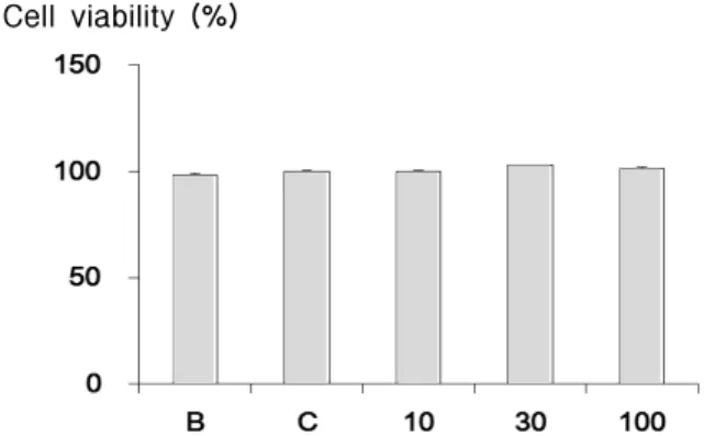
A. Cytotoxicity on HS68 human fibroblasts
In order to evaluate the cytotoxicity of ARHAE, samples were prepared at various concentrations and used to treat human fibroblasts (HS68). The results of this evaluation are shown in Fig. 1 at concentrations of 10, 30, 100 μg/ml. The cell viability was recalculated into 100% of control group. The cell viabilities of ARHAE 10 µg/ml treated, ARHAE 30 µg/ml treated, ARHAE 100 µg/ml treated are 100.1±0.5%, 103.0±0.0%, and 101.4±0.9%, respectively.
ARHAE showed no cytotoxicity up to the effective concentration for anti-wrinkle activity(less than 100 μg/ml) (Fig. 1).
Cell viability (%)
Fig. 1. Cell viability of ARHAE on HS68 human fibroblasts
B : blank, distilled water treated group without UVB irradiation.
C : control, distilled water treated group with UVB irradiation.
10, 30, and 100 : ARHAE 10, 30, and 100 µg/ml treated group.
Data are expressed as the mean ± SEM of three experiments.
B. Assay of collagen type I synthesis
To evaluate the amount of collagen type I synthesis that occurred upon exposure to the sample, collagen type I was quantitatively detected by using the previously described procollagen type I C-peptide assay kit. Collagens are synthesized as
precursor molecules, called procollagens. These molecules contain additional peptide sequences, usually referred to as ‘propeptides’, at both the amino- terminal end and the carboxy-terminal end. These propeptides are cleaved from the collagen triple-helix molecule during its secretion, after which the triple-helix collagens are polymerized into extracellular collagen fibrils. Thus, the amount of free propeptide stoichiometrically reflects the amount of collagen molecules synthesized8). The amounts of type I collagen synthesis of ARHAE were shown in Fig.
2. ARHAE increased the expression of type I collagen at a concentration of 100 µg/ml (18.7±8.1 ng/ml).
However, there was no significant difference. The collagen amounts of ARHAE 30 µg/ml and 10 µg/ml treated group did not increased (10.3±0.8 ng/ml and 8.4±1.0 ng/ml) (Fig. 2).
Collagen (ng/ml)
Fig. 2. Effect of ARHAE on collagen type I synthesis in human fibroblast cells
B : blank, distilled water treated group without UVB irradiation.
C : control, distilled water treated group with UVB irradiation.
10, 30, and 100 : ARHAE 10, 30, and 100 µg/ml treated group.
Data are expressed as the mean ± SEM of three experiments.
C. Assay of collagenase activity
To evaluate the collagenase activity, MMP-1 activity was quantitatively measured by using the previously described MMP-1 assay kit. The activities of MMP-1 of ARHAE treatment were recalculated into 100% of control group (Fig. 3). ARHAE signifi- cantly reduced the MMP-1 activity at concentrations
Collagenase activity (%)
Fig. 3. Effect of ARHAE on collagenase activity in human fibroblast cells
B : blank, distilled water treated group without UVB irradiation.
C : control, distilled water treated group with UVB irradiation.
10, 30, and 100 : ARHAE 10, 30, and 100 µg/ml treated group.
Data are expressed as the mean ± SEM of three experiments.
* : significantly different from the control, p<0.05.
of 10 µg/ml and 30 µg/ml (46.0±9.6% and 50.1±5.0%,
p<0.05). The collagenase activity of ARHAE 100
µg/ml treated group reduced to 75.5±7.7%, however this activity did not show the significance (Fig. 3).D. Tyrosinase activity assay
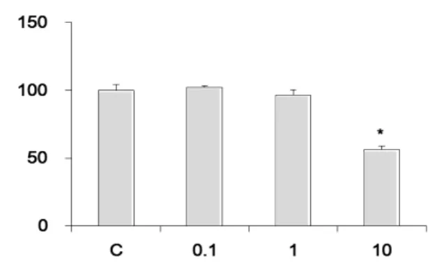
The activities of ARHAE on tyrosinase activity were recalculated into 100% of control group (Fig.
4). ARHAE significantly reduced the tyrosinase Tyrosinase activity (%)
Fig. 4. Effect of ARHAE on tyrosinase activity C : control, distilled water treated group.
0.1, 1, and 10 : ARHAE 0.1, 1, and 10 mg/ml treated group.
Data are expressed as the mean ± SEM of three experiments.
* : significantly different from the control, p<0.05.
activity at concentrations of 10mg/ml(56.4±2.7%,
p<0.05). The tyrosinase activity of ARHAE 0.1 and
1mg/ml treated groups did not show any signifi- cance(102.3±1.2% and 96.4±3.9%) (Fig. 4).E. L-DOPA oxidation
The activities of ARHAE on L-DOPA oxidation were recalculated into 100% of control group (Fig.
5). Although there was no significant difference, ARHAE reduced the L-DOPA oxidation activity at concentrations of 1 and 10 mg/ml (86.8±3.5% and 86.3±8.5%). ARHAE 0.1 mg/ml treated groups did not show any activity (112.9±6.4%) (Fig. 5).
L-DOPA oxidation (%)
Fig. 5. Effect of ARHAE on L-DOPA oxidation C : control, distilled water treated group.
0.1, 1, and 10 : ARHAE 0.1, 1, and 10 mg/ml treated group.
Data are expressed as the mean ± SEM of three experiments.
Ⅳ. Discussion and Conclusion
Recently, many people want to be looked younger than the actual age. Nowadays, aging seems to be considered as a disease to overcome rather than the natural phenomenon. There are two major theories of aging: First, the programmatic theory states that aging is an inherent genetic process. Second, the stochastic theory states that aging represents random environmental damage. Associated with cell damage and aging process of free radical production (a process
much enhanced after UV irradiation) and the number of errors during DNA replication to increase. Cellular own expression of the life of aging and reduce cell growth factors and may reflect the loss of cell receptors, growth inhibitors increase the response rate decreased the response of cells to growth signals. All of these results more pronounced in cells obtained from photodamaged skin9). It has been shown that UV irradiation leads to the formation of ROS that activate the MAP kinase pathway, which subsequently induces the expression and activation of MMPs in human skin in vivo1,2). MMPs are known to be upexpressed in human fibroblasts within hours after exposure to UV irradiation and are, therefore, considered key factors in the photoaging process. Therefore, agents with the ability to elevate extracellular matrix (ECM) protein levels or inhibit the major collagen-degrading enzymes like MMPs would prove to be useful in the development of effective anti-aging agents.
Astragali Radix is one of the famous Korean
medicinal herbs. It has been used for centuries as a primary tonic herb. Astragali Radix is widely used for it’s ‘Qi tonifying’ effect in Korean, Japan, and China10). It is used clinically for deficiency of the exterior leads to leakage of body fluids, resulting in spontaneous perspiration and other conditions of spleen deficiency including pale, sallow facial appearance, fatigue, tired extremities, decreased food intake, and diarrhea. It is also used to regulate water circulation and reduce edema. Its symptoms are facial edema, superficial edema, sensations of heaviness in the body, spontaneous sweating and intolerance of wind3,4).A variety of recent studies on the effect of
Astragali Radix has been done. Han et al revealed
the healing effect of Astragali Radix11), Kang et al studied the immuno modulatory effect of Astragali Radix12) and Choi et al reported the effect of Astragali Radix on collagen-induced arthritis13). About the antioidative effect of Astragali Radix Kim et al reported antioidative properties and whitening effect of the Astragali Radix, Atractylodis Rhizoma Alba and Acanthopanacis Cortex14).In order to evaluate the cytotoxicity of ARHAE, samples were prepared at various concentrations and used to treat human fibroblasts (HS68). There was no cytotoxicity in all treated concentrations.
Collagen is a group of naturally occurring proteins.
In nature, it is found exclusively in animals, especially in the flesh and connective tissues of mammals15). It is the main component of connective tissue, and is the most abundant protein in mammals, making up about 25% to 35% of the whole-body protein content16). Collagen, in the form of elongated fibrils, is mostly found in fibrous tissues such as tendon, ligament and skin, and is also abundant in cornea, cartilage, bone, blood vessels, the gut, and intervertebral disc. In muscle tissue it serves as a major component of endomysium. Collagen constitutes 1% to 2% of muscle tissue, and account for 6% of the weight of strong, tendinous muscles17).
Collagen occurs in many places throughout the body. So far, only 29 types of collagen have been identified and described. Over 90% of the collagen in the body, however, is of type I, II, III, and IV.
Among them, collagen type I is placed at skin, tendon, vascular, ligature, organs, and bone (main component of bone). Collagen-related diseases most commonly arise from genetic defects or nutritional deficiencies that affect the biosynthesis, assembly, postranslational modification, secretion, or other processes involved in normal collagen production.
Melanogenesis is a unique characteristic of melanocytes, and this process is regulated by melanogenic enzymes such as tyrosinase. Tyrosinase is a bifunctional enzyme which plays a pivotal role in the modulation of production, by catalyzing the hydroxylation of tyrosine to DOPA to DOPAquinine18). In this study, the amount of collagen type I was increased at a concentration of ARHAE 100 µg/ml.
However, there was no significant difference.
To evaluate the collagenase activity, MMP-1 activity was quantitatively measured. ARHAE significantly reduced the MMP-1 activity at concentrations of 10 µg/ml and 30 µg/ml.
The activities of ARHAE on tyrosinase activity
were significantly reduced at concentration of 10 mg/ml. The activities of ARHAE on L-DOPA oxidation were reduced concentration of 1 and 10 mg/ml, but there were no significant difference.
These results suggest that ARHAE may have potential as an anti-aging ingredient in cosmetic herbal acupuncture. I think further studies will be needed to unravel exactly under the molecular mechanisms.
Ⅴ. References
1. Fisher GJ, Datta SC, Talwar HS, Wang ZQ, Varani J, Kang S, Voorhees JJ. Molecular basis of sun-induced premature skin ageing and retinoid antagonism. Nature. 1996 Jan 25 ; 379(6563) : 335-9.
2. Shin JY, Hur W, Wang JS, Jang JW, Kim CW, Bae SH, Jang SK, Yang SH, Sung YC, Kwon OJ, Yoon SK. HCV core protein promotes liver fibrogenesis via up-regulation of CTGF with TGF-beta1. Exp Mol Med. 2005 Apr 30 ; 37(2) : 138-45.
3. Kim IR, Kim HC, Kuk YB, Park SJ, Park YK, Park JH, Seo BI, Seo YB, Song HJ, Shin MK, Lee YJ, Lee YC, Lee JH, Leem KH, Cho SI, Chung JK, Joo YS, Choi HY. Boncho-Hak.
Seoul : Young-Lim Press. 2007 : 576-8.
4. Chen JL, Chen TT. Chinese Medical Herbology and Pharmacology. CA : Art of Medicine Press.
2004 : 847-53.
5. Dan B and Andrew G. Chinese Herbal Medicine.
Materia Media. 1992 : 32-4.
6. Vanni A, Gastaldi D, Giunata G. Kinetic investi- gations on the double enzyme activity of the tyrosinase mushroom. Ann Chim. 1990 ; 80 : 35-60.
7. Joshi PC, Carraro C, Pathak MA. Involvement of reactive oxygen species in the oxidation of tyrosine and dopa to melanin and in skin tanning. Biochem Biophys Res Commun. 1987 Jan 15 ; 142(1) : 265-74.
8. Kim YH, Chung CB, Kim JG, Ko KI, Park SH, Kim JH, Eom SY, Kim YS, Hwang YI, Kim KH. Anti-wrinkle activity of ziyuglycoside I isolated from a Sanguisorba officinalis root extract and its application as a cosmeceutical ingredient.
Biosci Biotechnol Biochem. 2008 Feb ; 72(2) : 303-11.
9. Yaar M, Gilchrest BA. Cellular and molecular mechanisms of cutaneous aging. J Dermatol Surg Oncol. 1990 Oct ; 16(10) : 915-22.
10. Lee sena. A research for the whitening Effect of Astragali Radix. Semyung University : Thesis for the Degree of Master. 2008.
11. Han DO, Kim GH, Choi YB, Shim IS, Lee HJ, Lee YC, Kim JH, Chang GY, Hahm DH. Healing Effect of Astragali Radix Extracts on Experimental Open Wounds in Rats. Korea Journal of Oriental Physiology & Pathology. 2005 ; 19(1) : 92-7.
12. Kang H, Kim YB, Ahn KS. Immuno Modulatory Effect of Astragali Radix on OVA Induced Allergic Mouse Model. Korea Journal of Oriental Physiology
& Pathology. 2005 ; 19(3) : 612-7.
13. Choi BG, Cho MR, Kim JH, Ryu CR. Effect of Astragali Radix Herbal-acupuncture at ST36 on Collagen-induced Arthritis in Mice. The Journal of Korea Acupuncture & Moxibustion Society.
2008 ; 25(1) : 25-55.
14. Kim IC, Hur SS. Antioxidative Properties and whitening Effect of the Astragali Radix, Atractylodis Rhizoma Alba and Acanthopanacis Cortex. Journal of The Korean Oil Chemists’ Society. 2009 ; 26(2) : 110-6.
15. Müller WEG. The Origin of Metazoan Complexity:
Porifera as Integrated Animals. Integr Comp Biol. 2003 ; 43(1) : 3-10.
16. Di Lullo GA, Sweeney SM, Korkko J, Ala-Kokko L, San Antonio JD. Mapping the ligand-binding sites and disease-associated mutations on the most abundant protein in the human, type I collagen. J Biol Chem. 2002 Feb 8 ; 277(6) : 4223-31.
17. Sikorski ZE. Chemical and Functional Properties of Food Proteins. Boca Raton : CRC Press. 2001 : 242.
18. Hearing VJ and Jimenez M. Mammalian tyrosinase- the critical regulatory control point in melanocyte
pigmentation. Int J Biochem. 1987 ; 19 : 1141-7.