접수일 : 2014 년 4 월 12 일 , 게재승인일 : 2014 년 5 월 28 일 책임저자 : 임재영 , 경기도 성남시 분당구 구미로 173 번길 82
463-707, 분당서울대학교병원 재활의학과
Tel: 031-787-7730, Fax: 031-787-4051 E-mail: drlim1@snu.ac.kr
임상진료 지침을 이용한 이소성 골화증의 조기 진단 및 치료
증례 보고
분당서울대학교병원 재활의학과
김정길ㆍ이유경ㆍ임재영
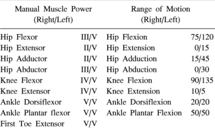
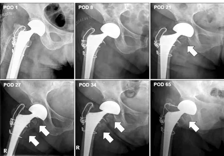
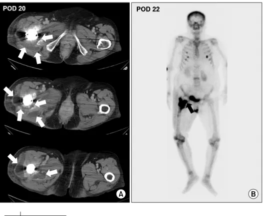
전체 글
증례 보고
김정길ㆍ이유경ㆍ임재영
수치
관련 문서
Modern Physics for Scientists and Engineers International Edition,
Since every classical or virtual knot is equivalent to the unknot via a sequence of the extended Reidmeister moves together with the forbidden moves, illustrated in Section 2,
웹 표준을 지원하는 플랫폼에서 큰 수정없이 실행 가능함 패키징을 통해 다양한 기기를 위한 앱을 작성할 수 있음 네이티브 앱과
_____ culture appears to be attractive (도시의) to the
with the experimental C versus t data. If the fit is unsatisfactory, another rate equation is guessed and tested. Integral method is especially useful for fitting simple
고관절 : 관골의 관골구(acetabulum)과 femur head sacroiliac joint: sacrum + ilium.. pubic symphysis : pubic + pubic
The index is calculated with the latest 5-year auction data of 400 selected Classic, Modern, and Contemporary Chinese painting artists from major auction houses..
1 John Owen, Justification by Faith Alone, in The Works of John Owen, ed. John Bolt, trans. Scott Clark, "Do This and Live: Christ's Active Obedience as the
