Introduction
Muscular dystrophies are genetic disorders caused by abnormalities in various genes (Aartsma-Rus et al, 2006; Dalkilic and Kunkel, 2003; Emery, 2002a;
Fokkema et al, 2005). Among them, a mutation of dystrophin gene coding for the protein dystrophin causes Duchenne muscular dystrophy (DMD), which affects 1 in 3,300∼6,000 boys (Emery, 1991; Emery, 2002b; Mendell et al, 2012). The dystrophin protein is an intricate part of the dystrophin-associated pro- tein complex and links subsarcolemmal cytoskeleton and extracellular matrix (Ibraghimov-Beskrovnaya et al, 1992; Ozawa et al, 2001; Rybakova et al, 1996;
Sutherland-Smith et al, 2003). It is also an essen- tial structural component of the muscle membrane, and provides mechanical stability in skeletal mus-
cle (Mizuno et al, 1994; Moens et al, 1993;
Ohlendieck et al, 1993; Petrof et al, 1993). That is, a mutation in the dystrophin gene breaks the dys- trophin-associated protein complex supporting in- tegrity of the skeletal muscle membrane and transmitting force. Additionally, the fragility caused by the absence of the dystrophin protein is ag- gravated by repeated intensive contraction because the muscle fibers are unable to endure eccentric contraction without structural integrity (Allen and Whitehead, 2011; Brussee et al, 1997; Deconinck and Dan, 2007; Petrof et al, 1993). An increase in skeletal muscle fatty tissue and severe muscle atrophy is observed as the child ages and then ambulatory loss is seen at around 8∼12 years (Beltran et al, 2015; Brooke et al, 1989; Bushby et al, 2010; Desguerre et al, 2009; Moxley et al, 2010;
Corresponding author: Woo-taek Lim [email protected]
Magnetic Resonance Imaging as a Biomarker for Duchenne Muscular Dystrophy
Woo-taek Lim, PhD, PT
Dept. of Physical Therapy, College of Health and Welfare, Woosong University Dept. of Sports Rehabilitation, College of Health and Welfare, Woosong University
Abstract
1)Muscular dystrophy is a hereditary musculoskeletal disorder caused by a mutation in the dystrophin gene. Duchenne muscular dystrophy (DMD) is one of the most common, and progresses relatively faster than other muscular dystrophies. It is characterized by progressive myofiber degeneration, muscle weakness and ultimately ambulatory loss. Since it is an X-linked recessive inheritance, DMD is mostly expressed in males and rarely expressed or less severe in females. The most effective measurement tool for DMD is magnetic resonance imaging (MRI), which allows non-invasive examination of longitudinal measurement. It can detect progressive decline of skeletal muscle size by measuring a maximal cross-sectional area of skeletal muscle. Additionally, other techniques in MRI, like T2-weighted imaging, assess muscle damage, including inflammation, by detecting changes in T2 relaxation time. Current MRI techniques even allow quantification of metabolic differences between affected and non-affected muscles in DMD. There is no current cure, but physical therapist can improve their quality of life by maintaining muscle strength and function, especially if treatment (and other forms of medical intervention) begins in the early stages of the disease.
Key Words: Duchenne muscular dystrophy; Eccentric exercise; Fatty infiltration; Inflammation;
Magnetic resonance imaging; Muscle damage; Muscular dystrophy.
Pardo et al, 2011). Ultimately, premature death oc- curs in the early twenties due to cardiac and res- piratory muscle failure (Fraser et al, 2012; Lovering et al, 2005). It is very important to detect patho- logical changes in skeletal muscle before functional declines so that intervention can begin as early as possible.
Invasive Measurement
Muscle biopsy today is a widely accepted assess- ment tool to diagnose and measure the disease pro- gression in DMD (Maunder-Sewry et al, 1980).
However, this invasive tool is limited to only a small amount of tissue at a time and narrow limits.
Additionally, it has been demonstrated that a DMD-affected muscle has different characteristics and progression varying by muscle region in a single muscle. The bigger issue is that the muscle biopsy may not be effective to evaluate the cardiopulmonary system, where monitoring is especially important in the late stages of the disease. Therefore, various non-invasive techniques involving X-rays, computed tomography (CT), and magnetic resonance imaging (MRI) are currently promoted as alternatives to the invasive method. These alternatives can be used without temporal and spatial limitations as they al- low longitudinal measurements covering the entire body.
Non-Invasive Measurements
Plain X-ray
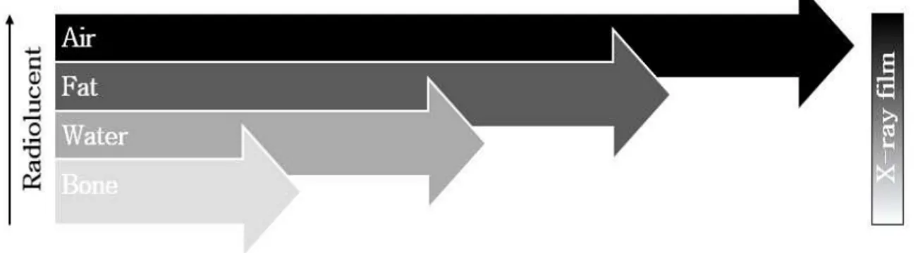
X-ray equipment is among the most frequently used, inexpensive, convenient, and old-fashioned tool in many clinical setting. The X-rays can penetrate objects, but the amount of X-rays reaching the de- tector varies according to the object being filmed (McKinnis, 2013) (Figure 1). For example, bones containing higher calcium absorb more X-rays than muscles and appear white on X-ray film while mus- cles show up in shades of gray. X-ray film provides enough resolution in shades of white and black to interpret pathological changes and abnormalities in bones and joints, but it is limited to an anatomic view of body structure. The limitations of X-rays in the diagnosis of disease led to the advent of CT, which provides a clear, well-contrasting cross-sec- tional image even between skeletal muscles in the body.
Computed tomography
CT, also known as X-ray CT, CT scanning, or computerized axial tomography scanning, provides a higher quality of tomographic images using many X-ray images (Heckmatt and Dubowitz, 1983). Unlike simple X-rays, CT shows cross-sectional images of a target object without overlapping various objects on a single image. Additionally, pathological changes in muscle can be cleary and easily verified on
Figure 1. Anatomy in four primary shades of grey.
high-resolution and high-contrast images. Recent ad- vances in technology allow 3-dimensional images of tumors, hemorrhaging, and even coronary arteries (Goodpaster et al, 2000; Jones et al, 1983; Liu et al, 1993; Termote et al, 1980). Due to the quality of the images, it has been used for much clinical research in preventive medicine as well. However, imaging tools based on X-ray penetration use ionizing radia- tion, which comes with the risk of undesirable side effects.
Magnetic resonance imaging
Unlike X-ray imaging tools, MRI does not expose the target to radiation. The magnetic resonance (MR) image was first introduced as a one-dimensional im- age in 1952 by Herman Carr, whereas current MRI provides the opportunity to quantify fatty infiltration, inflammation and edema in muscle using T1- and T2-weighted MR techniques (Carr and Purcell, 1954;
Garrood et al, 2009; Goodpaster et al, 2004;
Lamminen, 1990; Matsumura et al, 1988).
T1-weighted imaging has proven to be extensively in agreement with measurement of morphologic
changes in muscle (Akima et al, 2012; Mathur et al, 2010). Generally, the size of skeletal muscle is strong- ly related with muscle function. Thus, T1-weighted imaging provides cross-sectional area (CSA) imaging of muscle and has been invaluable in medical diagnosis. However, muscle size does not directly correlate to the muscle function in DMD. Wokke and colleagues have shown that in boys aged 8∼15 with DMD, the maximal CSA (CSAmax) of the triceps surae muscle (medial gastrocnemius, lateral gastro- cnemius, and soleus) was 52% larger, yet the boys had less functional ability and muscle strength than the age-matched controls (Wokke et al, 2014). This paradoxical finding is due to pseudohypertrophy re- sulting from fatty infiltration and replacement of fi- brotic tissue in muscle (Grindrod et al, 1983). When it comes to DMD, the T1-weighted technique is ap- propriate to measure the proportion of contractile tis- sue in muscle rather than quantitative measurements of muscle size (Akima et al, 2012).
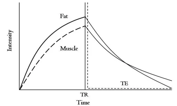
MRI is also useful to quantitatively measure mus- cle quality including biophysical and pathophysio- logical properties using the T2-weighted MR techni-
Figure 2. The example of T2 decay curve of fat and muscle (TR: repetition time, TE: echo time).
que (Arpan et al, 2013; Kim et al, 2010) (Figure 2).
Fleckenstein in 1989 found a relationship between muscle activity and subsequent increase in T2 relax- ation time (Fleckenstein et al, 1989). After physical exercise, T2 relaxation time was increased and lasted for a short time. An exercise-induced increase in T2
relaxation time does not usually last long if the physical exercise was not intense and/or not repeated. If the increase in T2 relaxation time is pro- longed, it might represent muscle inflammation or edema (Shellock et al, 1991). In the study of Mathur, T2 relaxation time was increased and lasted for 2 days after eccentric exercise in mdx mice, an animal model for DMD widely used in pre-clinical settings (Mathur et al, 2011). It is well known that muscle is more susceptible to damage (including inflammation
and edema) during eccentric exercise or intensive exercise (Lovering and Brooks, 2014; Petrof et al, 1993). Intensive muscle contraction leads to muscle damage due to an absence of the dystrophin protein, which provides structural integrity and stability to muscle membrane. The sensitivity of the T2-weighted MR technique for the differentiation of muscle dam- age makes it very useful in follow up on disease progression.
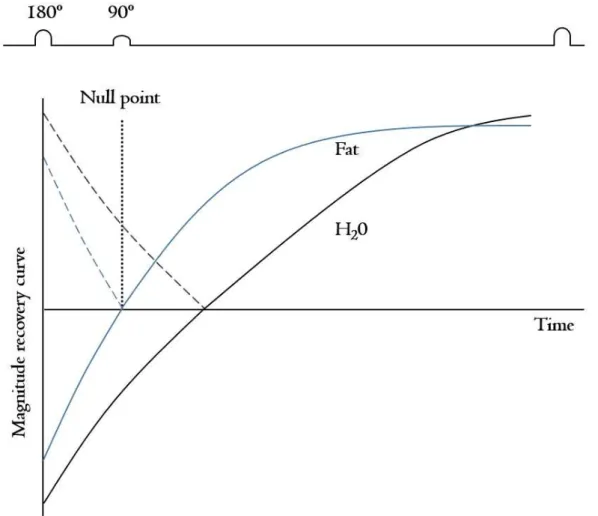
Pathological changes in muscle, including in- flammation or edema, might be more clearly detect- able with short-tau inversion recovery (STIR) imag- ing (Beltran et al, 2015; Tasca et al, 2012) (Figure 3). In young boys with DMD, the edema or in- flammation is often presented without any fatty infiltration. The STIR technique increases the sensi-
Figure 3. Magnititude T1 curve in the short tau inversion recovery (STIR) technique.
tivity of measurement of extracellular water content suggesting presence of edema and inflammation.
Marden found signal abnormalities from skeletal mus- cle appearing to be normal muscle on T1-weighted images using the STIR technique in two boys with DMD (Marden et al, 2005). Additionally, the STIR technique makes it easy to discriminate between fat and muscle by suppressing the signal from fatty tis- sue so that the examiner can exactly segment mus- cle CSAmax.
Recently, even lipid fraction and metabolic change in skeletal muscle have been measured by magnetic resonance spectroscopy (MRS) in conjunction with MRI (Felber et al, 2000; Lott et al, 2014; Torriani et al, 2012). As mentioned above, it is often difficult to diagnose young boys with DMD or discover pro- gression of the disease with conventional MRI. A re- cent study by Forbes reported higher lipid fraction, measured by MRS, in soleus and vastus lateralis muscles in young boys with DMD when compared with controls (Forbes et al, 2014). In Forbes’ study, MRS proved a very sensitive tool in determining lip- id fraction because it can provide MR signals from water and lipid separately and precisely. Additionally, MRS has been used to study skeletal metabolism in DMD. Metabolic changes such as glycolytic substrate glucose, glutamine, and glycolytic product lactate were significantly lower in those with DMD com- pared to controls (Sharma et al, 2003). Thus, MRS can be more powerful for evaluating patients with DMD when used in conjunction with MRI.
Conclusion
DMD is a life-threatening disease and worsens more quickly than other muscular dystrophies. There have been several clinical trials so far, but there is no current cure. Only glucocorticosteroids have been accepted as a pharmaceutical agent which can slow down symptoms of disease progression such as skel- etal muscle degeneration and loss of muscle tone.
Physical exercise with early medication, unless in- tensive eccentric exercise, can very helpful to main- tain functional activity and muscle strength. Since muscle damage in boys with DMD begins before functional declines, detecting changes in muscle is of the utmost importance so that physical therapy can begin as early as possible. MRI and/or MRS meas- urements have proven effective in detecting muscle damage even in the early stages of DMD.
References
Aartsma-Rus A, Van Deutekom JC, Fokkema IF, et al. Entries in the leiden duchenne muscular dys- trophy mutation database: An overview of mu- tation types and paradoxical cases that confirm the reading-frame rule. Muscle Nerve. 2006;34(2):
135-144.
Akima H, Lott D, Senesac C, et al. Relationships of thigh muscle contractile and non-contractile tis- sue with function, strength, and age in boys with duchenne muscular dystrophy. Neuromuscul Disord. 2012;22(1):16-25. http://dx.doi.org/10.1016/
j.nmd.2011.06.750
Allen DG, Whitehead NP. Duchenne muscular dys- trophy--what causes the increased membrane permeability in skeletal muscle? Int J Biochem Cell Biol. 2011;43(3):290-294. http://dx.doi.org/
10.1016/j.biocel.2010.11.005
Arpan I, Forbes SC, Lott DJ, et al. T2 mapping pro- vides multiple approaches for the character- ization of muscle involvement in neuromuscular diseases: A cross-sectional study of lower leg muscles in 5-15-year-old boys with duchenne muscular dystrophy. NMR Biomed. 2013;26(3):
320-328. http://dx.doi.org/10.1002/nbm.2851 Beltran E, Shelton GD, Guo LT, et al. Dystrophin-
deficient muscular dystrophy in a norfolk terrier.
J Small Anim Pract. 2015;56(5):351-354. http://
dx.doi.org/10.1111/jsap.12292
Brooke MH, Fenichel GM, Griggs RC, et al. Duchenne
muscular dystrophy: Patterns of clinical progression and effects of supportive therapy. Neurology. 1989;
39(4):475-481.
Brussee V, Tardif F, Tremblay JP. Muscle fibers of mdx mice are more vulnerable to exercise than those of normal mice. Neuromuscul Disord. 1997;
7(8):487-492.
Bushby K, Finkel R, Birnkrant DJ, et al. Diagnosis and management of duchenne muscular dys- trophy, part 1: Diagnosis, and pharmacological and psychosocial management. Lancet Neurol.
2010;9(1):77-93.
Carr HY, Purcell EM. Effects of diffusion on free pre- cession in nuclear magnetic resonance experiments.
Phys Rev. 1954;94(3):630-638.
Dalkilic I, Kunkel LM. Muscular dystrophies: Genes to pathogenesis. Curr Opin Genet Dev. 2003;13 (3):231-238.
Deconinck N, Dan B. Pathophysiology of duchenne muscular dystrophy: Current hypotheses. Pediatr Neurol. 2007;36(1):1-7.
Desguerre I, Christov C, Mayer M, et al. Clinical heterogeneity of duchenne muscular dystrophy (DMD): Definition of sub-phenotypes and pre- dictive criteria by long-term follow-up. PLoS One. 2009;4(2):e4347. http://dx.doi.org/10.1371/journal.
pone.0004347
Emery AE. Population frequencies of inherited neuro- muscular diseases--a world survey. Neuromuscul Disord. 1991;1(1):19-29.
Emery AE. Muscular dystrophy into the new millennium. Neuromuscul Disord. 2002a;12(4):
343-349.
Emery AE. The muscular dystrophies. Lancet. 2002b;
359(9307):687-695.
Felber S, Skladal D, Wyss M, et al. Oral creatine supplementation in duchenne muscular dys- trophy: A clinical and 31P magnetic resonance spectroscopy study. Neurol Res. 2000;22(2):
145-150.
Fleckenstein JL, Bertocci LA, Nunnally RL, et al. 1989 ARRS executive council award. Exercise-en-
hanced MR imaging of variations in forearm muscle anatomy and use: Importance in MR spectroscopy. AJR Am J Roentgenol. 1989;153(4):
693-698.
Fokkema IF, den Dunnen JT, Taschner PE. LOVD:
Easy creation of a locus-specific sequence var- iation database using an “LSDB-in-a-box”
approach. Hum Mutat. 2005;26(2):63-68.
Forbes SC, Willcocks RJ, Triplett WT, et al.
Magnetic resonance imaging and spectroscopy assessment of lower extremity skeletal muscles in boys with duchenne muscular dystrophy: A multicenter cross sectional study. PLoS One. 2014;
9(9):e106435. http://dx.doi.org/10.1371/journal.pone.
0106435
Fraser LK, Childs AM, Miller M, et al. A cohort study of children and young people with pro- gressive neuromuscular disorders: Clinical and demographic profiles and changing patterns of referral for palliative care. Palliat Med. 2012;
26(7):924-929.
Garrood P, Hollingsworth KG, Eagle M, et al. MR imaging in duchenne muscular dystrophy:
Quantification of T1-weighted signal, contrast uptake, and the effects of exercise. J Magn Reson Imaging. 2009;30(5):1130-1138. http://dx.
doi.org/10.1002/jmri.21941
Goodpaster BH, Kelley DE, Thaete FL, et al. Skeletal muscle attenuation determined by computed to- mography is associated with skeletal muscle lip- id content. J Appl Physiol (1985). 2000;89(1):
104-110.
Goodpaster BH, Stenger VA, Boada F, et al. Skeletal muscle lipid concentration quantified by mag- netic resonance imaging. Am J Clin Nutr. 2004;
79(5):748-754.
Grindrod S, Tofts P, Edwards R. Investigation of human skeletal muscle structure and composition by x-ray computerised tomography. Eur J Clin Invest. 1983;13(6):465-468.
Heckmatt JZ, Dubowitz V. Detecting the duchenne car- rier by ultrasound and computerized tomography.
Lancet. 1983;2(8363):1364.
Ibraghimov-Beskrovnaya O, Ervasti JM, Leveille CJ, et al. Primary structure of dystrophin-associated glycoproteins linking dystrophin to the ex- tracellular matrix. Nature. 1992;355(6362):696-702.
Jones DA, Round JM, Edwards RH, et al. Size and composition of the calf and quadriceps muscles in duchenne muscular dystrophy. A tomographic and histochemical study. J Neurol Sci. 1983;
60(2):307-322.
Kim HK, Laor T, Horn PS, et al. T2 mapping in duchenne muscular dystrophy: Distribution of disease activity and correlation with clinical assessments. Radiology. 2010;255(3):899-908. http://
dx.doi.org/10.1148/radiol.10091547
Lamminen AE. Magnetic resonance imaging of pri- mary skeletal muscle diseases: Patterns of dis- tribution and severity of involvement. Br J Radiol. 1990;63(756):946-950.
Liu M, Chino N, Ishihara T. Muscle damage pro- gression in duchenne muscular dystrophy eval- uated by a new quantitative computed tomog- raphy method. Arch Phys Med Rehabil. 1993;
74(5):507-514.
Lott DJ, Forbes SC, Mathur S, et al. Assessment of intramuscular lipid and metabolites of the lower leg using magnetic resonance spectroscopy in boys with duchenne muscular dystrophy.
Neuromuscul Disord. 2014;24(7):574-582. http://
dx.doi.org/10.1016/j.nmd.2014.03.013
Lovering RM, Brooks SV. Eccentric exercise in ag- ing and diseased skeletal muscle: Good or bad? J Appl Physiol (1985). 2014;116(11):1439-1445. http://
dx.doi.org/10.1152/japplphysiol.00174
Lovering RM, Porter NC, Bloch RJ. The muscular dystrophies: From genes to therapies. Phys Ther.
2005;85(12):1372-1388.
Marden FA, Connolly AM, Siegel MJ, et al.
Compositional analysis of muscle in boys with duchenne muscular dystrophy using MR imaging.
Skeletal Radiol. 2005;34(3):140-148.
Mathur S, Lott DJ, Senesac C, et al. Age-related
differences in lower-limb muscle cross-sectional area and torque production in boys with duch- enne muscular dystrophy. Arch Phys Med Rehabil. 2010;91(7):1051-1058. http://dx.doi.org/
10.1016/j.apmr.2010.03.024
Mathur S, Vohra RS, Germain SA, et al. Changes in muscle T2 and tissue damage after downhill running in mdx mice. Muscle Nerve. 2011;43(6):
878-886. http://dx.doi.org/10.1002/mus.21986 Matsumura K, Nakano I, Fukuda N, et al. Proton
spin-lattice relaxation time of duchenne dystrophy skeletal muscle by magnetic resonance imaging.
Muscle Nerve. 1988;11(2):97-102.
Maunder-Sewry CA, Gorodetsky R, Yarom R, et al.
Element analysis of skeletal muscle in duchenne muscular dystrophy using x-ray fluorescence spectrometry. Muscle Nerve. 1980;3(6):502-508.
McKinnis LN. Fundamentals of Musculoskeletal Imaging. 4th ed. Philadelphia, PA, F.A. Davis Co., 2013:10.
Mendell JR, Shilling C, Leslie ND, et al.
Evidence-based path to newborn screening for duchenne muscular dystrophy. Ann Neurol. 2012;
71(3):304-313. http://dx.doi.org/10.1002/ana.23528 Mizuno Y, Yoshida M, Nonaka I, et al. Expression of
utrophin (dystrophin-related protein) and dystro- phin-associated glycoproteins in muscles from patients with duchenne muscular dystrophy.
Muscle Nerve. 1994;17(2):206-216.
Moens P, Baatsen PH, Maréchal G. Increased sus- ceptibility of EDL muscles from mdx mice to damage induced by contractions with stretch. J Muscle Res Cell Motil. 1993;14(4):446-451.
Moxley RT 3rd, Pandya S, Ciafaloni E, et al. Change in natural history of duchenne muscular dys- trophy with long-term corticosteroid treatment:
Implications for management. J Child Neurol.
2010;25(9):1116-1129. http://dx.doi.org/10.1177/0883 073810371004
Ohlendieck K, Matsumura K, Ionasescu VV, et al.
Duchenne muscular dystrophy: Deficiency of dystrophin-associated proteins in the sarcolemma.
Neurology. 1993;43(4):795-800.
Ozawa E, Nishino I, Nonaka I. Sarcolemmopathy:
Muscular dystrophies with cell membrane defects. Brain Pathol. 2001;11(2):218-230.
Pardo AC, Do T, Ryder T, et al. Combination of steroids and ischial weight-bearing knee ankle foot orthoses in duchenne’s muscular dystrophy prolongs ambulation past 20 years of age--a case report. Neuromuscul Disord. 2011;21(11):
800-802. http://dx.doi.org/10.1016/j.nmd.2011.06.006 Petrof BJ, Shrager JB, Stedman HH, et al. Dystrophin protects the sarcolemma from stresses developed during muscle contraction. Proc Natl Acad Sci U S A. 1993;90(8):3710-3714.
Rybakova IN, Amann KJ, Ervasti JM. A new model for the interaction of dystrophin with f-actin. J Cell Biol. 1996;135(3):661-672.
Sharma U, Atri S, Sharma MC, et al. Skeletal mus- cle metabolism in duchenne muscular dystrophy (DMD): An in-vitro proton NMR spectroscopy study. Magn Reson Imaging. 2003;21(2):145-153.
Shellock FG, Fukunaga T, Mink JH, et al. Exertional muscle injury: Evaluation of concentric versus eccentric actions with serial MR imaging.
Radiology. 1991;179(3):659-664.
Sutherland-Smith AJ, Moores CA, Norwood FL, et al. An atomic model for actin binding by the
CH domains and spectrin-repeat modules of utrophin and dystrophin. J Mol Biol. 2003;329(1):
15-33.
Tasca G, Pescatori M, Monforte M, et al. Different molecular signatures in magnetic resonance imaging-staged facioscapulohumeral muscular dystrophy muscles. PloS One. 2012;7(6):e38779.
http://dx.doi.org/10.1371/journal.pone.0038779 Termote JL, Baert A, Crolla D, et al. Computed to-
mography of the normal and pathologic muscu- lar system. Radiology. 1980;137(2):439-444.
Torriani M, Townsend E, Thomas BJ, et al. Lower leg muscle involvement in duchenne muscular dystrophy: An MR imaging and spectroscopy study. Skeletal Radiol. 2012;41(4):437-445. http://
dx.doi.org/10.1007/s00256-011-1240-1
Wokke BH, van den Bergen JC, Versluis MJ, et al.
Quantitative MRI and strength measurements in the assessment of muscle quality in duchenne muscular dystrophy. Neuromuscul Disord. 2014;
24(5):409-416. http://dx.doi.org/10.1016/j.nmd.2014.
01.015
This article was received July 31, 2015, was re- viewed July 31, 2015, and was accepted September 2, 2015.