Introduction
A range of filling materials are used for perforation repair and root end-fillings in clinical endodontics, such as reinforced zinc oxide eugenol cement, inter- mediate restorative material (IRM), super ethoxy- benzoic acid (EBA) cement, calcium hydorixde, com- posite resins ,glass ionomer cements and mineral tri- oxide aggregate (MTA , Dentsply, Tulsa Dental, Tulsa, OK, USA). Among these, MTA, which is also called ProRoot MTA, has been used successfully in endodonctics for the past 10 years.1-3 More recently,
new products similar to MTA have been introduced into the market including MTA angelus4-6 and MTA- bio7 (Angelus Solucoes Odontologicas, Londrina, Brazil) and Bioaggregate8 (Innovative Bioceramix, Vancouver, Canada). Bioaggregate is sold exclusively in Canada. Its recommended applications are the same as those of MTA.
This new material has received less research atten- tion than other materials.9-12 Park et al. examined the chemical composition of BioAggregate and report- ed that it contained a significant amount of tantalum oxide instead of bismuth oxide.9 The major compo-
Biocompatibility of bioaggregate cement on human pulp and periodontal ligament (PDL) derived cells
Choo-Ryung Chung1, Euiseong Kim2, Su-Jung Shin2*
1Department of Orthodontics, 2Department of Conservative Dentistry, Yonsei University College of Dentistry, Seoul, Korea
Objectives: This study was performed to investigate the biocompatibility of newly introduced Bioaggregate on human pulp and PDL cells.
Materials and Methods: Cells were collected from human pulp and PDL tissue of extracted premolars. Cell culture plate was coated either with Bioaggregate or white MTA, then the same number of cells were poured to cell culture dishes. Cell attachment and growth was examined under a phase microscope after 1,3 and 7 days of seeding. Cell viability was measured and the data was analyzed using Student t-test and one way ANOVA.
Results: Both types of cells used in this study were well attached and grew healthy on Bioaggregate and MTA coated culture dishes. No cell inhibition zone was observed in Bioaggregate group. There was no sta- tistical difference of viable cells between bioaggreagte and MTA groups.
Conclusions: Bioaggregate appeared to be biocompatible compared with white MTA on human pulp and PDL cells. [J Kor Acad Cons Dent 2010;35(6):473-478.]
Key words:Bioaggregate; Biocompatibility; MTA; Periodontal ligament cell; Pulp cell
-Received 15 September 2010; revised 16 October 2010; accepted 17 October 2010- ABSTRACT
1Chung CR, DDS, PhD, Assistant Professor, Department of Orthodontics, Yonsei University College of Dentistry
2Kim E, DDS, MSD, PhD, Associate Professor; Shin SJ, DDS, MS, Assistant Professor, Department of Conservative Dentistry, Yonsei University, College of Dentistry, Seoul, Korea
*Correspondence to Su-Jung Shin, DDS, MS.
Assistant Professor, Department of Conservative Dentistry, GangNam Severance Dental Hospital, Yonsei University, Dogok-dong, Kangnam-gu, Seoul, Korea 135-270
TEL, +82-2-6279-6453; FAX, +82-2-3463-4052; E-mail, [email protected]
*This study was supported by a Grant from the Korean Health Care Technology R&D Project, Ministry of Health, Welfare & Family affairs, Republic of Korea (A084458).
nents were calcium silicate oxide and calcium silicate.
Zhang et al. reported that MTA and Bioaggregate powder have a similar bactericidal effects on E. fae- calis.8 More recently, Yuan et al. reported that Bioaggregate was nontoxic to mouse MC3T3-E1 osteoblast cells.12
Bioaggregate appears to be a modified or synthetic version of original MTA. Since the clinical indications of Bioaggregate are the same as those of MTA, it might be helpful to compare this new material with MTA, which has already been studied and proven to be effective and biocompatible over the past few years.13-15
This study examined the biocompatibility of BioAggregate compared to white ProRoot MTA on the cells derived from human dental pulp and periodon- tial tissue.
Materials and Methods Cell Culture
Human premolars planned to be extracted for orthodontic treatment were used. The institutional Review Board of Gangnam Severance Hospital, Yensei University approved both the protocol and informed consent. Written informed consent was obtained from each patient (No 3-2009-0069). After extraction, the teeth were stored in 50 ml polyethyl- ene tubes containing 10 ml alpha minimum essential medium (α-MEM)(Gibco/BRL, Grand Island, NY, USA) supplemented with 10% fetal bovine serum (FBS; Hyclone, Loga, UT, USA) and 1% penicillin.
After washing 3 times with α-MEM (3% antibiotics/
antimycotics), the tooth was held using sterilized gauze. The soft tissue on the middle 1/3 of the root was obtained by curettage with a No.15 blade. The tissue collected was kept in a 100 mm2 culture dish containing α-MEM and the culture plate was washed three times with the same culture media. A new cul- ture dish was used to apply the tissue, which was kept in the incubator at 37℃ in a humidified atmos- phere containing 5% CO2 in air until the plate became full with growing cells. The remaining tooth was split after making bucco-linugal grooves. The pulp tissue was removed using a sterilized spoon
excavator and cultured in a similar manner as peri- odontal tissue.
Preparation of a cement-coated tissue culture plate
White MTA (ProRoot) and Bioaggregate were mixed according to the manufacturer’s instructions.
A 6-well tissue culture dish was coated with the MTA paste prepared with sterile water (10 mg cement/ml of H2O per well). PDL and pulp cells were seeded on culture dishes coated with the freshly mixed cements to determine if the initial toxicity of the freshly mixed material can affect cell attachment and growth.
Optical Microscopy
The cells were plated on either an MTA coated or plastic surface (5.0×10⁵cells/well in a 6-well plate), and cultured for 1, 6, 48 and 72 hours before being examined by phase microscopy (Nikon TMS, Nikon Inc. Melville, NY, USA). Cells grown on cement coated dishes could not be visualized by this method since both cements used do not transmit light. Thus, the cells grown on plastic surface around cements were examined under the microscope and photos were taken.
Cell Viability test
The cells were grown for 3 days on a 12-well cul- ture plate coated with each type of cement. After 1×
105 cells were seeded, the cells grown on a regular plastic plate were used as the control. An EZ-Cytox cell viability assay kit (DAEILL LAB Service Co., Seoul, Korea), which measures the cell dehydroge- nase activity was used. This kit is similar to a MTT assay measuring the mitochondrial activity in viable cells. After adding 100 μL of the assay reagent, cell culture plate was kept in a 37℃ incubator for 1 hour.
Subsequently, 100 μL of supernatant was taken and added to a 96-well plate. The color density at 450 nm was read using an ELISA reader (Molecular Devices Inc/E-max, Washington, DC, USA). The data is presented as a percentage when the density
of the control was considered to be 100. The average of the triplicates was used for data analysis.
Statistical analysis
The data from the cell viability tests were analyzed using a Student’s t-test and one-way ANOVA. The control value was set to 100, and the results are expressed as the mean% ± standard deviation. A p value < 0.05 was considered significant.
Results Cell attachment
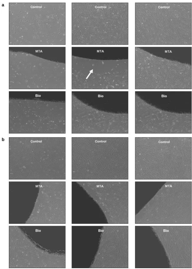
The effects of Bioaggregate cement on human pulp and PDL cell growth were examined by observing the cells grown on this cement using a phase microscope (Figure 1). The effects of MTA and Bioaggregate on cell growth were determined by examining the cells around the materials because cells grown on top of the material could not be visualized. Both types of cells on Bioaggregate showed no inhibition zones or gaps around the material at any time intervals.
However, although cells were fully grown around MTA, an inhibition zone was detected in the human pulp and PDL cell culture grown with MTA.
Cell viability test
Before performing the viability tests, each cell cul- ture plate was observed by optical microscopy to determine if the cell were growing healthily without dead floating cells.
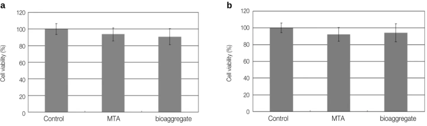
There were no significant differences in the human pulp and PDL cell cultures between control and experimental groups (Figure 2). There were 6.3 ± 8.02% and 9.25 ± 9.74% less viable cells in human PDL cells when cultured with MTA and Bioaggregate.
The similar result was shown in pulp cells.
Discussion
In this study, the biocompatibility of Bioaggregate was compared with white MTA since its chemical composition was different from that of MTA from our
previous study.9
Cell attachment and growth can be used as criteria to evaluate the biocompatibility or cytotoxicity of a material in a cell culture. MTA may not provide a favorable surface environment for cells while it sets because of the high pH generated during setting.
Many clinical reports and animal studies have sug- gested that this harmful effect is transient.16,17 The lack of an inhibition gap in the cell culture grown with Bioaggregate indirectly demonstrated its favor- able environment.
A cell viability test was performed using EZ-Cytox.
Compared to the MTT assay, this method employs a soluble terazolium salt, which is unnecessary for dis- solving formazan and removing the supernatant of the cell culture. There was no significant difference in the viable cell numbers between the MTA and Bioaggregate groups, which demonstrated both cements to be equally nontoxic to human pulp and PDL cells.
Our results are consistent with other previous studies, which demonstrated both cements were equally nontoxic to human pulp and PDL cells. De- Deus et al. showed in vitro biocompatibility compared to MTA when they tested this material using human mesenchymal cells.18
Also, the potentials that Bioaggregate can affect cell differentiation and/or mineralization have been shown in recent studies. Yan et al. found the increased level of alkaline phosphatase when cells were grown with Bioaggregate on 7 days.11They con- cluded that Bioaggregate may enhance PDL cell dif- ferentiation. Yuan et al. also found the increased mineralization when osteoblasts were grown with Bioaggregate.12
MTA has been proved as biocompatible and nontox- ic material in many previous studies. Recently, sev- eral new bioactive cements similar to MTA have been introduced. MTA angelus, MTA bio, and Bioaggreagte are the examples. These materials have been studied recently, and they are used in certain countries. In this study, white MTA was used as one of the experi- mental groups. By having MTA group as a reference, it was concluded that Bioaggregate might be compat- ible with MTA in terms with cell cytotoxicity and ini- tial cell growth.
Figure 1.Photos taken from an optical microscope (×40 magnification). The same number of cells (a, human pulp cells; b, PDL cells) were seeded to MTA or Bioaggregate coated cell culture dishes, then initial attachment was observed using a phase microscope.
PDL, periodontal ligament; MTA, mineral trioxide aggregate.
a
b
It is expected to have more new materials like MTA in the near future. At the same time, meticulous research should be needed before clinical applications.
Conclusions
Based on the findings of our study, Bioaggregate appeared to be compatible with MTA. It was found to be nontoxic to human pulp and PDL cells. There were no significant differences between Bioaggregate and MTA in terms of initial cell viability.
References
1. Koh ET, Torabinejad M, Pitt Ford TR, Brady K, McDonald F. Mineral trioxide aggregate stimulates a biological response in human osteoblasts. J Biomed Mater Res 1997;37(3):432-439.
2. Lee SJ, Monsef M, Torabinejad M. Sealing ability of a mineral trioxide aggregate for repair of lateral root per- forations. J Endod 1993;19(11):541-544.
3. Torabinejad M, Pitt Ford TR, McKendry DJ, Abedi HR, Miller DA, Kariyawasam SP. Histologic assess- ment of mineral trioxide aggregate as a root-end filling in monkeys. J Endod 1997;23(4):225-228.
4. Gomes-Filho JE, Rodrigues G, Watanabe S, Estrada Bernabe PF, Lodi CS, Gomes AC, et al. Evaluation of the tissue reaction to fast endodontic cement (CER) and Angelus MTA. J Endod 2009;35(10):1377-1380.
5. Hashem AA, Hassanien EE. ProRoot MTA, MTA- Angelus and IRM used to repair large furcation perfo- rations: sealability study. J Endod 2008;34(1):59-61.
6 Song JS, Mante FK, Romanow WJ, Kim S. Chemical analysis of powder and set forms of Portland cement, gray ProRoot MTA, white ProRoot MTA, and gray MTA-Angelus. Oral Surg Oral Med Oral Pathol Oral Radiol Endod 2006;102(6):809-815.
7. Lessa FC, Aranha AM, Hebling J, Costa CA. Cytotoxic effects of White-MTA and MTA-Bio cements on odonto-
blast-like cells (MDPC-23). Braz Dent J 2010;21(1):
24-31.
8. Zhang H, Pappen FG, Haapasalo M. Dentin enhances the antibacterial effect of mineral trioxide aggregate and bioaggregate. J Endod 2009;35(2):221-224.
9. Park JW, Hong SH, Kim JH, Lee SJ, Shin SJ. X-Ray diffraction analysis of white ProRoot MTA and Diadent BioAggregate. Oral Surg Oral Med Oral Pathol Oral Radiol Endod 2010;109(1):155-158.
10. Vivan RR, Zapata RO, Zeferino MA, Bramante CM, Bernardineli N, Garcia RB, et al. Evaluation of the physical and chemical properties of two commercial and three experimental root-end filling materials. Oral Surg Oral Med Oral Pathol Oral Radiol Endod 2010;110(2):250-256.
11. Yan P, Yuan Z, Jiang H, Peng B, Bian Z. Effect of bioaggregate on differentiation of human periodontal ligament fibroblasts. Int Endod J In press, 2010.
12. Yuan Z, Peng B, Jiang H, Bian Z, Yan P. Effect of bioaggregate on mineral-associated gene expression in osteoblast cells. J Endod 2010;36(7):1145-1148.
13. Chang S, Yoo H, Park D, Oh T, Bae K. Ingredients and cytotoxicity of MTA and 3 kinds of Portland cement. J Kor Acad Cons Dent 2008;33(4):364-376.
14. Kwon J, Lim S, Baek S, Bae K, Kang M, Lee W. The effect of mineral trioxide aggregate on the production of growth factors and cytokines by human periondontal ligament fibroblast. J Kor Acad Cons Dent 2007;32(3):
191-197.
15. Yoon Y, Yang I, Hwang Y, Hwang I, Choi H, Yoon S, Kim S, Oh W. Pulp response of mineral trioxide aggre- gate, calcium sulfate or calcium hydorixde. J Kor Acad Cons Dent 2007;32(2):95-101.
16. Apaydin W, Shanahang S, Torabinejad M. Hard-tissue healing after application of fresh or set MTA as root- end filling material. J Endod 2004;30(1):21-24.
17. Ford T, Torabinejad M, Abedi H, Bakland L, Kariyawasam S. Using mineral trioxide aggregate as a pulp-capping material. J Am Dent Assoc 1996;127(10) :1491-1494.
18. De-Deus G, Canabarro A, Alves G, Linhares A, Senne MI, Granjeiro JM. Optimal cytocompatibility of a bioce- ramic nanoparticulate cement in primary human mes- enchymal cells. J Endod 2009;35(10):1387-1390.
Figure 2.Cell viability tests. a, human pulp cells; b, PDL cells.
PDL, periodontal ligament; MTA, mineral trioxide aggregate.
a b
120 100 80 60 40 20 0
120 100 80 60 40 20 0
Cell viability (%)
Cell viability (%)
Control MTA bioaggregate Control MTA bioaggregate
국문초록
사람의 치수 및 치주인대 세포에 대한 Bioaggregate 시멘트의 생체적합성에 대한 연구
정주령1∙김의성2∙신수정2*
연세대학교 치과대학1치과교정과, 2치과보존과
연구목적: 본 연구는 인간의 치수 및 치근단 조직에서 유리된 세포를 이용하여 비교적 최근 소개된 Bioaggregate의 생체친 화성을 평가하는 데에 있다.
연구 재료 및 방법: 사람의 발거치로 부터 치수 및 치근단 조직에서 배양된 세포를 이용하였다. Bioaggregate와 white MTA를 혼합하여 세포배양판에 적용한 후 같은 수의 세포를 배양하였다. 1, 3, 그리고 7일 후 위상차현미경을 사용하여 세 포의 부착과 성장을 관찰하고 cell viability test를 시행하였다. 얻어진 결과는 Student t-test및 one way ANOVA를 이용 하여 분석하였다.
결과: 두가지 종류의 세포 모두 Bioaggregate와 MTA가 혼합된 배양판에서 잘 성장하였으며 Bioaggregate군에서는 inhi- bition zone이 관찰되지 않았다. Cell viability test에서 두 그룹간 통계적인 유의성 차이는 없었다.
결론: Bioaggreagete는 치수 및 치근단 세포에 대하여 MTA와 유사한 세포친화성을 보였다.
주요단어: 생체적합성; 치수세포; 치주인대세포; Bioaggregate; MTA