148 Copyright © 2010 Korean Neurological Association
Print ISSN 1738-6586 / On-line ISSN 2005-5013 10.3988/jcn.2010.6.3.148 CASE REPORT
J Clin Neurol 2010;6:148-151
Introduction
Frontotemporal lobar degeneration, progressive supranuclear palsy (PSP), and corticobasal degeneration (CBD) have over- lapping clinical and pathological features. PSP is a clinico- pathological entity that typically presents as an akinetic rigid syndrome with early postural instability, axial rigidity, and vari- able supranuclear gaze palsy. In particular, supranuclear verti- cal gaze palsy, moderate or severe postural instability, and falls during the first year after onset are the main characteristics of PSP and they affect the accuracy of the clinical diagnosis (9%
error according to logistic regression).1,2 We report a patholog- ically confirmed case of PSP that presented with behavioral changes including agitation and irritability, which eventually progressed to cardinal symptoms, such as frequent falls and su- pranuclear gaze palsy.
Case Report
In 2004, a 52-year-old Korean woman was evaluated for sleep
disturbance and behavioral changes over the previous two years. She walked around her neighborhood all day, meddling in other people’s affairs and starting arguments with them. Her neighbors did not want to spend time with her, and her inap- propriate behavior was worsening progressively. She visited a psychiatry clinic initiated by her husband, and was diagnosed with severe anxiety neurosis of unknown etiology. She was pre- scribed medicines for her insomnia and behavioral problems, but her symptoms worsened. Her husband reported that her memory had been impaired for 6 months before her first visit to us.
Her past medical and family histories were unremarkable.
She graduated from high school and then worked as a secre- tary for a small company until she got married. She was a very kind and careful housewife.
On neurological examination, she was alert and oriented.
However, she could not stay seated in a chair for more than a few minutes, instead walking around the outpatient clinic. Her speech was fluent and cranial nerve examinations produced normal results. The motor and sensory examinations were un-
Behavioral Changes as the Earliest Clinical Manifestation of Progressive Supranuclear Palsy
Hyun Jeong Han, MD, PhDa; Hyeyun Kim, MDa; Jong-Ho Park, MDa; Hyung-Woo Shin, MDa; Go Un Kim, MDa; Dong Sun Kim, MDa; Eun Ja Lee, MD, PhDb; Hwa Eun Oh, MD, PhDc; Seung-Hye Park, MD, PhDd; Yun Jung Kim, MD, PhDe
aDepartments of Neurology, bRadiology and cPathology, Myongji Hospital, College of Medicine, Kwandong University, Goyang, Korea
dDepartment of Pathology, Seoul National University Hospital, Seoul, Korea
eDepartment of Neurology, Hallym University, Ilsong Institute of Life Science, Pyongchon, Korea
Received March 17, 2009 Revised July 8, 2009 Accepted July 8, 2009 Correspondence Hyun Jeong Han, MD, PhD Department of Neurology, Myongji Hospital, College of Medicine, Kwandong University 697-24 Hwajeong-dong,
Deogyang-gu, Goyang 412-270, Korea Tel +82-31-810-5403
Fax +82-31-969-0500 E-mail [email protected]
BackgroundzzThe clinical and pathological heterogeneity of progressive supranuclear pal- sy (PSP) is well established. Even with a well-defined clinical phenotype and a thorough lab- oratory workup, PSP can be misdiagnosed, especially in its early stages.
Case ReportzzA 52-year-old woman, who we initially diagnosed with a behavioral variant of frontotemporal dementia developed parkinsonian features, which then progressed to gait instability and gaze abnormality.
ConclusionszzWe report herein a pathologically confirmed case of PSP presenting with behavioral changes including agitation and irritability, which eventually led to the cardinal symptoms of progressive supranuclear palsy.
J Clin Neurol 2010;6:148-151 Key Wordszz frontotemporal dementia, parkinsonism, progressive supranuclear palsy.
Han HJ et al.
www.thejcn.com 149 remarkable. She complained of mild lower back pain but no
gait disturbance.
She scored 23/30 on the Korean version of the Mini-Mental Status Examination3 and had a clinical dementia rating4 of 0.5.
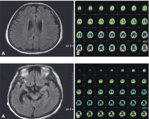
The Neuropsychiatric inventory5 revealed agitation, irritabil- ity, and disinhibition, and her husband reported her sleep dis- order. Her behavioral symptoms prevented the application of detailed neuropsychological tests because of her behavioral symptoms. The laboratory data, including thyroid function tests, were normal. Brain magnetic resonance imaging (MRI) showed diffuse atrophy changes (Fig. 1A) and brain 18F-fluoro- deoxyglucose positron emission tomography (18F-FDG PET) revealed subtly decreased glucose metabolism in the right pre- frontal and both posterior frontal areas (Fig. 1B).
We diagnosed this patient with a behavioral variant of frontotemporal dementia at that time. Six months later, she re- turned to our hospital due to severe lower back pain and gait disturbance. She underwent lumbar MRI in the orthopedic clinic, but no specific abnormalities were detected. Neurolog- ical examinations revealed mild features of Parkinson’s disease.
Her facial expression was slightly decreased and her speech was monotonous compared to at the previous examoination.
She had no tremor, but showed bradykinesia and mild symmet- ric rigidity. Limb apraxia and alien hand syndrome were not re- vealed. Her gait was short-stepped and slightly festinating, but without postural instability. Her extraocular movements showed no definite abnormalities. Our working diagnosis was FTD with parkinsonism. We requested a genetic study for FTD
and parkinsonism linked to chromosome 17 (FTDP-17), but this did not reveal a genetic mutation of microtubule associat- ed protein tau.
We prescribed the maximum allowable doses of the dopami- ne agonist and L-dopa, but thios resulted in her symptoms wa- xing and waning, rather than improving decisively. She grad- ually developed walking hesitation and was prone to falling.
Vertical gaze limitation was observed in a neurological exam- ination. We performed follow-up brain MRI and 18F-FDG PET studies. The brain MRI showed marked bilateral temporal and midbrain atrophy with ventricular dilatation, with the changes being greater in the right temporal lobe than in the left tempo- ral lobe (Fig. 2A). Brain 18F-FDG PET revealed decreased glu- cose metabolism in the bilateral frontotemporal and parietal ar- eas, with the decrease being in the right temporal lobe than in the left temporal lobe (Fig. 2B).
We made a diagnosis of probable PSP based on the Nation- al Institute of Neurological Disorders and Stroke and the Soci- ety for PSP Criteria.1 The patient started to develop difficulty swallowing and dysarthria, and these symptoms deteriorated relentlessly. Eventually, she became bedridden and required re- spiratory support with mechanical ventilation. Her family wanted confirmation of the diagnosis of her progressive disease, so we performed a brain biopsy and pathological evaluation.
The biopsy of the temporal and frontal cortices showed tau-pos- itive neurons (Fig. 3A) and reactive gliosis with tufted astrocy- tes (Fig. 3B).
Fig. 1. A: Brain magnetic resonance im- aging shows diffuse brain atrophy on flu- id-attenuated inversion recovery imag- es. B: Brain 18F-fluorodeoxyglucose posi- tron emission tomography reveals subtly decreased glucose metabolism in the right prefrontal and both posterior frontal
areas. A B
Fig. 2. A: Brain MRI shows marked bi- lateral temporal and midbrain atrophy with ventricular dilatation that is worse in the right temporal lobe than in the left temporal lobe on fluid-attenuated inver- sion recovery images. B: Brain 18F-fluo- rodeoxyglucose positron emission to- mography reveals decreased glucose metabolism in the bilateral frontotem- poral and parietal areas, with the de- crease being in the right temporal lobe
than in the left tempopral lobe. A B
Behavioral Changes in Progressive Supranuclear Palsy
150 J Clin Neurol 2010;6:148-151
Discussion
Most published reports on the behavior of patients with PSP emphasize symptoms related to depression or apathy, while psychotic or obsessive features are rarely described. Litvan et al.6 investigated the behavioral symptoms of PSP, Alzheim- er’s disease, and controls using the NPI and noted that apathy was the most frequent behavioral abnormality; moreover, the presence of high apathy and low agitation scale scores cor- rectly identified the patients with PSP.
In contrast, in our case, agitation and disinhibition were the earliest and most prominent behavioral symptoms. As shown previously, apathy and disinhibition are associated with pro- gressive dysfunction of different subcortical circuits and do not proceed in parallel, and neuropsychiatric disorders are in- dependent of cognitive dysfunction.
Five frontal-subcortical circuits unite regions of the frontal lobe with the striatum, globus pallidus, and thalamus, in func- tionally mediating motor activities, eye movements, cognition, and behavior. The five circuits originate in the supplementary motor area, frontal eye fields, dorsolateral prefrontal cortex, or- bitofrontal cortex, and anterior cingulate cortex, which respec- tively mediate volitional motor activities, saccadic eye move- ments, executive functions, social behavior, and motivation. All five of these circuits are affected in PSP, with corresponding functional and behavioral abnormalities being evident. Apathy is associated with dysfunction of the medial frontal-subcortical circuits and disinhibition is the behavioral correlate of the orbitofrontal-subcortical circuitry.6,7
We believe that the orbitofrontal cortex, rather than the me- dial frontal cortex, was initially involved in our patient because her behavioral symptoms of agitation and disinhibition devel- oped very early in her disease. The initial brain PET showed decreased glucose metabolism at right prefrontal and both pos- terior frontal cortices. To the best of our knowledge, agitation or disinhibition as an initial symptom of PSP is very rare, al- though psychosis has been reported as the presenting symptom in PSP in a few cases.8
PSP is a common cause of atypical parkinsonian syndrome with dementia. However, the diagnosis of PSP can be a diffi- cult one, and both false-positive and false-negative misdiag- noses may occur. Pathologically confirmed cases of cardiovas- cular disease, diffuse Lewy body disease, multiple system atro- phy, CBD, subcortical gliosis, Pick’s disease, Whipple’s disease, neurosyphilis, and prion disease have all been clinically misdi- agnosed as PSP.9 In fact, we initially diagnosed our case as FTD before the parkinsonian features did developed. Moreover, al- though parkinsonian features developed, we believed that she had FTD-P-17 and did not consider PSP because the clinical hall- marks such as a falling tendency or supranuclear gaze abnor- mality were not evident at that time. Litvan et al.1 noted that sev- eral features should raise suspicion of PSP, including early in- stability and falls and vertical supranuclear palsy particularly during the first year of symptom onset.
Williams et al., recently noted that pathologically confirmed cases show two clinical phenotypes based on factor analysis, which they named Richardson’s syndrome and PSP-parkin- sonism (PSP-P). RS, like classical PSP, is characterized by ear- ly-onset postural instability and falls, supranuclear gaze palsy, and cognitive decline. Conversely, PSP-P initially presents with parkinsonian features and is frequently confused with Parkin- son’s disease. They proposed that PSP-P represented a distinct clinical phenotype because different tau isoform deposition was seen in the basal pons, and this needs to be distinguished clinically from classical PSP.10 Based on previous reports and our case, gait instability and gaze abnormality are not absolute requirements as presenting symptoms for a diagnosis of prob- able PSP, and the initial clinical diagnosis criteria are not man- datory.
The tau-positive neurodegenerative diseases are character- ized by tau-positive inclusions and include PSP, CBD, Pick’s disease, multiple system atrophy, and FTDP-17.11 However, tauopathy-neurodegenerative diseases present some unique histopathologic features. Focal asymmetric cortical atrophy with ballooned neurons, nigral degeneration and astrocytic plaques in the affected cortex are characteristic features of CBD. On the other hand, the distinctive cytopathologic find-
Fig. 3. Brain biopsy of the temporal and frontal cortices. The immunohistochem- ical staining for (A) tau and (B) glial fibril- lary acidic protein shows tau-positive globose tangles (×400) and reactive glio- sis with tufted astrocytes (×400).
A B
Han HJ et al.
www.thejcn.com 151 ings of PSP are globose-type neurofibrillary tangles in the
brainstem, and substantia nigra, and tufted astrocytes, which are abundant in the precentral gyrus and striatum.12 The pathologic results in our case revealed globose tau-positive neurons and tufted astrocytes, which are findings more spe- cific to PSP, but not astrocytic plagues and ballooned neu- rons. Therefore, we consider that the pathological findings supported a diagnosis of PSP, but not CBD or FTDP-17 even though biopsy specimens were taken from the frontal and temporal cortices. We have reported here in the case of a pa- tient who presented with unusual behavioral symptoms, along with her clinical course with pathological data.
Conflicts of Interest
The authors have no financial conflicts of interest.
Acknowledgements
This study was supported by a grant of the Korea Health 21 R&D Project, Ministry of Health & Welfare, Republic of Korea (A050079).
REFERENCES
1. Litvan I, Agid Y, Calne D, Campbell G, Dubois B, Duvoisin RC, et al.
Clinical research criteria for the diagnosis of progressive supranucle- ar palsy (Steele-Richardson-Olszewski syndrome): report of the NINDS-SPSP international workshop. Neurology 1996;47:1-9.
2. Pearce JM. Progressive supranuclear palsy (Steele-Richardson-Ol- szewski syndrome): a short historical review. Neurologist 2007;13:302-
3. Kang Y, Na DL, Hahn S. A validity study on the Korean. Mini-mental 304.
state examination (K-MMSE) in dementia patients. J Korean Neurol Assoc 1997;15:300-307.
4. Choi SH, Na DL, Lee BH, Hahm DS, Jeong JH, Yoon SJ, et al. Esti- mating the validity of the Korean version of expanded clinical demen- tia rating (CDR) scale. J Korean Neurol Assoc 2001;19:585-591.
5. Cummings JL. The Neuropsychiatric inventory: assessing psychopa- thology in dementia patients. Neurology 1997;48:S10-16.
6. Litvan I, Mega MS, Cummings JL, Fairbanks L. Neuropsychiatric as- pects of progressive supranuclear palsy. Neurology 1996;47:1184- 1189.
7. Grafman J, Litvan I, Gomez C, Chase TN. Frontal lobe function in pro- gressive supranuclear palsy. Arch Neurol 1990;47:553-558.
8. Trzepacz PT, Murcko AC, Gillespie MP. Progressive supranuclear palsy misdiagnosed as schizophrenia. J Nerv Ment Dis 1985;173:377- 9. Murphy MA, Friedman JH, Tetrud JW, Factor S. Neurodegenerative 378.
disorders mimicking progressive supranuclear palsy: a report of three cases. J Clin Neurosci 2005;12:941-945.
10. Williams DR, de Silva R, Paviour DC, Pitman A, Watt HC, Kilford L, et al. Characteristics of two distinct clinical phenotypes in pathologi- cally proven progressive supranuclear palsy: Richardson’s syndrome and PSP-parkinsonism. Brain 2005;128:1247-1258.
11. Josephs KA, Petersen RC, Knopman DS, Boeve BF, Whitwell JL, Duffy JR, et al. Clinicopathologic analysis of frontotemporal and corticobasal degenerations and PSP. Neurology 2006;66:41-48.
12. Wakabayashi K, Takahashi H. Pathological heterogeneity in progres- sive supranuclear palsy and corticobasal degeneration. Neuropathol- ogy 2004;24:79-86.