Introduction
Myocardial ischemia is one of the most prevalent cause of acute heart failure (AHF).1)2) Ascertaining myocardial ischemia is essential to treatment and prognosis.3-5) Patients with isch- emic etiology may benefit from revascularization and show dif- ferent response to drug treatment.6)7) Furthermore, ischemic or- igin has a poorer prognosis and the mechanism of sudden death may differ between ischemic and nonischemic heart fail- ure patients,8) further emphasizing the potential importance of
accurate differential diagnosis between origins. Clinically, it of- ten is difficult to distinguish patients with and without under- lying coronary artery disease (CAD) as the cause of AHF in the absence of a prior history of CAD or typical electrocardiogram (ECG) changes and elevation of cardiac enzyme suggesting di- agnosis of acute coronary syndrome (ACS). Regional wall mo- tion abnormalities (RWMAs) on echocardiography are known to be useful to detect CAD, however, some patients show glob- al hypokinesis rather than RWMA.9) Although noninvasive ORIGINAL ARTICLE J Cardiovasc Ultrasound 2014;22(3):121-126
Myocardial Contrast Echocardiography for the Detection of Coronary Artery Disease in Patients with Global
Hypokinesis Admitted for First-Onset Acute Heart Failure: Pilot Study
Sung Eun Kim, MD, PhD1, Dae-Gyun Park, MD, PhD1, Ji Yeon Hong, MD2, Jun Hee Lee, MD, PhD1, Kyoo Rok Han, MD, PhD1, and Dong Jin Oh, MD, PhD1
1Cardiology Division, Kangdong Sacred Heart Hospital, Hallym University College of Medicine, Seoul, Korea
2Cardiology Division, KEPCO Medical Center, Seoul, Korea
Background: The non-invasive differentiation of ischemic and nonischemic acute heart failure (AHF) not resulting from acute myocardial infarction is difficult and has therapeutic and prognostic implications. The aim of this study was to assess whether resting myocardial contrast echocardiography (MCE) can detect coronary artery disease (CAD) in patients with decreased left ventricular (LV) systolic function and global hypokinesis presenting with AHF.
Methods: Twenty-one consecutive patients underwent low-power real-time MCE based on color-coded pulse inversion Doppler. Standard apical LV views were acquired during contrast IV infusion of Definity®. Following transient microbubbles destruction, the contrast replenishment rate (β), reflecting myocardial blood flow velocity, was derived by plotting signal intensity vs. time and fitting data to the exponential function: y (t) = A (1 - e-β(t-t0)) + C.
Results: Of the 21 (mean age 56.6 ± 13.6 years) patients, 5 (23.8%) demonstrated flow-limiting CAD (> 70% of luminal diameter narrowing). The mean ± standard deviation of LV ejection fraction was 29.6 ± 8.6%. Quantitative MCE analysis was feasible in 258 of 378 segments (68.3%). There were no significant difference in “β” and “Aβ” in patients without and with CAD (0.48 ± 0.27 vs. 0.45 ± 0.25, p = 0.453 for β and 2.99 ± 2.23 vs. 3.68 ± 3.13, p = 0.059 for Aβ, respectively). No contrast-related side effects were reported.
Conclusion: Resting quantitative MCE analysis in patients with AHF was feasible, however, the parameters did not aid in detecting of CAD.
KEY WORDS: Coronary disease · Diagnosis · Echocardiography · Heart failure.
• Received: May 31, 2014 • Revised: August 31, 2014 • Accepted: September 1, 2014
• Address for Correspondence: Dae-Gyun Park, Division of Cardiology, Kangdong Sacred Heart Hospital, Hallym University College of Medicine, 150 Seongan-ro, Gangdong-gu, Seoul 134-701, Korea Tel: +82-2-2224-2379, Fax: +82-2-488-0114, E-mail: [email protected]
• This is an Open Access article distributed under the terms of the Creative Commons Attribution Non-Commercial License (http://creativecommons.org/licenses/by-nc/3.0) which permits unrestricted non-commercial use, distribution, and reproduction in any medium, provided the original work is properly cited.
functional testing, i.e., stress radionuclide perfusion imaging, stress echocardiography testing, or cardiovascular magnetic res- onance imaging has been recommended to detect CAD,10) these tests cannot be done on bedside nor immediately after admission. Myocardial contrast echocardiography (MCE) uses intravenously administered contrast agent that traverses the microvasculature and several studies have shown accuracy of MCE in detecting flow-limiting CAD in patients with sus- pected CAD during vasodilator stress.11-13) However, MCE with vasodilator stress is also not feasible in acute setting in pa- tients with AHF. In one study, it took a mean of 9 ± 2 days af- ter admission to perform MCE with dipyridamole stress in patients with AHF.14) Therefore, early implementation of medications that usually not recommended in AHF patients such as antiplatelet agents or statins could be delayed.
A study with the blood perfusion imaging obtained in the basal condition by positron emission tomography have shown reduced blood flow distribution in patients with ischemic car- diomyopathy compared with normal blood flow in patients with idiopathic cardiomyopathy.15) We hypothesized that rest- ing MCE performed immediately after admission in patients with decreased LV ejection fraction (LVEF) may also be able to classify patients with myocardial ischemia. In this study, we sought to investigate the feasibility and usefulness of resting MCE for the early detection of flow-limiting CAD in patients presenting with first onset of AHF not caused by acute myo- cardial infarction (AMI).
Methods Patients
Consecutive patients presenting to the hospital with new- onset significant dyspnea and a clinical diagnosis of heart fail- ure based on Framingham criteria and whose baseline echocar- diography showed decreased LV systolic function (LVEF <
45%) without RWMA (i.e., global hypokinesis) were includ- ed in the study. Patients excluded from the study were those with typical ST elevation or ST depression on ECG with cre- atine kinase rise of more than twice the normal values, past history of documented AMI, history of significant valvular heart disease, myocardial revascularization, or ACSs. The study was approved by the Institutional Ethics Committee and conducted according to the Declaration of Helsinki.
Study design
A standard 12-lead ECG at the time of admission was re- corded. All patients underwent transthoracic echocardiogra- phy in standard apical and parasternal views (M3S probe, Viv- id 7 GE Healthcare, Horten, Norway) to assess RWMAs and global LVEF within 24 hours of admission. MCE was per- formed in patients who showed global hypokinesis at a mean 1 ± 1 days. For this pilot study, the data of 21 patients who underwent coronary arteriography before discharge from hos-
pital were analyzed, although resting MCE was performed in 33 individuals during study period.
MCE study
MCE was performed in the 3 apical views (i.e., apical 4-, 2-, and 3-chamber views) using low-power-continuous, power- modulation MCE at a mechanical index (MI) of 0.11. Back- ground gains were set so than minimal tissue signal was seen.
The color gains were then adjusted so that no Doppler signal was seen except at the mitral valve and proximal to apex. All patients underwent infusion of Definity (Lantheus Medical Imaging, Inc., North Billerica, MA, USA) at a rate of 240 mL/h, maintaining a homogenous contrast enhancement in the myocardium with no attenuation. Depletion-replenishment technique was used, in which a brief high-MI (1.2) “flash” im- pulse (duration 5–10 frames) was manually triggered, fol- lowed by low MI (MI = 0.11) imaging for up to 15 cardiac cycles. Nonstandard apical views were used, if required, to at- tempt to overcome basal attenuation artifacts. In large hearts, each myocardial wall (i.e., inferior and anterior walls) was im- aged separately when artifacts were observed in the peripheral fields.
Image analysis
The myocardial segments were assigned to the coronary ar- tery perfusion territories. A semiquantitative scoring system was used as follows: homogenous contrast perfusion = 2; par- tial/patchy contrast perfusion = 1; and absent contrast perfu- sion = 0.
Quantification of resting MCE images was performed off-line on a PC workstation. Analyses of the cineloops were performed blinded in random order using EchoPAC PC™ (GE Vingmed Ultrasound, Horten, Norway). Measurement of mean signal in- tensity (dB) was done in manually placed, equally sized and shaped regions of interest (ROI) in the 16 standard myocardi- al segments, plus the two apical segments of the apical long- axis view.16) When necessary, their position was slightly ad- justed to compensate for translation of the heart. The depth of the ROI was not changed. The myocardial signal intensity (SI) was plotted against time (t) and fitted to the exponential function: y (t) = A (1 - e-β(t-t0)) + C, where y is SI at any time during contrast replenishment, “A, dB” is the plateau SI cor- responding to the microvascular cross-sectional area or myo- cardial blood volume, “β, Sec-1” represented the rate of rise to the plateau reflecting myocardial blood velocity, “C” is the in- tercept at the origin reflecting the background intensity level.
The introduction of t0 simply reflects that the analysis soft- ware allowed one to choose where to set t = 0. To further com- pensate for possible non-zero initial value after flash, the con- stant C was added, implicating that the curve fitting was relatively independent of background myocardial SI. Segmen- tal values of A and β were derived from the replenishment cy- cles by careful frame-by-frame analysis. The product of A × β
reflected an index of myocardial blood flow (MBF).17) MCE images were analyzed separately by independent ob- servers who were blinded to clinical, angiographic, and other respective imaging data. Qualitative and quantitative MCE data were also analyzed separately by two different observers.
Coronary arteriography
Significant CAD was defined as the presence of > 70% lu- minal diameter narrowing of ≥ 1 major epicardial arteries or major branches.
Statistical analysis
Continuous variables were analyzed using Student’s t-test, and dichotomous variables were analyzed using the chi square test. Data showed as mean ± standard deviations (SD) or
number (%), and all variables that had a p value of 0.05 or less were considered statistically significant. We used statistical package SPSS version 19.0 (SPSS Inc., Chicago, IL, USA).
Results
Patient demography
Table 1 describes the study population. Of the 21 patients (mean age 56.6 ± 13.6 years, range 37–81), 5 (23.8%) dem- onstrated flow-limiting CAD (> 70% of luminal diameter narrowing). There were no significant differences in patient demography, coronary risk factors, and echocardiographic pa- rameters including LVEF (Table 2) in patients with and with- out CAD. The mean ± SD of LVEF and LV internal dimen- sions (diastole/systole) were 29.6 ± 8.6% and 60.8 ± 9.0/51.1
Table 1. Patients demographic data, heart rate, and BNP in patients without and with significant CAD (> 70% diameter stenosis)
Clinical characteristics No CAD (n = 16, 76.2%) CAD (n = 5, 23.8%) p value
Age (years) 55.2 ± 12.4 61.2 ± 18.0 0.515
Gender, male, n (%) 9 (56.3) 3 (60.0) 0.882
Hypertension, n (%) 9 (56.3) 4 (80.0) 0.340
Diabetes, n (%) 3 (18.3) 2 (40.0) 0.330
Smoking history, n (%) 5 (31.3) 2 (40.0) 0.717
Alcohol intake, n (%) 7 (43.8) 4 (80.0) 0.157
Coronary arteriography, n (overlapped)
LAD disease 4
RCA disease 2
LCx disease 3
Atrial fibrillation, n (%) 4 (25.0) 1 (20.0) 0.819
Heart rate (beats/min) 81.9 ± 12.4 77.2 ± 13.6 0.511
BNP (pg/dL) 1882.3 ± 1214.4 1947.8 ± 1779.2 0.942
BNP: B-type natriuretic peptide, CAD: coronary artery disease, LAD: left anterior descending artery, RCA: right coronary artery, LCx: left circumflex artery
Table 2. Echocardiographic parameters in patients without and with significant CAD
No CAD (n = 16, 76.2%) CAD (n = 5, 23.8%) p value
LV ejection fraction (%) 30.5 ± 9.5 26.8 ± 4.0 0.415
LV end-diastolic diameter (mm) 60.1 ± 9.6 63.0 ± 7.3 0.487
LV end-systolic diameter (mm) 50.2 ± 10.1 54.0 ± 7.5 0.388
IVSd (mm) 10.7 ± 1.5 10.2 ± 2.7 0.715
PWd (mm) 11.1 ± 2.0 10.6 ± 2.6 0.694
LV mass index (g/m2) 163.5 ± 50.5 173.2 ± 51.3 0.722
LA (mm) 47.6 ± 7.3 48.8 ± 5.7 0.762
LA volume index (mL/m2) 44.2 ± 9.7 43.7 ± 14.3 0.951
E (cm/s) 91.7 ± 28.6 88.0 ± 11.6 0.700
A (cm/s) 44.7 ± 9.4 31.3 ± 11.0 0.155
DT (cm/s) 120.5 ± 47.6 122.3 ± 9.3 0.905
E’ (cm/s) 4.8 ± 1.4 4.2 ± 2.3 0.825
A’ (cm/s) 5.3 ± 1.4 3.9 ± 0.9 0.046
S’ (cm/s) 3.9 ± 0.9 3.8 ± 0.5 0.823
E/E’ ratio 22.7 ± 9.2 29.8 ± 12.5 0.242
CAD: coronary artery disease, LV: left ventricle, IVS: interventricular septum, PW: posterior wall, LA: left atrium
± 9.5 mm, respectively, and the mean ± SD B-type natriuretic peptide level at admission was 1897.9 ± 1319.1 pg/dL.
MCE
Ultrasound contrast agents were administered to all 21 pa- tients. There were no immediate or short-term (≤ 24 h) ad- verse events from contrast infusion.
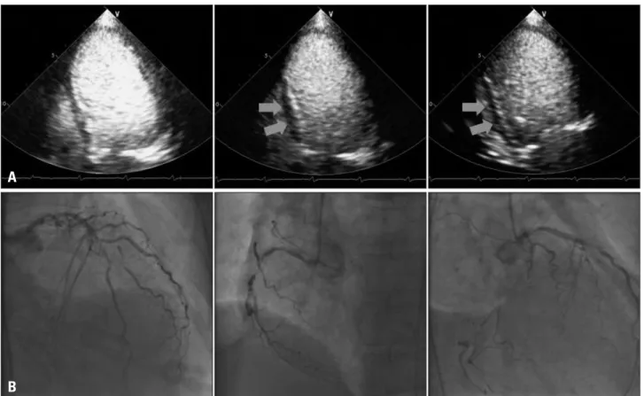
From qualitative MCE analysis, only one patient showed partial perfusion in basal inferior, mid-inferior, basal inferolat- eral and mid inferolateral wall whose coronary arteriography confirmed the total occlusion of right coronary artery (RCA) and left circumflex artery (LCx) with 70% stenosis in mid left anterior descending artery (LAD) (Fig. 1). The patient was an old lady (81 years old), and refused the coronary bypass graft surgery.
Quantitative MCE analysis was feasible in 258 of 378 seg- ments (68.3%) [97 of 126 (77.0%) segments in the 4-chamber view, 81 of 126 segments (64.3%) in the 2-chamber view, and
80 of 126 segments (63.5%) in the 3-chamber view]. The most frequent dropouts were observed in the basal segments of the lateral and anterior wall. According to coronary artery perfusion territories, quantitative analysis was feasible in 143 of 231 (61.9%) segments in the LAD territory, 56 of 84 (66.7%) of LCx territory, and 59 of 63 (93.7%) segments of RCA territory.
MCE parameters of A, β, and Aβ are shown in Table 3. “A”
value was significantly higher in patient with CAD (p = 0.010) and there were no significant difference in “β” and “Aβ” in pa- tients without and with CAD (0.48 ± 0.27 vs. 0.45 ± 0.25, p = 0.453 for β and 2.99 ± 2.23 vs. 3.68 ± 3.13, p = 0.059 for Aβ, respectively).
Discussion
We hypothesized that resting MCE may help to classify pa- tients with flow-limiting CAD presenting with an unknown cause of AHF with decreased LVEF. Resting quantitative MCE analysis was feasible, however, neither qualitative nor
Table 3. MCE parameters of A, β, and Aβ between patients with and without significant CAD
No CAD (n = 16, 76.2%) CAD (n = 5, 23.8%) p value
A (dB) 6.42 ± 7.64 7.64 ± 3.03 0.010
β (s-1) 0.48 ± 0.27 0.45 ± 0.25 0.453
Aβ (dB/s) 2.99 ± 2.23 3.68 ± 3.13 0.059
MCE: myocardial contrast echocardiography, CAD: coronary artery disease
Fig. 1. Case of 81-year-old female. Apical 4-chamber, 2-chamber, and 3-chamber view showed partial perfusion defect in inferior and inferolateral wall (arrows, A). Coronary arteriography showed 70% stenosis in mid left anterior descending artery, and total occlusion of right coronary artery and left circumflex artery (B).
A
B
quantitative data of MCE helped to differentiate patients with CAD from patients without CAD.
In our study, a quarter of patients presenting AHF showed significant coronary artery stenosis (> 70%). This is relatively small numbers, for in OPTIMIZE-HF study, 66% of patients with LV systolic dysfunction carried a diagnosis of CAD.7) We included only patients who had no known risk factors of indi- cating CAD (ischemic symptom, electrocardiographic change, rise of cardiac enzyme and RWMA in baseline echocardiogra- phy), because detection of CAD is most challenging in such patients. We found that many patients showed RWMA in baseline echocardiography or had elevated cardiac enzyme on admission, so only small number of patients matched the in- clusion criteria of the study. However, one may be able to as- sume that if baseline echocardiography in patients with no clinical evidence indicating CAD showed decreased LVEF and global hypokinesis, CAD is accompanied with relatively low probability, based on this result.
Quantitative MCE analysis was feasible in 68.3% of LV seg- ments. This low feasibility has been reported by other gro- ups.16)18) If the study subjects were not screened for echocar- diographic image quality, feasibility of quantitative analysis was around 70%, and this may reflect the real-world setting of performing MCE.
Resting MCE did not distinguish patients with CAD from patients without CAD. There could be two explanations for this negative result. One is that resting MBF velocity may be normal despite the presence of coronary artery stenosis because of coronary arteriolar vasodilation or collateral MBF from non- flow limiting coronary arteries.19)20) Vasodilator stress can help the detection of CAD based on the occurrence of reversible per- fusion defects, because myocardial blood flow velocity increases with vasodilator and MBF velocity reserve decreases with coro- nary artery stenosis.21) Many studies including of which aim was to differentiate ischemic cardiomyopathy from nonisch- emic cardiomyopathy showed that perfusion was also decreased in nonischemic cardiomyopathy.14)22)23) Abdelmoneim et al.24) proposed wall motion abnormality in Takotsubo cardiomyop- athy could be made by microvascular dysfunction as evi- denced by abnormal myocardial perfusion detected in MCE.
In our study, the cause of LV systolic dysfunction is not known yet, although hypertensive cardiomyopathy or idiopathic car- diomyopathy could be the most probable diagnosis. In acute stage of heart failure, myocardial perfusion became abnormal even without significant coronary artery stenosis, and that can be the other explanation of no differences of myocardial veloci- ty (β) and MBF (Aβ) between patients with and without CAD in our study.
The present study had several limitations. First, the popula- tion was small and thus, these negative findings could be caused by a low statistical power. Because some reports showed RWMA is not sensitive for detection of CAD in patients with dilated LV, exclusion of patients with RWMA could make
this study have less power. Second, only resting MCE was con- sidered in our study. Some reports showed diagnostic value of stress MCE for the detection of CAD, however, in acute set- ting, it is impossible to perform stress test in patients with AHF. So, this study was designed to perform resting MCE only, immediately after admission.
In conclusion, relatively small number of patients had CAD in patients with decreased LV systolic function with global hypokinesis. Resting quantitative MCE analysis was feasible in patients presenting as AHF, however, qualitative or quanti- tative data of MCE did not aid to differentiate patients with CAD from patients without CAD.
• Acknowledgements
This study was supported by a research grant from the Korean Society of Echocardiography.
References
1. Gheorghiade M, Sopko G, De Luca L, Velazquez EJ, Parker JD, Binkley PF, Sadowski Z, Golba KS, Prior DL, Rouleau JL, Bonow RO. Navigating the crossroads of coronary artery disease and heart failure.
Circulation 2006;114:1202-13.
2. Fox KF, Cowie MR, Wood DA, Coats AJ, Gibbs JS, Underwood SR, Turner RM, Poole-Wilson PA, Davies SW, Sutton GC. Coro- nary artery disease as the cause of incident heart failure in the population.
Eur Heart J 2001;22:228-36.
3. Bart BA, Shaw LK, McCants CB Jr, Fortin DF, Lee KL, Califf RM, O’Connor CM. Clinical determinants of mortality in patients with angio- graphically diagnosed ischemic or nonischemic cardiomyopathy. J Am Coll Cardiol 1997;30:1002-8.
4. Purek L, Laule-Kilian K, Christ A, Klima T, Pfisterer ME, Per- ruchoud AP, Mueller C. Coronary artery disease and outcome in acute congestive heart failure. Heart 2006;92:598-602.
5. Flaherty JD, Rossi JS, Fonarow GC, Nunez E, Stough WG, Abra- ham WT, Albert NM, Greenberg BH, O’Connor CM, Yancy CW, Young JB, Davidson CJ, Gheorghiade M. Influence of coronary angiog- raphy on the utilization of therapies in patients with acute heart failure syndromes: findings from Organized Program to Initiate Lifesaving Treat- ment in Hospitalized Patients with Heart Failure (OPTIMIZE-HF). Am Heart J 2009;157:1018-25.
6. Follath F, Cleland JG, Klein W, Murphy R. Etiology and response to drug treatment in heart failure. J Am Coll Cardiol 1998;32:1167-72.
7. Rossi JS, Flaherty JD, Fonarow GC, Nunez E, Gattis Stough W, Abraham WT, Albert NM, Greenberg BH, O’Connor CM, Yancy CW, Young JB, Davidson CJ, Gheorghiade M. Influence of coronary artery disease and coronary revascularization status on outcomes in patients with acute heart failure syndromes: a report from OPTIMIZE-HF (Orga- nized Program to Initiate Lifesaving Treatment in Hospitalized Patients with Heart Failure). Eur J Heart Fail 2008;10:1215-23.
8. Uretsky BF, Thygesen K, Armstrong PW, Cleland JG, Horowitz JD, Massie BM, Packer M, Poole-Wilson PA, Ryden L. Acute coro- nary findings at autopsy in heart failure patients with sudden death: results from the assessment of treatment with lisinopril and survival (ATLAS) tri- al. Circulation 2000;102:611-6.
9. Medina R, Panidis IP, Morganroth J, Kotler MN, Mintz GS. The value of echocardiographic regional wall motion abnormalities in detecting coronary artery disease in patients with or without a dilated left ventricle.
Am Heart J 1985;109:799-803.
10. Chrysohoou C, Greenberg M, Stefanadis C. Non-invasive methods in differentiating ischaemic from non-ischaemic cardiomyopathy. A review pa-
per. Acta Cardiol 2006;61:454-62.
11. Jeetley P, Hickman M, Kamp O, Lang RM, Thomas JD, Vannan MA, Vanoverschelde JL, van der Wouw PA, Senior R. Myocardial contrast echocardiography for the detection of coronary artery stenosis: a pro- spective multicenter study in comparison with single-photon emission comput- ed tomography. J Am Coll Cardiol 2006;47:141-5.
12. Firschke C, Andrássy P, Linka AZ, Busch R, Martinoff S. Adenosine myocardial contrast echo in intermediate severity coronary stenoses: a prospec- tive two-center study. Int J Cardiovasc Imaging 2007;23:311-21.
13. Senior R, Moreo A, Gaibazzi N, Agati L, Tiemann K, Shivalkar B, von Bardeleben S, Galiuto L, Lardoux H, Trocino G, Carrió I, Le Gu- ludec D, Sambuceti G, Becher H, Colonna P, Ten Cate F, Bramucci E, Cohen A, Bezante G, Aggeli C, Kasprzak JD. Comparison of sulfur hexafluoride microbubble (SonoVue)-enhanced myocardial contrast echocar- diography with gated single-photon emission computed tomography for detec- tion of significant coronary artery disease: a large European multicenter study. J Am Coll Cardiol 2013;62:1353-61.
14. Senior R, Janardhanan R, Jeetley P, Burden L. Myocardial contrast echocardiography for distinguishing ischemic from nonischemic first-onset acute heart failure: insights into the mechanism of acute heart failure. Cir- culation 2005;112:1587-93.
15. Boff GM, Zanco P, Della Valentina P, Cardaioli P, Thiene G, Chioin R, Dalla Volta S. Positron emission tomography is a useful tool in differen- tiating idiopathic from ischemic dilated cardiomyopathy. Int J Cardiol 2000;74:67-74; discussion 75-6.
16. Malm S, Frigstad S, Helland F, Oye K, Slordahl S, Skjarpe T. Quan- tification of resting myocardial blood flow velocity in normal humans using real-time contrast echocardiography. A feasibility study. Cardiovasc Ultra- sound 2005;3:16.
17. Wei K. Approaches to the detection of coronary artery disease using myocar- dial contrast echocardiography. Am J Cardiol 2002;90:48J-58J.
18. Abdelmoneim SS, Martinez MW, Mankad SV, Bernier M, Dhoble A, Pellikka PA, Chandrasekaran K, Oh JK, Mulvagh SL. Resting qualitative and quantitative myocardial contrast echocardiography to pre- dict cardiac events in patients with acute myocardial infarction and percuta- neous revascularization. Heart Vessels 2014. [Epub ahead of print]
19. Jayaweera AR, Wei K, Coggins M, Bin JP, Goodman C, Kaul S.
Role of capillaries in determining CBF reserve: new insights using myocardial contrast echocardiography. Am J Physiol 1999;277(6 Pt 2):H2363-72.
20. Janardhanan R, Burden L, Senior R. Usefulness of myocardial contrast echocardiography in predicting collateral blood flow in the presence of a per- sistently occluded acute myocardial infarction-related coronary artery. Am J Cardiol 2004;93:1207-11.
21. Kaul S. Myocardial contrast echocardiography: a 25-year retrospective.
Circulation 2008;118:291-308.
22. Watzinger N, Lund GK, Saeed M, Reddy GP, Araoz PA, Yang M, Schwartz AB, Bedigian M, Higgins CB. Myocardial blood flow in pa- tients with dilated cardiomyopathy: quantitative assessment with velocity- encoded cine magnetic resonance imaging of the coronary sinus. J Magn Re- son Imaging 2005;21:347-53.
23. Nitenberg A, Foult JM, Blanchet F, Zouioueche S. Multifactorial de- terminants of reduced coronary flow reserve after dipyridamole in dilated cardiomyopathy. Am J Cardiol 1985;55:748-54.
24. Abdelmoneim SS, Mankad SV, Bernier M, Dhoble A, Hagen ME, Ness SA, Chandrasekaran K, Pellikka PA, Oh JK, Mulvagh SL.
Microvascular function in Takotsubo cardiomyopathy with contrast echocar- diography: prospective evaluation and review of literature. J Am Soc Echo- cardiogr 2009;22:1249-55.