Impact of Tibial Tubercle Osteotomy on Final Outcome in Revision Total Knee Arthroplasty:
Our Experience and Technique in Pakistan
Abdul Rafay Qazi, MBBS, Faizan Iqbal, FCPS*, Syed Shahid Noor, FRCS, Nasir Ahmed, FCPS, Akram Ali Uddin, FCPS
†, Nouman Memon, FCPS, Naveed Memon, FCPS
‡Department of Orthopaedic Surgery, Liaquat National Hospital and Medical College, Karachi,
*Department of Orthopaedic Surgery, Patel Hospital, Karachi,
†Department of Orthopaedic Surgery, Fazaia Medical College, Karachi,
‡Department of Orthopaedic Surgery, Dow University of Health Sciences, Karachi, Pakistan
Background: Due to extensive fibrosis during revision surgery, adequate exposure is essential and it can be achieved with several extensile approach options, such as tibial tubercle osteotomy. Information regarding surgical exposure during revision arthroplasty is limited in developing countries, such as Pakistan, due to the lack of adequate data collection and follow-up. Therefore, the purpose of this study was to evaluate the impact of tibial tubercle osteotomy on final outcome of revision total knee arthroplasty (TKA).
Methods: A total of 231 revision TKAs were performed between January 2008 and December 2017. Twenty-nine patients under- went tibial tubercle osteotomy for adequate exposure during revision surgery. Of these, 27 patients with complete follow-up were included in our study. Factors examined include age at the time of revision surgery, gender, comorbidities, arthroplasty site (right or left), body mass index (BMI), and primary indications for the tibial tubercle osteotomy during revision TKA. Functional outcome was measured by using Knee Society score (KSS) at 3 months and the final follow-up. All statistical analysis was done using SPSS ver- sion 20.0 with a p-value < 0.05 considered significant.
Results: Out of 27 patients, 6 patients (22.2%) were men and 21 patients (77.7%) were women. Right knee revision arthroplasty was performed in 15 patients (55.5%), left knee revision arthroplasty was performed in 12 patients (44.4%), and bilateral revi- sion surgery was performed in only 1 patient (3.7%). The mean BMI was 29.2 kg/m2. We used a constrained condylar knee in 20 patients (74%), a rotating hinge knee in 5 patients (18.5%), and mobile bearing tray plus metaphyseal sleeves in 2 patients (7.4%).
The KSS was 52.21 ± 4.05 preoperatively, and 79.42 ± 2.2 and 80.12 ± 1.33 at 3 months and 12 months, respectively. Radiological union was achieved in all patients at 3 months. Of 27 patients, only 1 patient (3.7%) had proximal migration of the osteotomy site at 6 months: the patient was asymptomatic and union was also achieved and, therefore, no surgical intervention was performed.
Conclusions: Tibial tubercle osteotomy during revision TKA can be a safe and reliable technique with superior outcomes and minimal complication rates.
Keywords: Primary total knee arthroplasty, Prosthetic joint infection, Revision total knee arthroplasty, Tibial tubercle osteotomy, Knee Society score
Copyright © 2021 by The Korean Orthopaedic Association
This is an Open Access article distributed under the terms of the Creative Commons Attribution Non-Commercial License (http://creativecommons.org/licenses/by-nc/4.0) which permits unrestricted non-commercial use, distribution, and reproduction in any medium, provided the original work is properly cited.
Clinics in Orthopedic Surgery • pISSN 2005-291X eISSN 2005-4408 Received March 19, 2020; Accepted June 9, 2020
Correspondence to: Faizan Iqbal, FCPS
Department of Orthopaedic Surgery, Patel Hospital Karachi, St. 18, Block 4 Gulshan-e-Iqbal, Karachi, Sindh 75300, Pakistan Tel: +92-3402238126, Fax: +92-02134145094, E-mail: [email protected]
The number of revision total knee arthroplasty (TKA) is increasing day by day due to the rise in the number of primary TKA.1,2) In 6.6% to 38% of cases, revision TKA is performed due to prosthetic joint infection (PJI).3) Ad- equate exposure during revision TKA is one of the chal- lenges among arthroplasty surgeons because of underlying contracture of the extensor mechanism or limited mobil- ity, which might cause difficulty in eversion of the patella and could lead to a rupture of the patellar tendon from its insertion.4,5) There are various extensile approaches used during revision TKA in order to provide adequate exposure and allow easy implant removal such as the quadriceps snip approach (QS),6) quadriceps turndown technique,7) V-Y quadricepsplasty,8) and tibial tubercle osteotomy (TTO).9) Among all these approaches, the TTO and QS are commonly performed in complicated revision cases.10,11) Both approaches have some drawbacks. The QS is commonly associated with extensor mechanism scar- ring. Closure of TTO with screws or wires is associated with anterior knee pain, nonunion, proximal migration, and hardware-related problems.12) Information regard- ing TTO during revision surgery is limited in developing countries, such as Pakistan, due to the lack of adequate data collection and follow-up.
Therefore, the purpose of this study was to evaluate the impact of TTO on final outcome during revision TKA.
We hypothesized that TTO and its closure with a suture technique would be associated with superior outcomes and a minimal complication rate.
METHODS
This is a multicenter retrospective study conducted at the department of orthopedic surgery. The study was ap- proved by the ethics review committee of Liaquat National Hospital (No. 0162-2018). Patients who were operated for revision TKA were enrolled between January 2008 and December 2017. Data were principally collected from hospital records. A total of 231 revision arthroplasties were performed by a single experienced arthroplasty sur- geon (SSN). Twenty-nine patients, who underwent TTO for adequate exposure during revision arthroplasty, were identified from hospital records. Two patients were lost to follow-up after 3 months; therefore, 27 patients with com- plete follow-up were included in our study. The decision of TTO was made intraoperatively when the patella was un- able to be everted with the knee at 90° flexion. All patients who underwent revision TKA for any reason were includ- ed in our study. Patients who required tumor prosthesis or arthrodesis at any time during follow-up were excluded from our study. Factors examined include age the time of revision surgery, gender, comorbidities, site of arthro- plasty (right or left), body mass index (BMI), and primary indications for TTO during revision TKA. On admission for revision surgery, medical conditions of all patients were assessed and classified according to the American Society of Anesthesiologists (ASA) classification system.13) Radiographic assessments for the anteroposterior view, lateral view, and lower limb scanogram were performed in all patients who underwent revision TKA. All informa- tion regarding surgical procedures, final outcomes, and
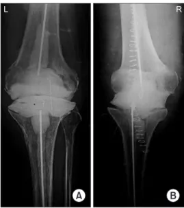
Fig. 1. Anteroposterior views of left (A) and right (B) knee joints with antibiotic cement spacers placed after adequate knee irrigation and debridement.
A B
L R
Fig. 2. Lateral views of left (A) and right (B) knee joints showing antibiotic cement spacers.
A B
L R
complications associated with TTO, such as migration of the TTO site, nonunion, infection (superficial or deep), anterior knee pain, and re-revision surgery, were obtained from hospital records. We used the Centers for Disease Control and Prevention criteria to diagnose prosthetic joint infection (PJI).14) Two-stage revision surgery was performed in all diagnosed cases of PJI as shown in Figs. 1 and 2. Revision surgery was performed 4 to 6 months later from primary surgery after excluding infection clinically and by laboratory workup.
We used a previous scar for exposure. The preferred approach was medial parapatellar in which an incision of the extensor retinaculum was started from the apex of the quadriceps tendon proximally to the tibial tubercle distally 6 to 8 cm from the joint line, exposing the whole tibial
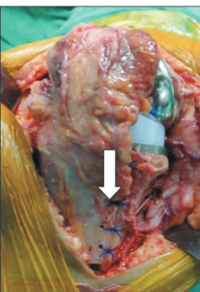
tubercle and patellar tendon. TTO was done on the whole length of the patellar tendon. With use of a mini saw, the osteotomy was performed initially from the medial cortex up to the lateral cortex, leaving the periosteal hinge on the lateral side. For proper closure, 1 cm of thickness of osteotomy should be left. After complete use of the saw, an osteotome was placed under the tibial tubercle to lever the osteotomy segment. This allowed for an excellent exposure of the proximal tibia for various purposes, such as removal of a previous implant, exposure of the femoral component by everting the patella and placing a new prosthesis under direct vision. Closure of the osteotomy site was very chal- lenging as it required proper placement of an osteotomy site in the previous location. With the help of Kocher, the osteotomy segment was brought to its original position and a 2-mm drill bit was used for holes first in the oste- otomy segment and then in the tibia. Care must be taken in order to prevent fracture of the segment by making a hole away from the edges. Usually, 4 holes were enough, 1 cm apart, depending on the length of osteotomy. A half circle taper cut needle of number 5 Ethibond suture was used in all cases because it was a braided polyester suture with high tensile strength. Intraoperative images of a trial implant and preparation of the tibia and femur is shown in Fig. 3. Suture was tied sequentially and knot was placed medially as shown in Fig. 4. The rest of the wound must be closed in a traditional manner. We used constrained condylar knees (CCK), rotating hinge knees (RHK), and mobile bearing tray (MBT) plus metaphyseal sleeves in revision TKAs. Revision surgery CCK is shown in Figs. 5 and 6. Postoperatively, the patient was encouraged to apply Fig. 3. Intraoperative images of the trial implant (A) and preparation of
the tibia and femur after tibial tubercle osteotomy (B).
A B
Fig. 4. An image showing closure of tibial tubercle osteotomy with Ethibond sutures. Also note the drill holes (arrow) through which sutures were passed to fix the osteotomy site.
L R
A B
Fig. 5. Anteroposterior views of left (A) and right (B) knee joints, showing constrained knee implants.
full weight-bearing with crutches and start range of mo- tion according to a protocol. Straight leg raise was allowed 6 weeks after surgery. A long-leg knee brace was used for 6 postoperative weeks.
Postoperative follow-up was performed at 2 weeks, 6 weeks, 3 months, 6 months, and 12 months. Postopera- tive X-rays were obtained immediately after surgery, at 6 weeks after surgery, and on each visit thereafter to look for osteotomy site healing, fracture at the osteotomy site, and obvious migration of the osteotomy site. Migration of the osteotomy site was evaluated by comparing the current position with the X-rays performed at 6 weeks, 3 months, and at 6 months. The osteotomy site was considered united when there was radiographic evidence of bridging callus on the lateral radiographs. Functional outcome was measured by using Knee Society score (KSS)15) at 3 months and the final follow-up. All statistical analysis was done using IBM SPSS ver. 20.0 (IBM Corp., Armonk, NY, USA) with a p- value < 0.05 considered as significant.
RESULTS
Out of 27 patients, 6 (22.2%) were men and 21 (77.7%) were women. The age of patients at revision surgery was 66.4 years (range, 51–74 years). Right knee revision ar- throplasty was performed in 15 patients (55.5%), left knee revision arthroplasty was performed in 12 patients (44.4%), and bilateral revision surgery was performed in only 1 pa- tient (3.7%). The mean BMI was 29.2 kg/m2. Out of the 27 patients, 7 (25.9%) were diabetic, 11 (40.7%) were hyper- tensive, 2 (7.4%) had ischemic heart disease, and 1 (3.7%)
had asthma and rheumatoid arthritis. Four patients (14.8%) had more than 2 comorbidities, and only 1 patient (3.7%) patient had no comorbidity. We used CCK in 20 patients (74%), RHK in 5 patients (18.5%), and MBT plus metaphy- seal sleeves in 2 patients (7.4%). We commonly encoun- tered patients with an ASA class 3 in our study. Detailed demographic characteristics are presented in Table 1.
A B
L R
Fig. 6. Lateral views of right (A) and left (B) knee joints, showing con- strained knee implants. Note union of the osteotomy site (white arrows).
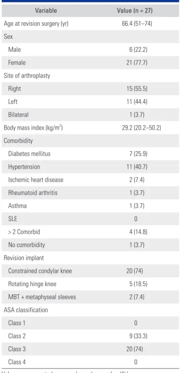
Table 1. Descriptive Statistics of Demographics
Variable Value (n = 27)
Age at revision surgery (yr) 66.4 (51–74) Sex
Male 6 (22.2)
Female 21 (77.7)
Site of arthroplasty
Right 15 (55.5)
Left 11 (44.4)
Bilateral 1 (3.7)
Body mass index (kg/m2) 29.2 (20.2–50.2) Comorbidity
Diabetes mellitus 7 (25.9)
Hypertension 11 (40.7)
Ischemic heart disease 2 (7.4)
Rheumatoid arthritis 1 (3.7)
Asthma 1 (3.7)
SLE 0
> 2 Comorbid 4 (14.8)
No comorbidity 1 (3.7)
Revision implant
Constrained condylar knee 20 (74)
Rotating hinge knee 5 (18.5)
MBT + metaphyseal sleeves 2 (7.4)
ASA classification
Class 1 0
Class 2 9 (33.3)
Class 3 20 (74)
Class 4 0
Values are presented as mean (range) or number (%).
SLE: systemic lupus erythematosus, MBT: mobile bearing tray, ASA:
American Society of Anesthesiologists.
Twenty-three patients (85.1%) had PJI, 5 (18.5%) had aseptic loosening, and 1 (3.7%) had instability. All patients who had PJI underwent primary TKA initially at another hospital and then were referred to our center for further management. The detailed indications for revision TKA are presented in Table 2.
KSS was measured in all patients who underwent TTO during revision TKA at 3 months and 12 months of follow-up. The KSS was 52.21 ± 4.05 preoperatively and 79.42 ± 2.2 and 80.12 ± 1.33 at 3 months and 12 months postoperatively, respectively. Functional score was 49.33 ± 3.24 preoperatively, whereas it was 80.28 ± 2.99 and 81.34
± 2.82 at 3 months and 12 months postoperatively. There was a major difference between clinical and function scores preoperatively and postoperatively (p < 0.05) (Table 3).
Radiological union was achieved at 3 months in all patients. Out of 27 patients, only 1 (3.7%) had proximal migration of the osteotomy site at 6 months after surgery.
The osteotomy site was united and the patient did well and was asymptomatic; therefore, no surgical intervention was performed for proximal migration of the osteotomy site.
Complications following revision TKA are presented in Table 4.
DISCUSSION
Surgical exposure during revision TKA is important to get good to excellent outcome.16) To the best of our knowl- edge, this study is the first from Pakistan to show results and complications after closing TTO with Ethibond su-
ture. The main indications for TTO during revision TKA in the current study were PJI of primary TKA, followed by aseptic loosening and instability. In 85% of cases, revision surgery was performed due to PJI, whereas in a study con- ducted by Haenle et al.,3) revision TKA was performed due to PJI in 6.6% to 38% of cases. Revision surgery after erad- ication of PJI remains challenging even for experienced arthroplasty surgeons due to underlying fibrosis and soft- tissue inflammation.17) The success of revision surgery de- pends on various factors such as operative techniques, as well as the postoperative rehabilitation protocol.18) There are various options related to closing TTO such as screws, cerclage wires, and absorbable sutures. Previous studies al- ready showed results of different options of closing TTO.19) We used Ethibond suture to close TTO in all patients. Et- hibond is a braided suture; therefore, it is long-lasting and has minimal soft-tissue reactivity. In this study, we did not compare our technique of closing TTO with other tech- niques but just showed our results and complications after closing osteotomy with Ethibond sutures.
Previous studies already showed results of different options of closing TTO.19,20) Segur et al.19) conducted a ret- rospective study of a prospective database of 26 patients, who underwent revision TKA for PJI. TTO was closed with two 18-guage monofilament stainless steel wires. The
Table 3. KSS Clinical and Function Scores
Variable Preoperative Postoperative 3 mo Postoperative 12 mo p-value*
Clinical score (out of 100) 52.21 ± 4.05 79.42 ± 2.2 80.12 ± 1.33 0.002
Function score (out of 100) 49.33 ± 3.24 80.28 ± 2.99 81.34 ± 2.82 0.001
Values are presented as mean ± standard deviation.
KSS: Knee Society score.
*p < 0.05 considered significant.
Table 4. Complications after Tibial Tubercle Osteotomy
Complication Patient (n = 27)
Migration of osteotomy site 1 (3.7)
Nonunion 0
Fracture at osteotomy site 0
Extension lag 0
Anterior knee pain 0
Re-infection (superficial or deep) 0
Re-revision surgery 0
Values are presented as number (%).
Table 2. Indications for Tibial Tubercle Osteotomy
Indication Patient (n = 27)
Aseptic loosening of primary TKA
(both femur and tibia) 5 (18.5)
Prosthetic joint infection 23 (85.1)
Instability 1 (3.7)
Values are presented as number (%).
TKA: total knee arthroplasty.
average clinical score was 59 (standard deviation [SD], 9.8), whereas functional score was 51 (SD, 23.4) preoperatively.
The average clinical score was 78 (SD, 15.5) and the aver- age functional score was 70 (SD, 16.6) at 12 months. In our series, the average clinical score was 52.21 ± 4.05 pre- operatively, whereas it was 79.42 ± 2.2 and 80.12 ± 1.33 at 3 and 12 months postoperatively, respectively. The average functional score was 49.33 ± 3.24 preoperatively, whereas it was 80.28 ± 2.99 and 81.34 ± 2.82 at 3 and 12 months postoperatively, respectively. These findings suggest that TTO during revision TKA was associated with superior outcomes, which was one of the hypotheses of our study.
The integrity of blood supply to the patella is impor- tant as well to prevent osteonecrosis.21) Previous studies compared different extensile approaches to get adequate exposure during revision TKA. TTO is performed distal to the extensor mechanism; therefore, patellar supply cannot be compromised and healing can occur at bone-to-bone interface. All other techniques such as V-Y quadriceps- plasty, patellar turn-down technique, and QS approach (combined with lateral release) are performed at the level of the extensor mechanism and can cause damage to blood supply of the patella with subsequent osteonecrosis.22)
The current study showed that closing TTO with Ethibond sutures is a simple and effective technique and results were also comparable with other standard tech- niques described in literature. Out of 27 patients, only 1 (3.7%) had migration of the osteotomy site at 6 months af- ter closing osteotomy with Ethibond suture. As radiologic union was achieved and the patient was asymptomatic, no surgical intervention was performed. Davis et al.23) per- formed TTO closure with two 4.5-mm cortical screws and 18-gauge stainless steel cerclage wire on 37 fresh frozen ca- davers. They actually created upper shelf at the osteotomy site to prevent migration later. In our series of patients, we did not make any shelf to prevent migration of the oste- otomy.
Closure of the osteotomy site with cortical screws or cerclage wiring is associated with anterior knee pain, ne-
cessitating a second surgery for hardware removal.10) None of the patients in the current study had anterior knee pain or underwent a second revision. The mean age of patients who underwent revision TKA and required TTO in this study was 66.4 years. Osteotomy closure with Ethibond suture not only avoids anterior knee pain but also prevents another surgery for hardware removal. Therefore, closing osteotomy with Ethibond suture was a safe and reliable technique to obtain good results with minimal complica- tion, which was another hypothesis of our study.
Previous studies demonstrated that fixation with screws was biomechanically stronger than suture fixa- tion.24) We found Ethibond suture is a stable and reliable technique for closing TTO. Although a comparative study is required between 2 techniques to assess stability, another aspect of closing osteotomy with either screws or cerclage wire is the prolonged operative time. Although we did not measure the operating time in our study, previous studies demonstrated additional 15 to 20 minutes was required for closing osteotomy with screws or cerclage wires.22)
Major limitations of this study were the limited number of patients and short follow-up period, which were not sufficient to evaluate the long-term outcome of this procedure. Our study revealed that TTO during revi- sion TKA could be a safe and reliable technique with supe- rior clinical outcomes and minimal complication rates.
CONFLICT OF INTEREST
No potential conflict of interest relevant to this article was reported.
ACKNOWLEDGEMENTS
We would like to express our special gratitude to Prof.
Syed Shahid Noor (Head of Department of Orthopedic Surgery, President of Pakistan Arthroplasty Society and Director of Pakistan National Joint Registry), who sup- ported us and offered deep insight into this project.
REFERENCES
1. Lee DH, Lee SH, Song EK, Seon JK, Lim HA, Yang HY.
Causes and clinical outcomes of revision total knee arthro- plasty. Knee Surg Relat Res. 2017;29(2):104-9.
2. Clement ND, MacDonald DJ, Hamilton DF, Burnett R. Pos- terior condylar offset is an independent predictor of func- tional outcome after revision total knee arthroplasty. Bone Joint Res. 2017;6(3):172-8.
3. Haenle M, Skripitz C, Mittelmeier W, Skripitz R. Economic impact of infected total knee arthroplasty. ScientificWorld- Journal. 2012;2012:196515.
4. Thienpont E. Revision knee surgery techniques. EFORT Open Rev. 2017;1(5):233-8.
5. Edwards PK, Levine M, Lowry Barnes C. Management of extensor mechanism during revision total knee arthroplasty.
In: Bono J, Scott R, eds. Revision total knee arthroplasty.
Cham: Springer; 2018. 205-21.
6. Abdel MP, Della Valle CJ. The surgical approach for revi- sion total knee arthroplasty. Bone Joint J. 2016;98(1 Suppl A):113-5.
7. Biggi S, Divano S, Tedino R, Capuzzo A, Tornago S, Cam- era A. Tibial tubercle osteotomy in total knee arthroplasty:
midterm results experience of a monocentric study. Joints.
2018;6(2):95-9.
8. Lombardi AV. Implant extraction in revision knee arthro- plasty. Orthop Proc. 2019;101(Supp_8):48.
9. DeHaan A, Shukla S, Anderson M, Ries M. Tibial tubercle osteotomy to aid exposure for revision total knee arthro- plasty. JBJS Essent Surg Tech. 2016;6(3):e32.
10. Sun Z, Patil A, Song EK, Kim HT, Seon JK. Comparison of quadriceps snip and tibial tubercle osteotomy in revision for infected total knee arthroplasty. Int Orthop. 2015;39(5):879- 85.
11. Glynn A, Huang R, Mortazavi J, Parvizi J. The impact of pa- tellar resurfacing in two-stage revision of the infected total knee arthroplasty. J Arthroplasty. 2014;29(7):1439-42.
12. Stambough JB, Clohisy JC, Barrack RL, Nunley RM, Keeney JA. Increased risk of failure following revision total knee re- placement in patients aged 55 years and younger. Bone Joint J. 2014;96(12):1657-62.
13. Daabiss M. American Society of Anaesthesiologists physical status classification. Indian J Anaesth. 2011;55(2):111-5.
14. Iqbal F, Shafiq B, Zamir M, et al. Micro-organisms and risk factors associated with prosthetic joint infection following primary total knee replacement-our experience in Pakistan.
Int Orthop. 2020;44(2):283-9.
15. Insall JN, Dorr LD, Scott RD, Scott WN. Rationale of the
Knee Society clinical rating system. Clin Orthop Relat Res.
1989;(248):13-4.
16. Abbas A, Razii N, Morgan-Jones R. Extensile exposure in revision total knee arthroplasty. In: Malhotra R, Gautam D, eds. Mastering orthopedic techniques: revision knee arthro- plasty. New Delhi: Jaypee Brothers; 2019. 63-9.
17. Bruckner C, Straube E, Petersen I, et al. Low-grade infec- tions as a possible cause of arthrofibrosis after total knee arthroplasty. Patient Saf Surg. 2019;13:1.
18. Divano S, Camera A, Biggi S, Tornago S, Formica M, Felli L.
Tibial tubercle osteotomy (TTO) in total knee arthroplasty, is it worth it? A review of the literature. Arch Orthop Trau- ma Surg. 2018;138(3):387-99.
19. Segur JM, Vilchez-Cavazos F, Martinez-Pastor JC, Macule F, Suso S, Acosta-Olivo C. Tibial tubercle osteotomy in septic revision total knee arthroplasty. Arch Orthop Trauma Surg.
2014;134(9):1311-5.
20. Padgett DE. Exposure options in revision total knee arthro- plasty. Orthop Proc. 2019;101(Supp_8):46.
21. DeBell H, Pinter Z, Pinto M, et al. Vascular supply at risk during lateral release of the patella during total knee ar- throplasty: a cadaveric study. J Clin Orthop Trauma. 2019;
10(1):107-10.
22. Zonnenberg CB, van den Bekerom MP, de Jong T, Nolte PA.
Tibial tubercle osteotomy with absorbable suture fixation in revision total knee arthroplasty: a report of 23 cases. Arch Orthop Trauma Surg. 2014;134(5):667-72.
23. Davis K, Caldwell P, Wayne J, Jiranek WA. Mechanical com- parison of fixation techniques for the tibial tubercle oste- otomy. Clin Orthop Relat Res. 2000;(380):241-9.
24. Halder AM. Tibial tubercle osteotomy. Oper Orthop Trau- matol. 2012;24(2):85-94.