Brief Report
114 Ann Dermatol
Received November 3, 2016, Revised December 14, 2016, Accepted for publication February 1, 2017
Corresponding author: Hyun-Chang Ko, Department of Dermatology, Pusan National University Yangsan Hospital, 20 Geumo-ro, Mulgeum-eup, Yangsan 50612, Korea. Tel: 82-55-360-1678, Fax: 82-55-360-1679, E-mail: hcko@pusan.ac.kr
This is an Open Access article distributed under the terms of the Creative Commons Attribution Non-Commercial License (http://creativecommons.org/
licenses/by-nc/4.0) which permits unrestricted non-commercial use, distribution, and reproduction in any medium, provided the original work is properly cited.
Copyright © The Korean Dermatological Association and The Korean Society for Investigative Dermatology 6. Barker A, Jones R, Jennison C. A prevalence study of age-
associated memory impairment. Br J Psychiatry 1995;167:
642-648.
7. Small GW. What we need to know about age related memory loss. BMJ 2002;324:1502-1505.
https://doi.org/10.5021/ad.2018.30.1.114
Congenital Linear Smooth Muscle Hamartoma with Hypertrichosis: Hair Density on Dermoscopy in Parallel with the Number of Smooth Muscle Bundles
Jin-Hwa Son
1, Hyunju Jin
1, Hyang-Suk You
1, Woo-Haing Shim
2, Jeong-Min Kim
2, Gun-Wook Kim
1, Hoon-Soo Kim
1, Byung-Soo Kim
1, Moon-Bum Kim
1, Hyun-Chang Ko
1,2,31Department of Dermatology, Pusan National University School of Medicine, Busan, 2Department of Dermatology and 3Research Institute for Convergence of Biomedical Science and Technology, Pusan National University Yangsan Hospital, Yangsan, Korea
Dear Editor:
Smooth muscle hamartoma (SMH) is a rare benign tumor of the skin, characterized by the proliferation of smooth muscle bundles within the reticular dermis1,2.
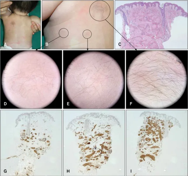
A 5-month-old girl presented with multiple skin-colored patches with hypertrichosis on the left upper back since birth (Fig. 1A). The lesions were composed of three patch- es with linear distribution (Fig. 1B). The mother of the in- fant had noted transient induration with piloerection of the lesions when exposed to cool air and rubbing, called a
“pseudo-Darier sign.” Dermoscopic findings showed dif- ferent types of hypertrichosis in the three patches (Fig.
1D∼F). Histopathological examination revealed numer- ous haphazardly arranged smooth muscle bundles in the dermis (Fig. 1C), and immunohistochemical staining showed diffusely stained smooth muscle actin (Fig. 1G∼I). These findings suggested a diagnosis of congenital SMH.
Gagné and Su3 suggested that hypertrichosis or prominent overlying hair in congenital SMH was usually present, but hair density was unchanged besides increased hair diame-
ter and length. However, we speculated that there is a re- lation between hypertrichosis including hair density and the amount of smooth muscle bundle. Interestingly, der- moscopy revealed hypertrichosis with varying densities at the different sites (Fig. 1D∼F), and histopathological exa- mination revealed different numbers of smooth muscle in the reticular dermis (Fig. 1G∼I). Thus, we measured the density of the hair (/mm3) in each lesion by using dermo- scopy and the area of smooth muscle in the reticular der- mis (%) by using a digital image analysis software (ImageJ 1.01 version; National Institutes of Health, Bethesda, MD, USA). The density of hair was 0.27/mm3 in the lateral hairy patch (Fig. 1D), 0.44/mm3 in the middle patch (Fig. 1E), and 1.40/mm3 in the medial patch (Fig. 1F), and the num- ber of smooth muscle bundles in the reticular dermis was 9.5%, 24.6%, and 31.0% in each patch (Fig. 1G∼I).
Therefore, we believed that the number of hair in multiple patches, as observed using dermoscopy, was in proportion with the number of smooth muscle bundles in each le- sion, as observed in the histopathological examination
Brief Report
Vol. 30, No. 1, 2018 115 Fig. 1. (A) Multiple linear scattered skin-colored patches with hypertrichosis on left upper back were observed. (B) Close-up view of left upper back. (C) Numerous smooth muscle bundles with various direction throughout dermis were shown (H&E, ×40). (D∼
F) In three sites, hypertrichosis with various density was shown on dermoscopy. (G∼I) Different amount of smooth muscles according to hypertrichosis was observed in the reticular dermis (smooth muscle actin, ×20).
Fig. 2. Correlation between hypertrichosis and the amount of smooth muscle bundle.
(Fig. 2).
In the literature, SMH probably represents aberrant devel- opment of the arrector pilorum during fetal maturation. In human skin, it may develop at the time of mesodermal ma- turation4. Koizumi et al.5 suggested that numerous CD34-po- sitive cells may be present in the stroma surrounding the smooth muscle bundles. It has been speculated that der- mal dendritic cells release growth factors or directly con- tact the epithelial cells of the bulge, a region considered to represent a reservoir of hair follicle stem cells.
We speculate that dermal dendritic cells situated in the SMH might have a role in the hypertrichosis. However, the definite relation between SMH and hypertrichosis re- mains to be established.
Brief Report
116 Ann Dermatol
Received September 7, 2016, Revised January 13, 2017, Accepted for publication February 6, 2017
Corresponding author: June Hyunkyung Lee, Department of Dermatology, Nowon Eulji Medical Center, Eulji University, 68 Hangeulbiseok-ro, Nowon-gu, Seoul 01830, Korea. Tel: 82-2-970-8580, Fax: 82-2-974-1577, E-mail: euljiderma@gmail.com
This is an Open Access article distributed under the terms of the Creative Commons Attribution Non-Commercial License (http://creativecommons.org/
licenses/by-nc/4.0) which permits unrestricted non-commercial use, distribution, and reproduction in any medium, provided the original work is properly cited.
Copyright © The Korean Dermatological Association and The Korean Society for Investigative Dermatology
Though it was focused one case to generalize the results, it is not possible to generalize the results. However, our case shows that a simple, non-invasive dermoscopic ex- amination to determine hair density could be an ancillary method to predict the number of smooth muscle bundle in the dermis.
CONFLICTS OF INTEREST
The authors have nothing to disclose.
REFERENCES
1. Kwon SB, Lee SJ, Kim DW, Jun JB. Congenital smooth
muscle hamartoma: a patchy follicular variant. Ann Dermatol 2000;12:231-234.
2. Gerdsen R, Lagarde C, Steen A, Steen KH, Uerlich M, Bieber T. Congenital smooth muscle hamartoma of the skin: clinical classification. Acta Derm Venereol 1999;79:408-409.
3. Gagné EJ, Su WP. Congenital smooth muscle hamartoma of the skin. Pediatr Dermatol 1993;10:142-145.
4. Huffman DW, Mallory SB. Congenital smooth muscle hamartoma. Am Fam Physician 1989;39:117-120.
5. Koizumi H, Kodama K, Tsuji Y, Matsumura T, Nabeshima M, Ohkawara A. CD34-positive dendritic cells are an in- trinsic part of smooth muscle hamartoma. Br J Dermatol 1999;140:172-174.
https://doi.org/10.5021/ad.2018.30.1.116
A Case of Atrophoderma Vermiculatum Showing a Good Response to Topical Tretinoin
Young Chae Lee, Sook-Ja Son, Tae Young Han, June Hyunkyung Lee
Department of Dermatology, Nowon Eulji Medical Center, Eulji University College of Medicine, Seoul, Korea
Dear Editor:
Atrophoderma vermiculatum (AV) is a rare, slowly pro- gressive, benign follicular disorder that affects primarily children1. AV is characterized by the development of in- flammatory, keratotic papules of the face that form pitted, atrophic, and depressed scars in a reticular or honeycomb pattern1,2.
A 12-year-old girl presented with sudden onset of atrophic scarring of both cheeks. Examination showed multiple, pitted, honeycomb scars on the both cheeks, temples, chin and neck. The lesions were similar regarding their size and morphology, were oval shaped, skin-colored, 1∼
2 mm in diameter, and approximately 1 mm deep (Fig.
1A, B). The lesions had developed several years prior and had gradually expanded centrifugally. Neither the eye- brows nor eyelashes were involved; no scarring alopecia was evident. The patient denied any subjective symptoms including itching or pain. Her medical history contained atopic dermatitis but no evidence of any physical trauma or inflammation prior to disease onset. The physical ex- amination and routine laboratory test showed all normal results. There was no relevant family history.
Histopathological examination of a punch biopsy speci- men of an atrophic keratotic papule on the cheek revealed follicular hyperkeratosis; aberrant, atrophic pilosebaceous units formed small finger-like projections into the sur-