Olmutinib Induced Lichen Planus Like Eruption
Vol. 30, No. 4, 2018 451
Received March 31, 2017, Revised August 7, 2017, Accepted for publication August 9, 2017
Corresponding author: Ji-Hye Park, Department of Dermatology, Samsung Medical Center, Sungkyunkwan University School of Medicine, 81 Irwon-ro, Gangnam-gu, Seoul 06351, Korea. Tel: 82-2-3410-6578, Fax:
82-2-3410-3869, E-mail: [email protected] ORCID: https://orcid.org/0000-0002-6699-5202
This is an Open Access article distributed under the terms of the Creative Commons Attribution Non-Commercial License (http://creativecommons.
org/licenses/by-nc/4.0) which permits unrestricted non-commercial use, distribution, and reproduction in any medium, provided the original work is properly cited.
Copyright © The Korean Dermatological Association and The Korean Society for Investigative Dermatology
pISSN 1013-9087ㆍeISSN 2005-3894
Ann Dermatol Vol. 30, No. 4, 2018 https://doi.org/10.5021/ad.2018.30.4.451
CASE REPORT
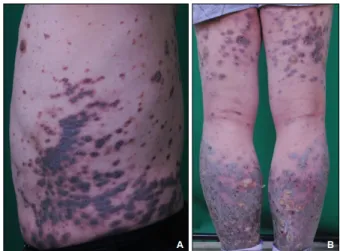
Fig. 1. (A) Scaly, violaceous to black-colored plaques and dusky brown macules on trunk. (B) Same lesions are at both thighs and calves. However, popliteal areas were spared.
Olmutinib Induced Lichen Planus Like Eruption
Seung Hwan Oh, Hyun Jeong Byun, Se Jin Oh, Ji-Young Jun, Ji-Hye Park, Jong Hee Lee, Dong-Youn Lee, Joo-Heung Lee, Jun-Mo Yang
Department of Dermatology, Samsung Medical Center, Sungkyunkwan University School of Medicine, Seoul, Korea
Drug induced lichen planus like eruption is an uncommon cutaneous adverse effect of several drugs. This appears sym- metric eruption of erythematous or violaceous plaques re- sembling lichen planus on the trunk and extremities. A 50-year-old male presented with scaly, violaceous plaques and dusky brown macules on whole body. For four months, the patient was treated with olmutinib, an oral, third-gen- eration epidermal growth factor receptor-tyrosine kinase inhibitor. In May 2016, olmutinib received its first global ap- proval in South Korea for the treatment of patients with lo- cally advanced or metastatic epidermal growth factor re- ceptor T790M mutation-positive non-small cell lung cancer.
The biopsy specimen from the patient showed features of li- chen planus. We diagnosed him with olmutinib-induced li- chen planus like eruption. He was treated with oral methyl- prednisolone and topical desoxymethasone 0.25% ointment.
At the same time, olmutinib dose was decreased to three- fourths of this patient’s starting dose. After that, the cutaneous lesions improved. (Ann Dermatol 30(4) 451∼453, 2018) -Keywords-
Drug eruptions, EGFR tyrosine kinase inhibitor, Lichen planus, Olmutinib
INTRODUCTION
Olmutinib is an oral, third-generation epidermal growth factor receptor-tyrosine kinase inhibitor (EGFR-TKI) for lo- cally advanced or metastatic EGFR T790M mutation-pos- itive non-small cell lung cancer. This received its first global approval in South Korea in May 20161. We present a case of lichen planus (LP) like eruption in a man after ol- mutinib treatment. To our knowledge, this is the first re- ported case of olmutinib induced LP like eruption.
CASE REPORT
A 50-year-old male presented with scaly, violaceous pla- ques and dusky brown macules on whole body for one month (Fig. 1A). Flexural areas were spared (Fig. 1B).
There was no mucous membrane lesion. The patient had been treated with olmutinib for four months because of non-small cell lung cancer. Pleural and brain metastasis
SH Oh, et al
452 Ann Dermatol
Fig. 2. Histopathological images of skin biopsy. (A) Orthokeratosis, wedge-shaped hypergranulosis, saw-toothed irregular elongated rete ridges in the epidermis. Max-Joseph spaces are shown (blue arrows). Lichenoid lymphocytic infiltration in the dermis (H&E, ×40).
(B) Dyskeratotic cells in the epidermis (red arrow) (H&E, ×200).
were found at the time of diagnosis. Therefore, clinical tri- al with olmutinib for palliative chemotherapy was per- formed with patient consent. No other medication was used to this patient within three months. Skin biopsy specimen of his dorsum of hand demonstrated focal ortho- keratosis, wedge-shaped hypergranulosis, saw-toothed ir- regular elongated rete ridges, and dermal lichenoid lym- phocytic infiltration with scanty eosinophils. Max-Joseph space and dyskeratotic cells were also shown in the speci- men (Fig. 2). These histologic features resembled those of LP. However, the clinical features were different from typi- cal features of idiopathic LP, and the cutaneous lesions ap- peared after olmutinib therapy. Consequently, we diag- nosed this olmutinib induced LP like eruption. He was treated with oral methylprednisolone 20 mg per day initially. Every three days, the dosage of methylprednisolone was reduced in half than before. Topical desoxymetha- sone 0.25% ointment was also applied to his lesions. At the same time, olmutinib dosage was decreased to three- fourths of this patient’s starting dose (800 mg per day to 600 mg per day), because the oncologist considered this cutaneous reaction as grade III adverse event. After three weeks, the body surface area of cutaneous lesions de- creased to about a half. After the discontinuation of sys- temic corticosteroid therapy, he was treated with oral anti- histamine, topical steroid and tacrolimus ointments for
several weeks because of itch. Although the dosage of ol- mutinib has been maintained 600 mg per day for four months, new lesions have not appeared.
DISCUSSION
Drug induced LP like eruption is an uncommon cutaneous adverse event of several drugs such as antimalarial, be- ta-blockers, gold salts, methyldopa, or quinidine2. Latency period varies from months to a year or more. It depends upon the class of drug, dose, host reaction, and con- current medications3. Typical cutaneous lesion of drug in- duced LP like eruption is symmetric eruption of eryth- ematous or violaceous plaques on the trunk and ex- tremities resembling idiopathic LP. However, drug in- duced LP like eruption rarely shows the flexural dis- tribution which is common in idiopathic LP. Furthermore, mucous membrane involvement is less common in drug induced LP like eruption4.
In this case, skin lesions developed after the initiation of olmutinib therapy and improved after decreasing of dos- age of olmutinib. Flexural areas were spared. Furthermore, mucous membranes were not involved. The histopatho- logic features show those of idiopathic LP. Therefore, we called this case as ‘olmutinib induced LP like eruption.’
There are several reports of cutaneous adverse events as-
Olmutinib Induced Lichen Planus Like Eruption
Vol. 30, No. 4, 2018 453 sociated with biologics or targeted therapy. For example,
tumor necrosis factor-α inhibitors are well known as the one of agents causing LP like eruption. EGFR-TKIs can have major skin toxic effects including acneiform erup- tion, pruritus, xerosis, and paronychia5,6. However, we cannot find the literatures about LP, LP like eruption, or li- chenoid drug eruption associated with the first- and sec- ond-generation EGFR-TKIs, gefitinib, erlotinib, and afatinib.
There is no reported case of LP like eruption related to EGFR inhibitors in PubMed. On the other hand, other TKIs except EGFR-TKIs can cause LP or LP like eruption, with 2 literatures, 9 cases reported to date in PubMed7,8. Some authors hypothesized that signal inhibition of plate- let-derived growth factor receptor, c-kit receptor or Src family kinase by TKIs may lead to keratinocytic apoptosis and lymphocytic inflammation7. However, TKIs used in their literature are first-, second-, or third-generation TKIs such as imatinib, nilotinib, dasatinib, nilotinib, or ponati- nib7,8.
There is no reported literature of cutaneous adverse events of olmutinib. Pathomechanism of olmutinib induced LP like eruption is also uncertain. Tyrosine kinases can inhibit not only mutant EGFR, but also wild-type EGFR9. Olmuti- nib is an irreversible kinase inhibitor, binding to a cysteine residue near the kinase domain, and known as having lit- tle effect on cell lines with wild-type EGFR10. Theoretically, olmutinib should have minimal effect to normal skin, but LP like eruption was occurred in our case. Serious cuta- neous adverse events of olmutinib, toxic epidermal nec- rolysis and Stevens-Johnson syndrome, also became an is- sue a few months ago. Therefore, we claim that clinicians should be aware of the risk of cutaneous adverse events of olmutinib.
CONFLICTS OF INTEREST
The authors have nothing to disclose.
REFERENCES
1. Kim ES. Olmutinib: first global approval. Drugs 2016;76:
1153-1157.
2. Ellgehausen P, Elsner P, Burg G. Drug-induced lichen planus. Clin Dermatol 1998;16:325-332.
3. Brauer J, Votava HJ, Meehan S, Soter NA. Lichenoid drug eruption. Dermatol Online J 2009;15:13.
4. Goldsmith LA, Katz SI, Gilchrest BA, Paller AS, Leffell DJ, Wolff K, et al. Fitzpatrick's dermatology in general medicine.
8th ed. New York: McGraw-Hill Medical, 2012:304.
5. Agero AL, Dusza SW, Benvenuto-Andrade C, Busam KJ, Myskowski P, Halpern AC. Dermatologic side effects asso- ciated with the epidermal growth factor receptor inhibitors.
J Am Acad Dermatol 2006;55:657-670.
6. Lacouture ME, Schadendorf D, Chu CY, Uttenreuther- Fischer M, Stammberger U, O'Brien D, et al. Dermatologic adverse events associated with afatinib: an oral ErbB family blocker. Expert Rev Anticancer Ther 2013;13:721-728.
7. Patel AB, Solomon AR, Mauro MJ, Ehst BD. Unique cutaneous reaction to second- and third-generation tyrosine kinase inhibitors for chronic myeloid leukemia. Dermatology 2016;232:122-125.
8. Pretel-Irazabal M, Tuneu-Valls A, Ormaechea-Perez N.
[Adverse skin effects of imatinib, a tyrosine kinase inhibitor].
Actas Dermosifiliogr 2014;105:655-662. Spanish.
9. Liao BC, Lin CC, Lee JH, Yang JC. Update on recent preclinical and clinical studies of T790M mutant-specific irreversible epidermal growth factor receptor tyrosine kinase inhibitors. J Biomed Sci 2016;23:86.
10. Lee KO, Cha MY, Kim M, Song JY, Lee JH, Kim YH, et al.
Abstract LB-100: Discovery of HM61713 as an orally available and mutant EGFR selective inhibitor. Cancer Res 2014;74(19 Suppl):LB-100.