Rebleeding after Initial Endoscopic Hemostasis in Peptic Ulcer Disease
Endoscopic hemostasis is the first-line treatment for upper gastrointestinal bleeding (UGIB).
Although several factors are known to be risk factors for rebleeding, little is known about the use of antithrombotics. We tried to verify whether the use of antithrombotics affects rebleeding rate after a successful endoscopic hemostasis for peptic ulcer disease (PUD).
UGIB patients who underwent successful endoscopic hemostasis were included. Rebleeding was diagnosed when the previously treated lesion bled again within 30 days of the initial episode. Of 522 UGIB patients with PUD, rebleeding occurred in 93 patients (17.8%). The rate of rebleeding was higher with aspirin medication (P = 0.006) and after a long endoscopic hemostasis (P < 0.001). Of all significant variables, procedure time longer than 13.5 min was related to the rate of rebleeding (OR, 2.899; 95% CI, 1.768-4.754;
P < 0.001) on the logistic regression analysis. The rate of rebleeding after endoscopic hemostasis for PUD is higher in the patients after a long endoscopic hemostasis.
Endoscopic hemostasis longer than 13.5 min is related to rebleeding after a successful endoscopic hemostasis for PUD.
Keywords: Hemostasis; Endoscopic; Peptic Ulcer; Rebleeding; Upper Gastrointestinal Bleeding
Mi Jin Hong, Sun-Young Lee, Jeong Hwan Kim, In-Kyung Sung, Hyung Seok Park, Chan Sup Shim, and Choon Jo Jin
Department of Internal Medicine, Konkuk University School of Medicine, Seoul, Korea
Received: 20 March 2014 Accepted: 7 July 2014 Address for Correspondence:
Sun-Young Lee, MD
Department of Internal Medicine, Konkuk University School of Medicine, 120-1 Neungdong-ro, Gwangjin-gu, Seoul 143-729, Korea
Tel: +82.2-2030-7747, Fax: +82.2-2030-7748 E-mail: [email protected]
Funding: This work was supported by the National Research Foundation of Korea funded by the Korean Government (NRF 2012K2A2A4010622).
http://dx.doi.org/10.3346/jkms.2014.29.10.1411 • J Korean Med Sci 2014; 29: 1411-1415
INTRODUCTION
Upper gastrointestinal bleeding (UGIB) remains one of the more common emergencies, despite recent advances in endoscopic techniques. The first-line therapy for UGIB is endoscopic he- mostasis. However, even if this treatment is successful, subse- quent rebleeding is not a rare event. Studies carried out to de- termine the predictors of rebleeding in patients with nonvari- ceal UGIB have revealed the following influential factors such as persistence of endoscopic stigmata, a large ulcer, failure to use a proton pump inhibitor (PPI) after the hemostasis, epi- nephrine monotherapy, postprocedure use of heparin, and liv- er cirrhosis (1-3). In addition, a recent Korean study found that the significant risk factors were a lower hemoglobin level (≤ 9 g/dL), a relatively inexperienced therapeutic endoscopist (i.e., a career of < 2 yr), injection of large volumes of epinephrine (> 15 mL), epinephrine monotherapy, and comorbidities such as chronic renal disease or liver cirrhosis (4). However, some le- sions rebleed despite the absence of any of these above factors.
While little is known about the effect of antithrombotics on the rate of rebleeding, the worldwide increase in the elderly po- pulation means that their use has become an important issue in gastrointestinal (GI) endoscopy. A Japanese study has shown that GI bleeding occurs more frequently in Japanese patients who take aspirin, ticlopidine, and/or warfarin than in those with-
out these antithrombotics (5). That study found that the bleed- ing occurred in the esophagus or stomach, but not in the lower GI tract. Aspirin is known to irreversibly inhibit the action of cy- clooxygenase-1, suppress both tissue prostaglandin synthesis and platelet production of thromboxane A2, and increase the risk of bleeding (6). In addition, warfarin increases the rate of major extracranial hemorrhage, especially when there is a his- tory of GI bleeding, concurrent use of antiplatelet or nonsteroi- dal anti-inflammatory drugs, genetically different warfarin me- tabolism, a high international normalized ratio (INR), comor- bid illnesses, or a long duration of medication (7). The aim of this study was to determine the risk factors for rebleeding after successful endoscopic hemostasis therapy for peptic ulcer dis- ease (PUD) relative to the use of antithrombotics.
MATERIALS AND METHODS Patients
UGIB patients due to PUD who underwent successful endo- scopic hemostasis between August 2005 and September 2012 at our center were included in this study. Exclusion criteria were the failure of endoscopic hemostasis, cause of bleeding other than PUD, uncertain endoscopic findings, patients under 18 yr- old, or lack of the follow-up data. Bleedings from malignancy were also excluded. The data were collected on the patient’s past
medical history and types of medication at the time of UGIB.
All of the patients provided written informed consent prior to undergoing the procedure, and were followed up for more than 30 days after the hemostasis.
Upper gastrointestinal endoscopic procedure
Endoscopic procedures were performed by 10 GI faculties (staffs more than 3 yr of career on therapeutic endoscopy) and train- ees (GI fellows) using endoscope (GIF-H260; Olympus, Tokyo, Japan) at our center. Endoscopic procedures performed between 8:30 am and 6:00 pm from Monday to Friday were defined as a routine procedure. Others were classified as an emergency pro- cedure.
Types of endoscopic hemostasis
The method of endoscopic hemostasis was solely decided by the attending GI endoscopist according to one’s own preference.
The types of endoscopic hemostasis of our center consist of epinephrine injection, electrocauterization, hemoclipping, and others (histoacryl injection, endoscopic band ligation, etc.).
Epinephrine injection was performed using 0.9% NaCl (9 mL) and 1:1,000 epinephrine (1 mL) mixtures. Electrocauterization was performing using a coagrasper (Olympus, CD-120 U, To- kyo, Japan) under the power of 80W intensity. Clipping was per- formed using a hemoclip (135-degree angle, Hx-610-135; Olym- pus Optical Co., Ltd., Tokyo, Japan). After the procedure, intra- venous PPI (pantoprazole) were administered continuously for 72 hr as recommended by the guideline (8). Patients were hos- pitalized for at least 72 hr after the endoscopic hemostasis, and were discharged with oral PPI medication. PPIs were continued at least 4-8 weeks with the cessation of antithrombotics.
Rebleeding after endoscopic hemostasis
Second look endoscopy was carried out when there was a sus- picious sign of rebleeding such as hematemesis, melena, or a reduction in hemoglobin level by more than 2.0 g/dL per day.
Rebleeding was diagnosed when the lesion bled again within 30 days of the initial endoscopic hemostasis. In cases of rebleed- ing, endoscopic hemostasis was performed according to the preference of the attending endoscopist. The types of endoscop- ic hemostasis for rebleeding were similar to those of the initial methods such as epinephrine injection, electrocauterization, hemoclipping, and others (histoacryl injection, endoscopic band ligation, etc.).
Statistical analysis
Patients with and without rebleeding were compared with re- spect to demographic, clinical, and endoscopic variables using the chi-square test for categorical variables and t-test for con- tinuous variables. When the data were not normally distributed or when the sample size was small, the Mann-Whitney U-test or
Fisher’s exact test was used instead of t-test or chi-square test.
Median value with ranges was shown using the Mann-Whitney test for two groups. The rate of rebleeding was assessed by case- control analysis. A receiver operating characteristic (ROC) curve was constructed by plotting sensitivity (true-positive rate) against 1-specificity (false-positive rate) over all possible threshold lev- els of procedure time which is related to rebleeding. Logistic re- gression analysis was performed to verify the significance of al- lergic disease according to rebleeding using odds ratio (OR) and 95% of CI (confidence interval). A P-value of less than 0.05 was considered statistically significant.
Ethics statement
The study was approved by institutional review board (IRB) of Konkuk University School of Medicine which confirmed that the study was in accordance with the ethical guidelines of the Helsinki Declaration (KUH1010448). After the IRB approval, this study was registered as ClinicalTrials.gov ID: KCT0000640 (https:
//cris.nih.go.kr/cris).
RESULTS
Characteristics of the patients with respect to the presence of rebleeding
Of the 522 UGIB patients due to PUD who fulfilled the inclusion criteria, 122 patients (23.4%) were taking antithrombotics. Re- bleeding occurred in 93 (17.8%) of the 522 patients. Proportions of underlying diseases did not differ between the patients with and without rebleeding (Table 1).
Of the 30 patients on antithrombotics who experienced re- bleeding, only a 73 yr-old male was taking clopidogrel alone without aspirin. The other 29 patients (96.7%) were taking aspi- rin including 4 patients with a combination of aspirin and clop- idogrel. All of the aspirin-taking patients were taking 100 mg a day. According to the Mann-Whitney test, there was no differ- ence in the onset of rebleeding after the initial endoscopic he- mostasis between the patients who taking aspirin at the time of UGIB (median of 3.5 days ranging from 1-18 days) and patients without aspirin (median of 2.5 days ranging from 1-26 days, P = 0.209).
In 19 of these 30 patients (63.3%), hematemesis was main symptom of the rebleeding. Melena was main symptom of the rebleeding in other 10 patients, whereas one patient showed decreased hemoglobin level more than 2.0 g/dL per day. All ex- cept one of the subjects required transfusion of red blood cells (RBCs). The exception was a 72 yr-old patient with a current history of ischemic heart disease whose hemoglobin level was stable (13.2 g/dL). Since he showed a sudden reduction in he- moglobin level from 17.5 to 13.2 g/dL within 24 hr, RBC was trans- fused to prevent any perfusion defect on the recently damaged cardiac muscles.
Link between the use of antithrombotics and rebleeding The most commonly used antithrombotic medication was as- pirin in both groups. Of the 93 patients who experienced re- bleeding, 25 were taking one antithrombotic and five were tak- ing two different antithrombotics at the time of the initial UGIB.
Of the 429 patients who did not experience rebleeding after the initial endoscopic hemostasis, 84 were taking one antithrom- botic and 11 were taking two antithrombotics. The proportion of patients taking antithrombotics was higher among those with rebleeding (P = 0.032). Among the antithrombotics being used, the rate of rebleeding was higher among the patients tak- ing aspirin medication (P = 0.006).
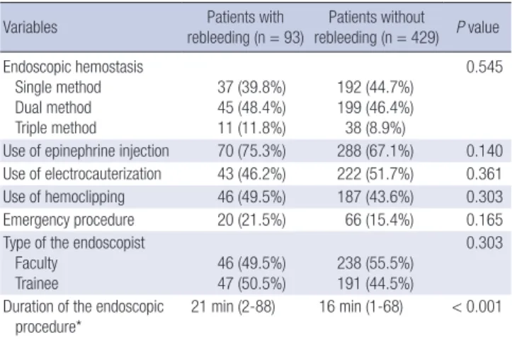
Rate of rebleeding relative to the endoscopic procedure There was no significant difference between the patients with and without rebleeding with regard to the method of endosco- pic hemostasis, emergency endoscopy, and the amount of ex- perience of the attending endoscopist (Table 2). The procedure time was significantly longer in those with rebleeding than in those without rebleeding (P < 0.001).
The optimal cut-off value with regard to the duration of en- doscopic hemostasis was 13.5 min with area under the ROC curve of 0.644 (P < 0.001). Of all significant variables, procedure time longer than 13.5 min was significantly related to rebleed- ing (OR, 2.899; 95% CI, 1.768-4.754; P < 0.001) on the logistic regression analysis (Table 3).
Rate of rebleeding according to the endoscopist
There was no significant difference in the rate of rebleeding (P = 0.574) among the patients of different GI endoscopists. The du- ration of endoscopic hemostasis was significantly higher when performed by trainee (P < 0.001). Median procedure time of trainee was 17.1 min (2-88 min), while that of experienced en- doscopist was 11.3 min (1-60 min). The type of organ differed significantly between the endoscopists (P < 0.001), because some of them were major in pancreaticobiliary diseases and some received calls from intensive care units.
The preferences with regard to the use of epinephrine injec- tion (P < 0.001) and electrocauterization (P < 0.001) differed significantly among the GI endoscopists, resulting in different proportion of single or combination therapies (P < 0.001) and in the duration of the endoscopic hemostasis procedure (P <
0.001). Although it was not statistically significant, one endos- copist who had a low preference for electrocauterization (22.2%) showed the highest rebleeding rate (27.8%) among all endosco- pists. The other endoscopist who preferred single method (75.7%) without epinephrine injection showed shortest duration of pro- cedure (7 min).
Table 1. Basal characteristics of the patients according to the presence of rebleeding
Variables Patients with
rebleeding (n = 93) Patients without rebleeding (n = 429) P value Age (yr-old, mean ± SD) 64.4 ± 15.6 61.6 ± 15.8 0.123
Gender (male, %) 72 (77.4%) 322 (75.1%) 0.691
Location of bleeding ulcer Esophagus
Stomach Duodenum
1 (1.1%) 62 (66.6%) 30 (32.3%)
20 (4.7%) 292 (68.1%) 117 (27.3%)
0.208
Forrest classification Ia
Ib IIa IIb IIc
7 (7.5%) 42 (45.2%) 39 (41.9%) 5 (5.4%) 0 (0%)
24 (5.6%) 205 (47.8%) 187 (43.6%) 9 (2.1%) 4 (0.9%)
0.334
Underlying disease Cerebral disease Cardiac disease Diabetes mellitus Hypertension
9 (9.7%) 11 (11.8%) 20 (21.5%) 37 (39.8%)
22 (5.1%) 50 (11.6%) 76 (17.7%) 144 (33.6%)
0.142 1.000 0.379 0.280 Numbers of antithrombotics
None One Two
63 (67.7%) 25 (26.9%) 5 (5.4%)
337 (78.5%) 84 (19.6%) 8 (1.9%)
0.032
Current medication Aspirin Clopidogrel Warfarin NSAIDs
29 (31.2%) 4 (4.3%) 0 (0%) 2 (2.2%)
76 (17.7%) 10 (2.3%) 10 (2.3%) 4 (0.9%)
0.006 0.289 0.222 0.291 SD, standard deviation; NSAID, non-steroidal anti-inflammatory drug.
Table 2. Link between rebleeding and the type of endoscopic hemostasis
Variables Patients with
rebleeding (n = 93) Patients without rebleeding (n = 429) P value Endoscopic hemostasis
Single method Dual method Triple method
37 (39.8%) 45 (48.4%) 11 (11.8%)
192 (44.7%) 199 (46.4%) 38 (8.9%)
0.545
Use of epinephrine injection 70 (75.3%) 288 (67.1%) 0.140 Use of electrocauterization 43 (46.2%) 222 (51.7%) 0.361
Use of hemoclipping 46 (49.5%) 187 (43.6%) 0.303
Emergency procedure 20 (21.5%) 66 (15.4%) 0.165
Type of the endoscopist Faculty
Trainee 46 (49.5%)
47 (50.5%) 238 (55.5%) 191 (44.5%)
0.303
Duration of the endoscopic
procedure* 21 min (2-88) 16 min (1-68) < 0.001
*Median with ranges was shown using the Mann-Whitney test since the data were not normally distributed.
Table 3. Significant variables related to rebleeding
Variables Odds ratio 95% confidence
interval P value Use of antithrombotics
Yes
No 1.296
1 (reference) 0.038-2.304 0.245 Aspirin medication
Yes
No 6.460
1 (reference) 0.808-11.652 0.079 Procedure time
> 13.5 min
< 13.5 min 2.899
1 (reference) 1.768-4.754 < 0.001 Data were analyzed by logistic regression analysis.
DISCUSSION
The rate of rebleeding after endoscopic hemostasis for UGIB is higher in the patients after a long endoscopic hemostasis in this study. Notably, the rate of rebleeding did not differ significantly between the GI endoscopists who performed the hemostasis, while it did differ with use of epinephrine injection and electro- cauterization leading to a significant difference in the duration of the endoscopic hemostasis procedure.
Interesting finding of the present study is that the duration of endoscopic hemostasis was significantly related to the rate of rebleeding. It may be that the duration of endoscopic hemosta- sis can be influenced by the nature of the bleeding lesion, lead- ing to a difficult procedure. In addition, most of the endosco- pists in the present study had a preferred hemostatic method, regardless of the patient’s condition, and the site, location, and characteristics of the bleeding lesion. The preferences for the use epinephrine injection and electrocauterization were signif- icantly different among GI endoscopists, although there was no significant difference in the rate of rebleeding. This can be ex- plained by our previous study showing that the preference for a specific method depends more on the personal experience and prior mentoring of the clinician than on the published guide- lines (9).
Of the antithrombotics being used by the patients in the pres- ent study, aspirin was the most frequently prescribed drugs, and as such, most GI endoscopists are aware of its impact on GI ble- eding. We previously showed that Eastern endoscopists do not typically perform endoscopic biopsy procedures while patients are receiving warfarin, and do not perform polypectomies on patients who are currently taking aspirin due to the risk of bleed- ing (10). In that study, we found that Eastern endoscopists tend to believe that aspirin increases the risk of bleeding in Asians than the Caucasians. This is consistent with Japanese studies showing that the incident rates of gastroduodenal mucosal in- jury and bleeding are significantly higher among Asians who are taking antiplatelet medication (5, 11).
A recent Korean study showed that warfarin increases the frequencies of both rebleeding and thromboembolic events (12). Our study is consistent with a previous study showed that mild to moderate anticoagulation medication does not increase the rate of rebleeding following endoscopic therapy for nonvar- iceal UGIB (13). In that study, the INR was not a predictor of re- bleeding for UGIB, the need for RBC transfusion, requirement for surgery, length of hospitalization, or death. On the other hand, in the present study it was not possible to determine why the patients taking clopidogrel did not show a higher rate of re- bleeding. Although it is well known that combination medica- tion with aspirin and a PPI is superior for preventing rebleeding than clopidogrel alone (14), it remains to be established wheth- er monotherapy with clopidogrel can induce more severe drug-
induced ulcers that might lead to frequent UGIB.
The most up-to-date safety and efficacy data have to led to the current recommendation for hemostasis in UGIB of me- chanical or ablation therapy, with or without epinephrine in- jection (15). More recently, monotherapy using either mechan- ical or ablation therapy has replaced the combination method, as shown in the present study. Monotherapy was used in 77.9%
of cases in a recent study, whereas combination therapy was used for only 21.5% with a heat probe being the most commonly used device for endoscopic hemostasis (16). Yet another study revealed a changing trend in the favored endoscopic hemosta- sis procedure between the periods 1995-2000 and 2006-2009 (17), such that while injection and intravenous H2 blocker were predominantly used during the former period, hemoclipping and intravenous PPI were the preferred procedures during the latter. The greater number of cases of severe GI bleeding in the 2006-2009 resulted in the outcome of endoscopic hemostasis not differing between two times periods despite the interven- ing advances in medical procedure.
There are several limitations in our study including the lack of details on ulcer characteristics and various types of oral PPI medication used after the discharge. Since this study was based on the medical chart review, exact size of the bleeding ulcer or exact body mass index were missing. However, we assume that these factors would not have affected the result of our study, since even the Forrest classification did not differ between two groups. Recent studies have shown that the amount of bleeding is related to the rate of rebleeding (18, 19). It has been demon- strated that the initial presentation of either hematemesis or fresh red blood through a nasogastric tube are high risk factors for rebleeding (18), and that RBC transfusion is significantly re- lated to subsequent rebleeding in nonvariceal UGIB patients (19). Most of the patients on antithrombotics who experienced rebleeding in the present study required RBC transfusion, and in most patients, the initial manifestations included hemateme- sis. Together these results suggest that these factors should be considered significant for predicting rebleeding, especially in patients after a long endoscopic hemostasis procedure.
In conclusion, a long endoscopic procedure time influence the occurrence of rebleeding after successful endoscopic he- mostasis for UGIB. Procedure time longer than 13.5 min should be considered more carefully in terms of rebleeding than other disease-related factors or GI endoscopist-related factors. Care- ful monitoring is required after the long hemostasis procedure for PUD.
DISCLOSURE
The authors declare that there is nothing to disclose except the acknowledgment that the study was supported by the National Research Foundation of Korea.
ORCID
Mi Jin Hong http://orcid.org/0000-0002-2984-7171 Sun-Young Lee http://orcid.org/0000-0003-4146-6686 Jeong Hwan Kim http://orcid.org/0000-0002-2503-2688 In-Kyung Sung http://orcid.org/0000-0002-3848-5571 Hyung Seok Park http://orcid.org/0000-0003-3141-4858 Chan Sup Shim http://orcid.org/0000-0002-3300-4417 Choon Jo Jin http://orcid.org/0000-0001-7569-4076
REFERENCES
1. Seo YS, Kim YH, Ahn SH, Yu SK, Baik SK, Choi SK, Heo J, Hahn T, Yoo TW, Cho SH, et al. Clinical features and treatment outcomes of upper gastrointestinal bleeding in patients with cirrhosis. J Korean Med Sci 2008; 23: 635-43.
2. Chiu PW, Joeng HK, Choi CL, Kwong KH, Ng EK, Lam SH. Predictors of peptic ulcer rebleeding after scheduled second endoscopy: clinical or en- doscopic factors? Endoscopy 2006; 38: 726-9.
3. Travis AC, Wasan SK, Saltzman JR. Model to predict rebleeding follow- ing endoscopic therapy for non-variceal upper gastrointestinal hemor- rhage. J Gastroenterol Hepatol 2008; 23: 1505-10.
4. Suk KT, Kim HS, Lee CS, Lee IY, Kim MY, Kim JW, Baik SK, Kwon SO, Lee DK, Ham YL. Clinical outcomes and risk factors of rebleeding fol- lowing endoscopic therapy for nonvariceal upper gastrointestinal hem- orrhage. Clin Endosc 2011; 44: 93-100.
5. Suehiro T, Yakeishi Y, Sakai F, Matsuzaki K, Sanefuji K, Toyokawa T, Shio- shita K, Sugie Y, Okudaira Y, Kano T, et al. Gastrointestinal bleeding as- sociated with antithrombotic therapy in the elderly in Japan. Hepato- gastroenterology 2012; 59: 774-7.
6. Sostres C, Lanas A. Gastrointestinal effects of aspirin. Nat Rev Gastroen- terol Hepatol 2011; 8: 385-94.
7. Hylek EM. Complications of oral anticoagulant therapy: bleeding and nonbleeding, rates and risk factors. Semin Vasc Med 2003; 3: 271-8.
8. Chung IK, Lee DH, Kim HU, Sung IK, Kim JH; Korean College of Heli- cobacter and Upper Gastrointestinal Research; Korean Association of Gastroenterology. Guidelines of treatment for bleeding peptic ulcer dis- ease. Korean J Gastroenterol 2009; 54: 298-308.
9. Tang SJ, Lee SY, Hynan LS, Yan J, Riley FC, Armstrong L, Rodriguez-Frias E, Xu L, Pruna E, Lara LF, et al. Endoscopic hemostasis in nonvariceal
upper gastrointestinal bleeding: comparison of physician practice in the East and the West. Dig Dis Sci 2009; 54: 2418-26.
10. Lee SY, Tang SJ, Rockey DC, Weinstein D, Lara L, Sreenarasimhaiah J, Choi KW; Korean Association for the Study of Intestinal Disease. Man- aging anticoagulation and antiplatelet medications in GI endoscopy: a survey comparing the East and the West. Gastrointest Endosc 2008; 67:
1076-81.
11. Yamamoto T, Sanaka M, Nagasawa K, Abe K, Fukami M, Nakayama S, Tsuchiya A, Ishii T, Kuyama Y. Gastroduodenal mucosal injury in pa- tients on antiplatelet therapy. Thromb Res 2007; 120: 465-9.
12. Lee JK, Kang HW, Kim SG, Kim JS, Jung HC. Risks related with with- holding and resuming anticoagulation in patients with non-variceal upper gastrointestinal bleeding while on warfarin therapy. Int J Clin Pract 2012; 66: 64-8.
13. Wolf AT, Wasan SK, Saltzman JR. Impact of anticoagulation on rebleed- ing following endoscopic therapy for nonvariceal upper gastrointestinal hemorrhage. Am J Gastroenterol 2007; 102: 290-6.
14. Chan FK, Ching JY, Hung LC, Wong VW, Leung VK, Kung NN, Hui AJ, Wu JC, Leung WK, Lee VW, et al. Clopidogrel versus aspirin and esome- prazole to prevent recurrent ulcer bleeding. N Engl J Med 2005; 352: 238- 44.
15. Barkun AN, Bardou M, Kuipers EJ, Sung J, Hunt RH, Martel M, Sinclair P;
International Consensus Upper Gastrointestinal Bleeding Conference Group. International consensus recommendations on the management of patients with nonvariceal upper gastrointestinal bleeding. Ann Intern Med 2010; 152: 101-13.
16. Endo M, Higuchi M, Chiba T, Suzuki K, Inoue Y. Present state of endo- scopic hemostasis for nonvariceal upper gastrointestinal bleeding. Dig Endosc 2010; 22: S31-4.
17. Kawamura T, Yasuda K, Morikawa S, Itonaga M, Nakajima M. Current status of endoscopic management for nonvariceal upper gastrointestinal bleeding. Dig Endosc 2010; 22: S26-30.
18. Maggio D, Barkun AN, Martel M, Elouali S, Gralnek IM; Reason Investi- gators. Predictors of early rebleeding after endoscopic therapy in patients with nonvariceal upper gastrointestinal bleeding secondary to high-risk lesions. Can J Gastroenterol 2013; 27: 454-8.
19. Restellini S, Kherad O, Jairath V, Martel M, Barkun AN. Red blood cell transfusion is associated with increased rebleeding in patients with non- variceal upper gastrointestinal bleeding. Aliment Pharmacol Ther 2013;
37: 316-22.