44 www.kjlm.org
Plant Root Hair in Tap Water: A Potential Cause for Diagnostic Confusion
Sadia Shakoor, M.D.1, Mohammad Wasay, M.D.2, Afia Zafar, FRCPath.1, and Mohammad Asim Beg, Ph.D.1 Departments of Microbiology1 and Medicine2, the Aga Khan University Hospital, Karachi, Pakistan
Plant root hairs are commonly found artifacts in parasitology specimens and may be confused with helminthes by an untrained eye. We report a case of brain tuberculoma where the tissue sample was contaminated with root hair derived from tap water; the presence of this root hair, which mimicked a larva, led to diagnostic confusion. Therefore, tap water should be considered a source of root hair and vegetable matter.
Key Words: Helminthes, Artifacts, Water supply
Received: July 28, 2010 Manuscript No: KJLM-10-120 Revision received: August 22, 2010
Accepted: November 16, 2010
Corresponding author: Sadia Shakoor, M.D.
Department of Clinical Microbiology, Aga Khan University Hospital, Stadium Road, Karachi 74800, Pakistan
Tel: 92-21-3486-1608, Fax: 92-21-3486-4924, Email: [email protected] ISSN 1598-6535 © The Korean Society for Laboratory Medicine.
This is an Open Access article distributed under the terms of the Creative Commons Attribution Non-Commercial License (http://creativecommons.org/licenses/by-nc/3.0) which permits unrestricted non-commercial use, distribution, and reproduction in any medium, provided the original work is properly cited.
Korean J Lab Med 2011;31:44-46 DOI 10.3343/kjlm.2011.31.1.44
Case Report
Clinical Microbiology
KJLM
INTRODUCTION
Root hair from plants and vegetable matter are commonly seen in inherently contaminated clinical samples such as stool [1] and sputum [2]. Sterile samples such as aseptically collected pus or tissues are free of such artifacts. However, the presence of artifacts in samples may be due to the rare instances of basal laboratory media contamination during tissue processing. We present a cautionary report stating that the contamination of brain tissue smears with unfil- tered tap water may lead to unnecessary further processing and investigation.
CASE REPORT
A 57-yr-old man under treatment for tuberculous men- ingitis and hydrocephalus was admitted to a tertiary care hospital for the investigation of drowsiness. He had been receiving first-line anti-tubercular treatment (ATT) and 10
mg/day of prednisone for the past 5 months. The initial workup included a head computed tomography scan, which showed multiple small space-occupying lesions in both the cerebellar hemispheres. The baseline laboratory workup in- cluded complete blood count, urinalysis, and stool exami- nation, the results of which were normal. A craniotomy and biopsy were also performed, and the perioperative findings were unremarkable. The tissue samples were sent for histo- pathological examination and bacterial and mycological culturing. Microscopic examination of a potassium hydrox- ide (KOH)-treated tissue sample showed a single 2-mm structure resembling a “worm,” with tapering ends. The same tissue sample was re-examined for this structure by teasing the tissue with a sterile scalpel and suspending it in sterile saline; similar segments were seen (Fig. 1), and a di- agnostic dilemma arose as to the identification of these structures. The initial histopathological findings had not re- vealed similar structures. Therefore, we re-examined vari- ous areas in the tissue by repeated sectioning. Meanwhile, the attending physician was alerted to the possibility of Central Nervous System (CNS) helminthosis, and an em- piric treatment with 200 mg/day of albendazole was initi- ated, while additional clues to a disseminated helminthic infection were sought. However, the peripheral smear ex- amination did not reveal eosinophilia, and the 3 stool ex- aminations were negative for larvae. No such helminth-like structure was seen on examination of the different sections of the tissue, although minimal inflammatory cells and re- active glial cells were noted. Finally, parasitic infestation was ruled out, the albendazole treatment was discontinued, and
Shakoor S, et al. • Plant Root Hair Mimics a Helminth
www.kjlm.org 45
DOI 10.3343/kjlm.2011.31.1.44
KJLM
the patient was tentatively diagnosed with a tuberculoma.
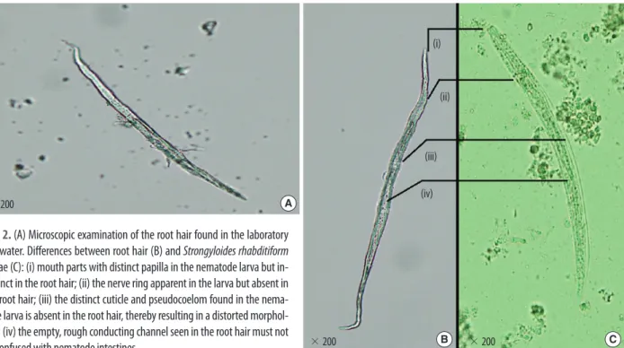
As a follow-up of the initial diagnostic confusion, saline and KOH blanks were examined for similar structures. Struc- tures with an internal canal and tapering ends of lengths varying between 2 mm and 5 mm were found in the media and in tap water from the microbiology laboratory. These structures were distinct root hair artifacts (Fig. 2). Gram- stained slides that had been washed under laboratory tap water showed similar structures, which had been disregarded
as artifacts by microscopists. On further investigation, it was found that the laboratory filtration unit had malfunc- tioned. However, the hospital tap water and water in other laboratories in the hospital were free of such root hair. We thought that construction work that involved the laying of water pipes near the microbiology laboratory might have led to the appearance of such the root hair in tap water. Sur- prisingly enough, the plant material persisted even after au- toclaving. When the unit was repaired, the water was free of
A B
Fig. 1. Wet mount preparation with saline of 2 separate sections of the brain tissue. The structures shown created a diagnostic dilemma as such artifacts are not common: (A) shows 2 distorted structures that have a definite outer covering but no pseudocoelom, (B) shows a tapering structure with a central refractile core, more suggestive of a root hair.
×200 A
×200 ×200
(i)
(ii)
(iii) (iv)
B C
Fig. 2. (A) Microscopic examination of the root hair found in the laboratory tap water. Differences between root hair (B) and Strongyloides rhabditiform larvae (C): (i) mouth parts with distinct papilla in the nematode larva but in- distinct in the root hair; (ii) the nerve ring apparent in the larva but absent in the root hair; (iii) the distinct cuticle and pseudocoelom found in the nema- tode larva is absent in the root hair, thereby resulting in a distorted morphol- ogy; (iv) the empty, rough conducting channel seen in the root hair must not be confused with nematode intestines.
46 www.kjlm.org
Shakoor S, et al. • Plant Root Hair Mimics a Helminth
DOI 10.3343/kjlm.2011.31.1.44
KJLM
these structures.
DISCUSSION
Root hairs are commonly found artifacts in parasitology laboratories [3]. The presence of root hair in clinical mate- rial is known to create diagnostic confusion, because the hair strands mimic nematode larvae [4]. However, medical and botanical literature is devoid of reports citing tap water as a source of plant root hair. In this paper, we report tap water as the source of plant root hair. Although parasites themselves also contaminate tap water, the morphological appearance of the structures in our case is representative of root hair and not nematode larvae (Fig. 2B). Worms have both internal and external structures, such as an intestine and mouthparts, which differentiate them from root hair [2]. When the patient’s tissue was carefully observed for the doubtful structures, an elongated central refractile core sug- gestive of a root hair was seen (Fig. 1B).
In general, all basal media in microbiology laboratories are autoclaved prior to use. Although we ensured sterile processing, 2 factors led to the contamination of the sample.
First, the malfunctioning of the filtration unit, and second, the homogenization of the sample with saline during its processing. Although saline suspension of tissue samples is not recommended, we carried it out since the sample re- ceived by the laboratory was dry and its quantity was insuf- ficient for all the tests requested. To prevent such occur- rences, clinicians sending tissues to a laboratory for testing must ensure adequate tissue quantity and suspend the tis- sue in an appropriate medium while the laboratory staff must strive to minimize contamination of basal media and samples to prevent errors. Fortunately, since the problem was pursued and remedied in time in our laboratory, we did
not experience similar problems with the other samples.
In our case, a provisional diagnosis of CNS helminthosis was considered only because of the patient’s unusual history and physical examination findings, including a slow response to ATT, cerebrospinal fluid shunting, and steroid therapy, and the absence of focal neurological findings. The tissue structures seen in Fig. 1 were not diagnostic; however, the possibility of other infections such as disseminated strongy- loidiasis and angiostrongylus remained.
Additional investigations and histopathological sections stained and examined at a different laboratory in the hospi- tal, which had a separate water supply, aided us in excluding the diagnosis of CNS helminthosis.
In conclusion, root hair from plant and vegetable matter are artifacts frequently encountered in laboratory practice.
Other than clinical material, unfiltered tap water should also be considered an important source of root hair. Fur- ther, laboratories are advised to use filtered distilled water in all wet-lab experiments.
Authors’ Disclosures of Potential Conflicts of Interest No potential conflict of interest relevant to this article was reported.
REFERENCES
1. Paul Y. Undigested food presenting as bizarre objects in stool. In- dian Pediatr 1994;31:1130-1.
2. Martínez-Girón R, González-López JR, Esteban JG, García-Mi- ralles MT, Alvarez-de-los-Heros C, Ribas-Barceló A. Worm-like artifacts in exfoliative cytology. Diagn Cytopathol 2006;34:636-9.
3. Isenberg HD, ed. Clinical microbiology procedures handbook. 2nd ed. Washington DC: ASM Press, 2004.
4. Garcia LS and Bruckner DA, eds. Diagnostic medical parasitology.
3rd ed. Washington DC: ASM Press, 1997:749.