ISSN 2234-3806 • eISSN 2234-3814
Ann Lab Med 2012;32:289-293
http://dx.doi.org/10.3343/alm.2012.32.4.289
MYC Rearrangement Involving a Novel
Non-immunoglobulin Chromosomal Locus in Precursor B-cell Acute Lymphoblastic Leukemia
Ja-Young Seo, M.D.1*, Soo Hyun Lee, M.D.2*, Hee-Jin Kim, M.D.1, Keon Hee Yoo, M.D.2, Hong Hoe Koo, M.D.2, Yong Gon Cho, M.D.3, Sam Im Choi, M.D.3, and Sun-Hee Kim, M.D.1
Departments of Laboratory Medicine and Genetics1 and Pediatrics2, Samsung Medical Center, Sungkyunkwan University School of Medicine, Seoul;
Department of Laboratory Medicine3, Chonbuk National University Medical School, Jeonju, Korea
MYC rearrangement, a characteristic cytogenetic abnormality of Burkitt lymphoma and several subsets of other mature B-cell neoplasms, typically involves an immunoglobulin gene partner. Herein, we describe a case of precursor B-cell lymphoblastic leukemia har- boring a MYC rearrangement with a novel non-immunoglobulin partner locus. The patient was a 4-yr-old Korean boy with ALL of the precursor B-cell immunophenotype. At the time of the second relapse, cytogenetic analyses revealed t(4;8)(q31.1;q24.1) as a clonal evolu- tion. The MYC rearrangement was confirmed by FISH analysis. He died 3 months after the second relapse without achieving complete remission. To our knowledge, this is the first report of a case of MYC rearrangement with a non-immunoglobulin partner in precursor B-cell lymphoblastic leukemia.
Key Words: Precursor B-cell acute lymphoblastic leukemia, MYC gene rearrangement, Non-immunoglobulin partner
Received: June 9, 2011 Revision received: July 11, 2011 Accepted: February 13, 2012 Corresponding author: Hee-Jin Kim Department of Laboratory Medicine and Genetics, Samsung Medical Center, Sungkyunkwan University School of Medicine, 81 Irwon-ro, Gangnam-gu, Seoul 135-710, Korea
Tel: +82-2-3410-2701 Fax: +82-2-3410-2719 E-mail: heejinkim@skku.edu
*These authors contributed equally to this work.
© The Korean Society for Laboratory Medicine.
This is an Open Access article distributed under the terms of the Creative Commons Attribution Non-Commercial License (http://creativecom- mons.org/licenses/by-nc/3.0) which permits unrestricted non-commercial use, distribution, and reproduction in any medium, provided the original work is properly cited.
INTRODUCTION
Chromosomal rearrangements involving the MYC gene, located on band 8q24, are well known characteristic of Burkitt lym- phoma, and are also found in subsets of mature B-cell neo- plasms [1]. The MYC rearrangement results in dysregulation of the MYC proto-oncogene and plays a key role in the pathogene- sis and progression of disease, by juxtaposition of the MYC gene to immunoglobulin genes in particular. The major cytogenetic abnormality is the MYC-immunoglobulin heavy chain gene (IGH) rearrangement t(8;14)(q24;q32), which is followed by MYC-rear- rangements t(8;22)(q24;q11) and t(2;8)(p12;q24). Although MYC
rearrangements are primarily found in mature B-cell lymphoid neoplasms, rare cases of precursor B-cell ALL carrying the MYC rearrangement have also been reported [2-9]. The majority of these cases had leukemic blasts morphologically reminiscent of Burkitt lymphoma, but had a precursor B-cell immunopheno- type (positive for terminal deoxynucleotidyl transferase [TdT]).
All of these cases had MYC rearrangements that involved im- munoglobulin genes.
Herein we report a pediatric case of precursor B-cell ALL with a MYC rearrangement involving a novel non-immunoglobulin partner locus. To our knowledge, this is the first report of a case of a MYC rearrangement with a non-immunoglobulin partner in
ISSN 2234-3806 • eISSN 2234-3814
precursor B-cell ALL.
CASE REPORT
A 4-yr-old boy was diagnosed with B-cell ALL at an outside hos- pital. The leukemic blasts were positive for CD19, CD20, CD22, and HLA-DR and were negative for CD10 and CD34 (Table 1).
The leukemic blasts co-expressed the T-lymphoid marker CD5 and myeloid markers CD13, CD14, and CD33. Immunohisto- chemistry showed that a clot section was positive for TdT. Both flow cytometry and immunohistochemistry showed that blasts
were negative for myeloperoxidase (MPO). A cytogenetic study revealed del(22)(q11.2). The cerebrospinal fluid (CSF) was neg- ative for leukemic blasts. The patient was enrolled in the high- risk Children’s Cancer Group (CCG)-1882 protocol and received induction chemotherapy followed by double-delayed intensifica- tion. He attained complete remission and was receiving mainte- nance therapy.
Fifteen months after the initial diagnosis, the patient was trans- ferred to our institution for evaluation of nausea and vomiting and was diagnosed with isolated central nervous system (CNS) relapse of the disease. In the CSF specimen, the white blood cell
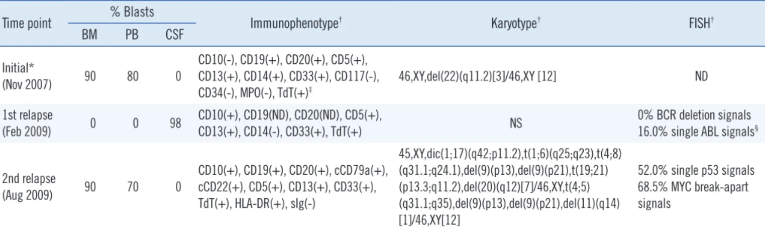
Table 1. Summary of the patient’s hematologic, immunophenotypic, and cytogenetic data Time point % Blasts
Immunophenotype† Karyotype† FISH†
BM PB CSF
Initial*
(Nov 2007) 90 80 0
CD10(-), CD19(+), CD20(+), CD5(+), CD13(+), CD14(+), CD33(+), CD117(-),
CD34(-), MPO(-), TdT(+)‡ 46,XY,del(22)(q11.2)[3]/46,XY [12] ND
1st relapse
(Feb 2009) 0 0 98 CD10(+), CD19(ND), CD20(ND), CD5(+),
CD13(+), CD14(-), CD33(+), TdT(+) NS 0% BCR deletion signals
16.0% single ABL signals§
2nd relapse
(Aug 2009) 90 70 0
CD10(+), CD19(+), CD20(+), cCD79a(+), cCD22(+), CD5(+), CD13(+), CD33(+), TdT(+), HLA-DR(+), sIg(-)
45,XY,dic(1;17)(q42;p11.2),t(1;6)(q25;q23),t(4;8) (q31.1;q24.1),del(9)(p13),del(9)(p21),t(19;21) (p13.3;q11.2),del(20)(q12)[7]/46,XY,t(4;5) (q31.1;q35),del(9)(p13),del(9)(p21),del(11)(q14) [1]/46,XY[12]
52.0% single p53 signals 68.5% MYC break-apart signals
*Data from another hospital; †Data from BM aspirate specimens at initial diagnosis and 2nd relapse and from CSF specimen at 1st relapse; ‡Data obtained by immunohistochemistry on a clot section; §The nature of the ABL deletion signal could not be determined because the cytogenetic analysis was not successful.
Abbreviations: BM, bone marrow; PB, peripheral blood; CSF, cerebrospinal fluid; MPO, myeloperoxidase; TdT, terminal deoxynucleotidyl transferase; ND, not done; NS, not successful.
Fig. 1. Morphology of the leukemic blasts in the cerebrospinal fluid at first relapse (A) and in the bone marrow at second relapse (B) (Wright-Giemsa stain, ×1,000).
B A
count was 1,450/µL with 98% leukemic blasts (Table 1 and Fig.
1A). The leukemic blasts were positive for CD10, CD5, CD13, CD33, and nuclear TdT and were negative for CD14 and CD34. Cytogenetic analyses of leukemic blasts in the CSF failed due to the poor quality of the specimen. FISH analyses for del(22) (q11.2) using the Vysis LSI BCR/ABL Dual Color, Dual Fusion Translocation Probe (Abbott Molecular Inc., Des Plaines, IL, USA) showed no interphase cells with BCR (22q11.2) signal deletion and 16.0% of cells with a single ABL (9q34) signal. There was no evidence of leukemic blasts in the peripheral blood or bone marrow. A cytogenetic study showed no abnormal clones in the bone marrow samples. The patient was treated with the CCG- 1882 protocol, along with whole brain irradiation (24 Gy divided into 12 fractions) and whole spine irradiation (6 Gy divided into 3 fractions) during the consolidation period.
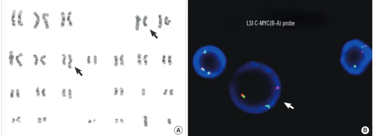
Four months from the initial relapse, the patient experienced a second relapse with ≥90% leukemic blasts in the bone marrow (Fig. 1B). Immunophenotypically, the blasts were positive for CD19, CD10, CD20, cytoplasmic CD79a, cytoplasmic CD22, HLA- DR, and TdT, with co-expression of CD5, CD13, and CD33 (Ta- ble 1). Cytogenetic analysis revealed complex structural abnor- malities, including t(4;8)(q31.1;q24.1) (Fig. 2A). FISH analysis using the Vysis LSI MYC Dual Color, Break Apart Rearrangement Probe (Abbott Molecular Inc.) revealed MYC rearrangement in 68.5% of interphase cells (Fig. 2B). There was no IGH/MYC fu- sion signal in a FISH study using the Vysis LSI IGH/MYC/CEP8 Tri-Color Dual Fusion probe (Abbott Molecular Inc.). In addition, a FISH study using the Vysis LSI p53 probe (Abbott Molecular
Inc.) showed deletion of the p53 (17p13.1) signal in 52.0% of interphase cells, which was compatible with the presence of dic(1;17)(q42;p11.2) observed in conventional cytogenetics (Ta- ble 1). Despite aggressive reinduction chemotherapy, the patient died 3 months after the second relapse of disease.
DISCUSSION
The MYC rearrangement is considered the hallmark of Burkitt lymphoma; however, it also arises in other subsets of mature B-cell neoplasms. In particular, the MYC rearrangement was reported to be a critical event in the progression of follicular lym- phoma to higher-grade lymphoma or leukemia [10, 11]. Biologi- cally, the c-myc protein has a central role in the transcriptional regulation of various processes, including cell growth, cell cycle progression, and apoptosis [12]. Translocation of one MYC allele into the vicinity of an immunoglobulin heavy chain gene on chro- mosome 14q32, or less commonly, the kappa and lambda light chain genes on chromosomes 2p12 and 22q11, respectively, leads to deregulated expression of c-myc and cell proliferation.
According to the experience of the Pediatric Oncology Group, the MYC rearrangement accounts for 0.1% (5/5,280) of cases of pediatric ALL with the precursor B-cell phenotype [9]. Our litera- ture review of precursor B-cell ALL cases showed that all the partners involved in MYC rearrangements were immunoglobulin genes [2-9] (Table 2). The most common partner was t(8;14), followed by 2p12 and 22q11. In contrast, the partner chromo- somal locus of the MYC rearrangement in our case was not a
Fig. 2. Cytogenetic analysis showing complex chromosomal abnormalities including t(4;8)( q31.1;q24.1). Black arrows indicate the rear- ranged chromosomes 4 and 8 (A). FISH analysis using the Vysis LSI MYC Dual Color, Break Apart Rearrangement Probe (Abbott Molecular Inc.), which showed the MYC gene rearrangement. White arrow indicates a interphase cell with 1 fusion and 1 break apart signal (B).
LSI C-MYC(B-A) probe
B A
conventional immunoglobulin loci, but 4q31. Band 4q31 is a chromosomal locus that has been rarely involved in hematologic malignancies; t(4;5)(q31;q31) and t(4;21)(q31;q22) were reported in MDS/AML and T-cell ALL [13-16]. SH3D19 is the only gene identified as involved in t(4;21)(q31;q22) in AML [14]. Although no genes in 4q31 have been reported to be involved in precursor B-cell ALL, another candidate gene is MAML3, a coactivator of the notch signaling pathway [17].
The MYC rearrangement was detected in leukemic cells in the bone marrow during the second relapse. We speculated that the leukemic cells underwent serial genetic evolution from the first through the second relapse. Unfortunately, we could not ascertain whether the MYC rearrangement was absent at initial diagnosis, because the MYC FISH of the initial bone marrow sample was not successful. However, complex cytogenetic ab- normalities, including the translocation involving the MYC locus, which was detected at the second relapse, provided evidence of genetic evolution.
The presence of the MYC rearrangement warrants intensive treatment due to the highly proliferative nature of the neoplastic cells. In the Pediatric Oncology Group experience, 5 patients with precursor B-cell ALL received intensive chemotherapy, be- ing considered the presence of the MYC rearrangement, and 4 achieved long-term survival [9]. The patient in the present re- port attained a complete response (CR) for less than 6 months after the first relapse, and he never achieved a CR after the sec- ond relapse. He was treated based on the protocol for high-risk precursor B-cell ALL, since the MYC gene rearrangement was Table 2. Precursor B-cell ALL cases with MYC rearrangements
Reference Age/sex FAB morphology Karyotype*
Kaneko et al., 1980 [5] 10/M L1 46,XY,-8,del(3)(q21q25),t(8;14)(q24;q32),+der(8)(1q;8q)(cen;cen) Mufti et al., 1983 [8] 21/M L2 46,XY,t(8;14)(q23;q32),t(14;18)(q32;q21)
De Jong et al., 1988 [2] 44/M L1 46,XY,del(3)(p),del(7)(q),t(8;14),t(14;18),-19,-21,+mar 1,+mar 2
Navid et al., 1999 [9] 4/F Atypical L3 46,XX,t(8;14)(q24;q32)[11]/47,idem,+1(q10)[7]/46,idem,der(22)t(1;22)(q11;p11)[2]
Navid et al., 1999 [9] 14/M L3 47,XY,+i(1)(q10),t(8;14)(q24;q32)[6]/46,XY[10]
Navid et al., 1999 [9] 13/F L3 46,XX,t(8;14)(q24;q32)[11]/46,idem,inv(2)(p11q12)[33]/46,XX[11]
Navid et al., 1999 [9] 14/F L3 46,XX,dup(1)(q21q44),t(8;14)(q24;q32)[4]/46,X,der(X)t(X;1)(p22;q23),t(8;14)(q24;q32) [6]/47,XX,+i(1)(q10),t(8;14)(q24;q32)[10]
Navid et al., 1999 [9] 6/M L3 46,XY,t(8;14)(q24;q32),t(14;17)(q32;q21),[cp4]/46,idem,dup(1)(q32q21)[2]/46,XY[4]
Loh et al., 2000 [7] 2/F L2 t(2;8)(p12;q24)*
Komrokji et al., 2003 [6] 45/M L1/L2 47,XY,+i(1)(q10),t(8;14)(q24;q32)
Gupta et al., 2004 [3] 8/F L2/L3 t(8;22)(q24.1;q11.2)*
Hassan et al., 2007 [4] 4/M L3 t(8;14)(q24;q32)*
MYC rearrangements are shown in bold. *Complete karyotype information is not available.
Abbreviation: FAB, French-American-British.
unexpectedly detected during the second relapse. This suggested that the protocol for high-risk precursor B-cell ALL (CCG-1882) might have been less effective in our patient. In addition to the MYC gene rearrangement, p53 deletion from dic(1;17)(q42;p11.2) was observed at the second relapse of the disease. P53 deletion is a recurrent genetic aberration in Burkitt lymphoma/leukemia [18], and p53 inactivation was reportedly associated with predis- position to oncogenic translocations in B lineage lymphomas and poor prognosis [19]. Immunophenotypically, the leukemic blasts in our patient expressed not only B-lymphoid antigens but also myeloid and T-lymphoid antigens, including CD5. CD5- positive precursor B-cell ALL is extremely rare, and only 4 such cases have been reported in the literature [20-22]. All 4 patients with CD5-positive precursor B-cell ALL were adolescents, and had aggressive disease courses and poor outcomes. From this perspective, the aberrant CD5 expression in our patient in com- bination with the cytogenetic abnormality might have been a poor prognostic factor.
In summary, we described the first case of precursor B-cell ALL with a MYC rearrangement involving a novel non-immuno- globulin partner locus. The outcome of this case suggested that the presence of the MYC rearrangement in ALL with the precur- sor B-cell phenotype warrants intensive treatment, regardless of the partner gene. Further identification of patients with this cyto- genetic abnormality will allow us to expand our knowledge re- garding its prognostic significance and the optimal treatment for this rare subgroup of patients.
Authors’ Disclosures of Potential Conflicts of Interest
No potential conflicts of interest relevant to this article were re- ported.
REFERENCES
1. Boerma EG, Siebert R, Kluin PM, Baudis M. Translocations involving 8q24 in Burkitt lymphoma and other malignant lymphomas: a historical review of cytogenetics in the light of todays knowledge. Leukemia 2009; 23:225-34.
2. De Jong D, Voetdijk BM, Beverstock GC, van Ommen GJ, Willemze R, Kluin PM. Activation of the c-myc oncogene in a precursor-B-cell blast crisis of follicular lymphoma, presenting as composite lymphoma. N Engl J Med 1988;318:1373-8.
3. Gupta AA, Grant R, Shago M, Abdelhaleem M. Occurrence of t(8;22) (q24.1;q11.2) involving the MYC locus in a case of pediatric acute lym- phoblastic leukemia with a precursor B cell immunophenotype. J Pedi- atr Hematol Oncol 2004;26:532-4.
4. Hassan R, Felisbino F, Stefanoff CG, Pires V, Klumb CE, Dobbin J, et al.
Burkitt lymphoma/leukaemia transformed from a precursor B cell: clini- cal and molecular aspects. Eur J Haematol 2008;80:265-70.
5. Kaneko Y, Rowley JD, Check I, Variakojis D, Moohr JW. The 14q+ chro- mosome in pre-B-ALL. Blood 1980;56:782-5.
6. Komrokji R, Lancet J, Felgar R, Wang N, Bennett JM. Burkitt’s leuke- mia with precursor B-cell immunophenotype and atypical morphology (atypical Burkitt’s leukemia/lymphoma): case report and review of liter- ature. Leuk Res 2003;27:561-6.
7. Loh ML, Samson Y, Motte E, Moreau LA, Dalton V, Waters S, et al. Trans- location (2;8)(p12;q24) associated with a cryptic t(12;21)(p13;q22) TEL/
AML1 gene rearrangement in a child with acute lymphoblastic leuke- mia. Cancer Genet Cytogenet 2000;122:79-82.
8. Mufti GJ, Hamblin TJ, Oscier DG, Johnson S. Common ALL with pre-B- cell features showing (8;14) and (14;18) chromosome translocations.
Blood 1983;62:1142-6.
9. Navid F, Mosijczuk AD, Head DR, Borowitz MJ, Carroll AJ, Brandt JM, et al. Acute lymphoblastic leukemia with the (8;14)(q24;q32) transloca- tion and FAB L3 morphology associated with a B-precursor immuno- phenotype: the Pediatric Oncology Group experience. Leukemia 1999;
13:135-41.
10. Au WY, Horsman DE, Gascoyne RD, Viswanatha DS, Klasa RJ, Connors JM. The spectrum of lymphoma with 8q24 aberrations: a clinical, path- ological and cytogenetic study of 87 consecutive cases. Leuk Lympho- ma 2004;45:519-28.
11. Young KH, Xie Q, Zhou G, Eickhoff JC, Sanger WG, Aoun P, et al. Trans- formation of follicular lymphoma to precursor B-cell lymphoblastic lym- phoma with c-myc gene rearrangement as a critical event. Am J Clin Pathol 2008;129:157-66.
12. O’Neil J and Look AT. Mechanisms of transcription factor deregulation in lymphoid cell transformation. Oncogene 2007;26:6838-49.
13. Mikhail FM, Serry KA, Hatem N, Mourad ZI, Farawela HM, El Kaffash DM, et al. A new translocation that rearranges the AML1 gene in a pa- tient with T-cell acute lymphoblastic leukemia. Cancer Genet Cytogenet 2002;135:96-100.
14. Nguyen TT, Ma LN, Slovak ML, Bangs CD, Cherry AM, Arber DA. Iden- tification of novel Runx1 (AML1) translocation partner genes SH3D19, YTHDf2, and ZNF687 in acute myeloid leukemia. Genes Chromosomes Cancer 2006;45:918-32.
15. Van Limbergen H, Poppe B, Michaux L, Herens C, Brown J, Noens L, et al. Identification of cytogenetic subclasses and recurring chromosom- al aberrations in AML and MDS with complex karyotypes using M-FISH.
Genes Chromosomes Cancer 2002;33:60-72.
16. Veldman T, Vignon C, Schrock E, Rowley JD, Ried T. Hidden chromo- some abnormalities in haematological malignancies detected by multic- olour spectral karyotyping. Nat Genet 1997;15:406-10.
17. Lin SE, Oyama T, Nagase T, Harigaya K, Kitagawa M. Identification of new human mastermind proteins defines a family that consists of posi- tive regulators for notch signaling. J Biol Chem 2002;277:50612-20. 18. Preudhomme C, Dervite I, Wattel E, Vanrumbeke M, Flactif M, Lai JL, et
al. Clinical significance of p53 mutations in newly diagnosed Burkitt’s lymphoma and acute lymphoblastic leukemia: a report of 48 cases. J Clin Oncol 1995;13:812-20.
19. Rowh MA, DeMicco A, Horowitz JE, Yin B, Yang-Iott KS, Fusello AM, et al. Tp53 deletion in B lineage cells predisposes mice to lymphomas with oncogenic translocations. Oncogene 2011;30:4757-64.
20. Ahmed D, Ahmed TA, Ahmed S, Tipu HN, Wiqar MA. CD5-positive acute lymphoblastic leukemia. J Coll Physicians Surg Pak 2008;18:310-1. 21. Peterson MR, Noskoviak KJ, Newbury R. CD5-positive B-cell acute lym-
phoblastic leukemia. Pediatr Dev Pathol 2007;10:41-5.
22. Subirá D, Roman A, Jiménez-Garófano C, Prieto E, Martínez-Delgado B, Aceituno E, et al. Brief report. CD19/CD5 acute lymphoblastic leukemia.
Med Pediatr Oncol 1998;31:551-2.