Cosmetic selection of skin incision for resection of choledochal cyst in young female patients
Shin Hwang1, Jong-Woo Choi2, Tae-Yong Ha1, Gi-Won Song1, and Dong-Hwan Jung1
Departments of 1Surgery and 2Plastic Surgery, Asan Medical Center, University of Ulsan College of Medicine, Seoul, Korea
Backgrounds/Aims: Open surgery for choledochal cyst has a disadvantage of skin incision scar from operative wound, which can be a definite disadvantage especially in young female patients. This study focused on the cosmetic aspect of skin incision for resection of choledochal cyst in young female patients. Methods: During a 2-year study period, 11 adult female patients aged less than 40 years underwent primary resection of choledochal cyst by a single surgeon.
The cosmetic effect of two types of skin incision was evaluated. Results: The patients underwent mini-laparotomy through either a right subcostal incision (n=8) or an upper midline incision (n=3). The mean length of skin incision was 10 cm for right subcostal incisions and 9 cm for upper midline incisions. It took approximately 1 hour to repair the operative wound meticulously in both groups. At the 6 month to 1 year follow-up, a slight bulge on the skin scar was observed in 3 (37.5%) patients of the right subcostal incision group and 1 (33.3%) patient of the upper midline incision group. Conclusions: The results of this preliminary study support the claim that cosmetic effect of the upper midline incision for CCD surgery appears to be non-inferior to that of the right subcostal incision if the incision is placed accurately and repaired very meticulously. (Korean J Hepatobiliary Pancreat Surg 2016;20:127-132)
Key Words: Choledochal cyst; Skin incision; Cosmetics; Young female; Upper midline incision
Received: May 23, 2016; Revised: May 30, 2016; Accepted: June 5, 2016 Corresponding author: Shin Hwang
Department of Surgery, Asan Medical Center, University of Ulsan College of Medicine, 88 Olympic-ro 43-gil, Songpa-gu, Seoul 05505, Korea Tel: +82-2-3010-3930, Fax: +82-2-3010-6701, E-mail: [email protected]
Copyright Ⓒ 2016 by The Korean Association of Hepato-Biliary-Pancreatic Surgery
This is an Open Access article distributed under the terms of the Creative Commons Attribution Non-Commercial License (http://creativecommons.org/
licenses/by-nc/4.0) which permits unrestricted non-commercial use, distribution, and reproduction in any medium, provided the original work is properly cited.
Korean Journal of Hepato-Biliary-Pancreatic Surgery ∙ pISSN: 1738-6349ㆍeISSN: 2288-9213
INTRODUCTION
Choledochal cyst disease (CCD) is a pathologic con- dition characterized by congenital dilatation of the biliary system, including the common, intrahepatic, and intra- pancreatic bile ducts. It is common in Asian patients, es- pecially in females and infants. Due to recent advances in diagnostic imaging techniques, CCD can be diagnosed at any age, from the antenatal period to adult life.1-6 CCD diagnosis in adult age has been made more frequently over time than before. Surgery in adult patients has been performed to resolve symptoms and to prevent or treat CCD-associated malignancies. There are several points of concern regarding surgical treatment such as surgical complications, remnant cystic lesions, malignant changes and minimal-incision approach.
Laparoscopic or robotic resection of choledochal cyst in adult patients has been occasionally reported, but its
long-term outcomes especially with respect to anastomotic stenosis and incomplete cyst resection appear to have not yet been fully assessed.6-9 Thus, until now, open surgery has been regarded as the standard procedure for resection of CCD in adult patients. Open surgery has a definite dis- advantage of skin incision scar from operative wound, which can cause psychological and emotional stress espe- cially in young female patients. Therefore, this study fo- cused on the cosmetic aspect of two skin incisions for re- section of CCD in young female patients.
MATERIALS AND METHODS
This study included 11 adult female patients with CCD who were less than 40 years of age (age range: 18-38 years). They underwent primary open surgery for CCD by a single surgeon (SH) within 2 years between January 2014 and December 2015. Patients who had undergone
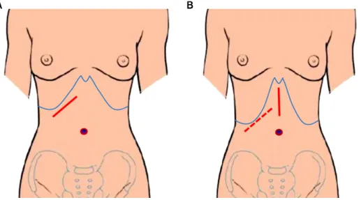
Fig. 1. Selection of incision for mini-laparotomy in female young adult patients with choledochal cyst. In patients with a relatively wide epigastrium (A), a right subcostal incision is more appropriate. In contrast, an upper midline incision appears to be more suitable for patients with a relatively narrow epigastrium (B).
prior upper abdominal surgery such as CCD operation in childhood were excluded. In addition, patients who under- went a concurrent surgical procedure such as combined hepatectomy for intrahepatic lesions were also excluded.
CCDs were classified according to the Todani mod- ification of the original Alonso-Lej classification as type I in 8 patients and type IVa in 3 patients.10 Thus, all 11 study patients had naïve CCD, which was treated by mini-laparotomy. Detailed surgical procedures have been described previously.1 All patients recovered uneventfully after surgery and were regularly followed up to date;
therefore, this study focused on cosmetic selection of skin incision and postoperative scar management. This study was approved by the Institutional Review Board of the Asan Medical Center.
In young female patients, a mini-laparotomy incision of around 9 cm in length is usually performed. The right subcostal incision is preferred because it is placed oblique to the transverse skin creases on the upper abdomen.
Another surgical option is to perform an upper midline incision, but it crosses the transverse skin creases. The ad- vantages and disadvantages of these two skin incisions were compared in this study.
RESULTS
During the first year of the study period, all five pa- tients underwent mini-laparotomy via the right subcostal incision. In contrast, during the second year, 6 patients un- derwent mini-laparotomy via either the right subcostal in-
cision (n=3) or the upper midline incision (n=3).
In 9 patients with the right subcostal incision, the mean length of skin incision was 10 cm (range: 8-12 cm), which was dependent on the depth of the subcutaneous fat and location of the right costal cartilages. After skin marking over the location of bilateral costal cartilages and xiphoid process, the skin incision was made about 2 cm away from the right costal cartilage margins (Fig. 1A). The up- permost segment of the right rectus muscle was transected. The incision was extended by using two re- tractors, one retractor each from the right side and left side. No additional incision was performed because it might result in malalignment of the extended skin incision line, leading to disadvantage in wound cosmetics. Many surgical gauze sponges (5-8 sheets) were placed in the right subphrenic area to displace the liver towards the cau- dal side, so that the hepatic hilum would be beneath the operative wound. After completing the surgical procedure, the operative wound was repaired with interrupted sutures in two layers (peritoneum, transversalis and internal obli- que muscle; and external oblique muscle and fascia) using absorbable suture materials. A Jackson-Pratt drain measur- ing 3.2 mm in outer diameter was inserted under the inter- rupted sutures on Scarpa’s fascia. The skin was closed with interrupted subcuticular monofilament sutures along with topical application of adhesive sterile strips. None of the patients developed wound problem. These patients were advised to apply a silicone gel sheet (Cica-Care, Smith & Nephew, London, UK) from 6 weeks after sur- gery until 1 year. At the 1 year follow-up, a slight bulge
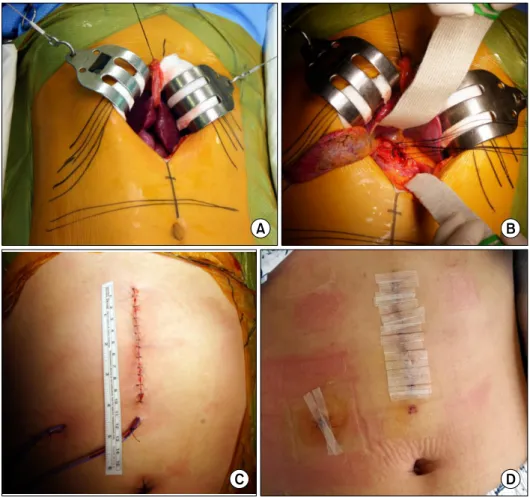
Fig. 2. Operative photographs of upper midline mini-laparotomy in a 32-year-old female patient.
(A) After skin marking of the bi- lateral subcostal margins, a 9.5 cm-long upper midline incision is made. (B) Hepatic hilum is displaced toward the midline af- ter gauze packing at the right lat- eral side. (C) The wound is re- paired meticulously and then ad- hesive tapes are applied. (D) The small skin stitches and wound drain are removed at 5 days after surgery.
on the skin scar was observed in 3 patients (37.5%) and the remaining 5 patients showed a flat skin scar. None of the patients showed noticeable atrophy of the ipsilateral rectus muscle. None of the patients complained of skin discomfort or paresthesia over the denervated skin area.
An upper midline incision was made in 3 selected pa- tients with a narrow epigastric area between the costal cartilages due to a slim body dimension (Fig. 1B). The length of skin incision uniformly measured as 9 cm. The incision was extended by using two retractors, one re- tractor each from the right side and left side (Fig. 2A).
No additional incision extension was performed. Many surgical gauze sponges (7-10 sheets) were placed in the right lateral area to shift the liver towards the midline, so that the hepatic hilum was placed under the operative wound. After completing the surgical procedure, the oper- ative wound was repaired with interrupted sutures in two layers (peritoneum and posterior sheath of the rectus mus- cles; anterior sheath of the rectus muscles) using absorb- able suture materials. A Jackson-Pratt drain measuring 3.2 mm in outer diameter was inserted under the interrupted
sutures on Scarpa’s fascia. The skin was closed with inter- rupted subcuticular monofilament sutures and both ends of skin incision were additionally repaired with 5-0 Nylon to reinforce skin approximation (Fig. 2B). Finally, adhe- sive sterile strips were attached. None of the patients de- veloped wound problem. They were also advised to apply a silicone gel sheet. At the 6 month to 1 year follow-up, a slight bulge on the skin scar was observed in 1 patient (33.3%) and the other 2 patients showed a flat skin scar.
DISCUSSION
After resection of choledochal cyst in adult patients, anastomotic stenosis of hepaticojejunostomy is the most common and most serious complication unless the patients do not have accompanying malignancy. We have pre- viously reported that the late liver-related complications such as intrahepatic stone formation, recurrent cholangitis and overt anastomotic stricture occurred in at least 16 of the 102 patients (15.7%) with CCD type I and 15 of the 72 patients (20.8%) with CCD type IVa.1
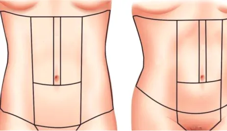
Fig. 3. Aesthetic subunits of the abdominal region and adjacent areas. The upper midline in- cision is located at the upper midline aesthetic subunit which overlies the midline of the abdomen.14
The anastomotic stricture may be closely associated with technical factors, although its risk factors were re- ported as CCD type IVa, large cyst size, short symptom duration and high grade infiltration of inflammatory cells.11 All portions of choledochal cysts should be re- moved, but in practice, residual proximal cyst walls may be left to facilitate biliary anastomosis.1,12 The proximal end of the cyst extending from the confluence of the hep- atic duct to the junction of the common duct is often diffi- cult to define clearly. Therefore, we used a customized procedure including the initial incision-opening the cyst wall and then identifying the luminal appearance grossly to determine the proximal transection line. Although this technique occasionally resulted in some cyst remnants in the hepatic ducts, it effectively reduced the risk of intract- able anastomotic stricture. The size of the anastomosis and the blood supply to the bile duct stump also seem to be important factors related to the occurrence of anasto- motic stricture. If there is a stenotic portion around the hilar bile ducts, it should be widened through in- dividualized ductoplasty. If the hepatic duct openings ap- pear to be relatively small or hypoplastic, a wider anasto- mosis must be beneficial, after leaving some cyst wall remnant.
Such an individualized approach requires a consid- erable experience with CCD surgery and biliary reconstruction. Secure biliary reconstruction in CCD sur- gery is often demanding in adult patients; therefore, a higher incidence of surgical complications is anticipated after laparoscopic or robotic resection than after open sur- gery, unlike in pediatric patients.6-9 Considering that we
experienced a transferred case requiring reoperation due to intractable anastomotic stricture after robotic surgery as well as we observed unreported surgical complications following laparoscopic approach, the indications for mini- mal-incision approach should be prudently selected as well as the surgical technique should be further refined.
As shown in the results of this study, we recognized that the cosmetic effect of both right subcostal and upper midline mini-laparotomy incisions may be similar. We have performed the upper midline incision more than 1,000 times for various hepatobiliary operations, but we have rarely performed it in young female patients except for living liver donors.13 Based on the preliminary results of this study, the upper midline approach may be permis- sible for CCD surgery from the standpoint of wound cosmetics. After placing several surgical gauze sponges in the right lateral area to shift the liver towards the midline, the hepatic hilar structures are readily exposed. If the right subcostal incision had been used in our 3 selected patients with upper midline incision, the incision in these patients might have been elongated and its right lower end might have been lowered to the umbilical level. Regarding the relaxed skin tension line, subcostal incision is usually 30-40o oblique and upper midline incision is perpendicu- lar, thus the latter is more disadvantageous. In contrast, it is reasonable to consider preservation of the rectus mus- cle function, especially in young patients with vigorous physical activity. Thus, regarding the rectus muscle func- tion, the upper midline incision is more advantageous.
We think that the indications for upper midline incision include a narrow epigastrium with slender body dimension
and special conditions requiring full rectus muscle func- tion due to occupation-specific physical activity. In such patients, selection of the upper midline incision would easily facilitate CCD surgery as well as help in wound cosmetics.
To ensure that the skin wound is cosmetically sound, two points should be emphasized. First, for upper midline incision, this incision should be placed exactly at the mid- line, thus resulting in complete symmetry. Based on the anatomical characteristics and the typical fat deposition pattern, the abdominal area can be considered to have 10 aesthetic subunits including the abdominal region and ad- jacent areas that significantly affect the aesthetic appear- ance of the midsection (Fig. 3).14 The upper midline in- cision is located at the upper midline aesthetic subunit which overlies the midline of the abdomen extending from the xyphoid sternum to the umbilicus. Since these aesthetic subunits give aesthetic silhouette to the body, incisions should be placed to show symmetry. Second, meticulous time-consuming surgical techniques are mandatory.15-17 It is important to incise the skin sharply with a knife with low-power electrocauterization because thermal burns in- volving the epidermis and dermis lead to delayed for- mation of hypertrophic scar. During surgery, excessive skin tension from wound traction should be avoided be- cause it also induces ischemia of the skin wound edge, which also leads to hypertrophic scar. A slightly longer incision would be cosmetically better than a very tightly retracted small-in-size skin incision. We usually spend 1 hour for the repair of mini-laparotomy incision measuring approximately 9-10 cm in young female patients. It in- cludes two interrupted layer-by-layer abdominal wall clo- sures, wound drain insertion, Scarpa’s fascia repair, inter- rupted subcuticular sutures, reinforcement of edge skin su- tures, and adhesive tape application.
After surgery, we also suggest application of a silicone gel sheet up to 1 year, but more than a half of our patients did not comply with the application, probably due to in- convenience and ineffectiveness.17-20 For patients showing low compliance to silicone gel sheet, it would be recom- mended to use silicone gel since 3 months after surgery.
Self-drying silicone gel is appealing because it is effec- tive, no fixation is required and it is invisible when dry.21 In conclusion, the results of this preliminary study sup- port the claim that cosmetic effect of the upper midline
incision for CCD surgery appears to be non-inferior to that of the right subcostal incision if the incision is placed accurately and repaired very meticulously.
REFERENCES
1. Cho MJ, Hwang S, Lee YJ, Kim KH, Ahn CS, Moon DB, et al. Surgical experience of 204 cases of adult choledochal cyst disease over 14 years. World J Surg 2011;35:1094-1102.
2. Wiseman K, Buczkowski AK, Chung SW, Francoeur J, Schaeffer D, Scudamore CH. Epidemiology, presentation, diag- nosis, and outcomes of choledochal cysts in adults in an urban environment. Am J Surg 2005;189:527-531.
3. Singham J, Schaeffer D, Yoshida E, Scudamore C. Choledochal cysts: analysis of disease pattern and optimal treatment in adult and paediatric patients. HPB (Oxford) 2007;9:383-387.
4. Dhupar R, Gulack B, Geller DA, Marsh JW, Gamblin TC. The changing presentation of choledochal cyst disease: an incidental diagnosis. HPB Surg 2009;2009:103739.
5. Lipsett PA, Pitt HA. Surgical treatment of choledochal cysts. J Hepatobiliary Pancreat Surg 2003;10:352-359.
6. Hwang DW, Lee JH, Lee SY, Song DK, Hwang JW, Park KM, et al. Early experience of laparoscopic complete en bloc excision for choledochal cysts in adults. Surg Endosc 2012;26:3324-3329.
7. Duan X, Mao X, Jiang B, Wu J. Totally laparoscopic cyst ex- cision and Roux-en-Y hepaticojejunostomy for choledochal cyst in adults: a single-institute experience of 5 years. Surg Laparosc Endosc Percutan Tech 2015;25:e65-e68.
8. Senthilnathan P, Patel ND, Nair AS, Nalankilli VP, Vijay A, Palanivelu C. Laparoscopic management of choledochal cyst- technical modifications and outcome analysis. World J Surg 2015;39:2550-2556.
9. Margonis GA, Spolverato G, Kim Y, Marques H, Poultsides G, Maithel S, et al. Minimally invasive resection of choledochal cyst: a feasible and safe surgical option. J Gastrointest Surg 2015;19:858-865.
10. Todani T, Watanabe Y, Narusue M, Tabuchi K, Okajima K.
Congenital bile duct cysts: Classification, operative procedures, and review of thirty-seven cases including cancer arising from choledochal cyst. Am J Surg 1977;134:263-269.
11. Kim JH, Choi TY, Han JH, Yoo BM, Kim JH, Hong J, et al.
Risk factors of postoperative anastomotic stricture after excision of choledochal cysts with hepaticojejunostomy. J Gastrointest Surg 2008;12:822-828.
12. Trinidad-Hernandez M, Rivera-Perez VS, Hermosillo-Sandoval JM. Adult choledochal cyst. Am J Surg 2007;193:221-222.
13. Suh SW, Lee KW, Lee JM, Choi Y, Yi NJ, Suh KS. Clinical outcomes of and patient satisfaction with different incision meth- ods for donor hepatectomy in living donor liver transplantation.
Liver Transpl 2015;21:72-78.
14. Parashar S. Art of abdominal contouring: advanced liposuction.
New Delhi: Jaypee Brothers Medical Publisher, 2016.
15. Mackeen AD, Berghella V, Larsen ML. Techniques and materi- als for skin closure in caesarean section. Cochrane Database Syst Rev 2012;11:CD003577.
16. Macpherson N, Lee S. Effect of different suture techniques on tension dispersion in cutaneous wounds: a pilot study. australas J Dermatol 2010;51:263-267.
17. Wall PD, Deucy EE, Glantz JC, Pressman EK. Vertical skin in- cisions and wound complications in the obese parturient. Obstet Gynecol 2003;102:952-956.
18. Li-Tsang CW, Lau JC, Choi J, Chan CC, Jianan L. A pro-
spective randomized clinical trial to investigate the effect of sili- cone gel sheeting (Cica-Care) on post-traumatic hypertrophic scar among the Chinese population. Burns 2006;32:678-683.
19. Eishi K, Bae SJ, Ogawa F, Hamasaki Y, Shimizu K, Katayama I. Silicone gel sheets relieve pain and pruritus with clinical im- provement of keloid: possible target of mast cells. J Dermatolog Treat 2003;14:248-252.
20. Nikkonen MM, Pitkanen JM, Al-Qattan MM. Problems asso- ciated with the use of silicone gel sheeting for hypertrophic scars in the hot climate of Saudi Arabia. Burns 2001;27:498-501.
21. Puri N, Talwar A. The efficacy of silicone gel for the treatment of hypertrophic scars and keloids. J Cutan Aesthet Surg 2009;2:
104-106.