ISSN 2234-3806 • eISSN 2234-3814
https://doi.org/10.3343/alm.2021.41.5.463
Determination of Clinical Characteristics of
Mycobacterium kansasii-Derived Species by Reanalysis of Isolates Formerly Reported as M. kansasii
Young-gon Kim , M.D.1, Hong Yeul Lee , M.D.2, Nakwon Kwak , M.D.2, Jae Hyeon Park , M.D.1, Taek Soo Kim , M.D.1, Man Jin Kim , M.D.1, Jee-Soo Lee , M.D.1, Sung-Sup Park , M.D., Ph.D.1, Jae-Joon Yim , M.D., Ph.D.2,3, and
Moon-Woo Seong , M.D., Ph.D.1
1Department of Laboratory Medicine, Seoul National University Hospital, Seoul, Korea; 2Division of Pulmonary and Critical Care Medicine, Department of Internal Medicine, Seoul National University Hospital, Seoul, Korea; 3Department of Internal Medicine, Seoul National University College of Medicine, Seoul, Korea Background: Seven genotypic subtypes of Mycobacterium kansasii were recently demon-
strated to represent distinct species based on phylogenomic analysis. Mycobacterium kansasii sensu stricto (formerly known as subtype 1) is most frequently associated with human diseases; only a few studies have compared the diverse clinical characteristics of M. kansasii subtypes, including their drug susceptibilities. We determined the actual inci- dence of infections caused by each subtype of M. kansasii and identified their clinical characteristics.
Methods: We subtyped isolates identified as M. kansasii over the last 10 years at a tertiary care hospital. Percent identity score of stored sequencing data was calculated using cu- rated reference sequences of all M. kansasii subtypes. Clinical characteristics were com- pared between those classified as subtype 1 and other subtypes. Student’s t-test, Wil- coxon rank-sum test, and Fisher’s exact test were used for comparisons.
Results: Overall, 21.7% of the isolates were identified as species distinct from M. kansasii.
The proportion of patients with subtype 1 M. kansasii infection who received treatment was significantly higher than that of patients with other subtype infections (55.3% vs.
7.7%, P =0.003). Only patients with subtype 1 infection received surgical treatment. Non- subtype 1 M. kansasii isolates showed a higher frequency of resistance to ciprofloxacin and trimethoprim/sulfamethoxazole.
Conclusions: Non-subtype 1 M. kansasii isolates should be separately identified in routine clinical laboratory tests for appropriate treatment selection.
Key Words: Mycobacterium kansasii, subtypes, subtype 1 M. kansasii, Non-subtype 1 M.
kansasii
Received: September 14, 2020 Revision received: November 1, 2020 Accepted: March 20, 2021
Corresponding author:
Moon-Woo Seong, M.D., Ph.D.
Department of Laboratory Medicine Seoul National University Hospital, 101 Daehak- ro, Jongno-gu, Seoul 03080, Korea Tel: +82-2-2072-4180
Fax: +82-2-747-0359 E-mail: [email protected]
© Korean Society for Laboratory Medicine This is an Open Access article distributed under the terms of the Creative Commons Attribution Non-Commercial License (https://creativecom- mons.org/licenses/by-nc/4.0) which permits unrestricted non-commercial use, distribution, and reproduction in any medium, provided the original work is properly cited.
INTRODUCTION
Mycobacterium kansasii is a common non-tuberculous myco- bacterial (NTM) species with relatively high pathogenicity [1, 2].
It can cause severe lung diseases, similar to that caused by My- cobacterium tuberculosis [3, 4]. Seven subtypes of M. kansasii
have been described, based on restriction fragment length poly- morphism analysis of hsp65 [5-7]. PCR-restriction enzyme analysis of rpoB and tuf has also been used to distinguish sub- types [8, 9]. Among these subtypes, M. kansasii sensu stricto is considered the most pathogenic and is isolated most frequently [4, 10-13]. Reports of human diseases caused by subtype 2 are
2017-03-16 https://crossmark-cdn.crossref.org/widget/v2.0/logos/CROSSMARK_Color_square.svg
rare, demonstrating its higher association with immunosuppres- sion than subtype 1 [7, 14]. There is a consensus that the other subtypes (subtypes 3–7) are not human pathogens, although this opinion is controversial owing to the limited evidence caused by the paucity of isolates of these subtypes [4]. “Myco- bacterium kansasii complex” has been proposed as an inclusive term comprising all subtypes of M. kansasii and M. gastri, which is a closely related species but indistinguishable from M. kansa- sii by 16S rRNA sequencing [4, 15].
Although the difference in the pathogenicity of M. kansasii subtypes has been recognized, these subtypes have recently been differentiated into distinct species [15, 16]. Unlike sub- species, subtypes are not a part of the standard taxonomic clas- sification system, and reporting of subtypes in mycobacterial identification by clinical laboratories is not mandatory [17, 18].
However, the recently published M. kansasii-derived species, which were formerly classified as M. kansasii subtypes, have not been included as target species of commercial kits for NTM identification [15]. Genotyping of at least one discriminatory tar- get, such as rpoB and hsp65, is required for accurate species identification.
The detection frequency of M. kansasii subtypes in clinically relevant populations have been reported; however, only a few studies have compared the clinical characteristics among infec- tions caused by M. kansasii subtypes [4, 13]. Several studies on the clinical relevance of M. kansasii have not separately ana- lyzed the subtypes, resulting in substantial variability (17%–
88%) in the reported rate of pulmonary diseases caused by M.
kansasii isolates and diversity in radiological findings [1, 2, 19–24].
To fill this knowledge gap, we reanalyzed the sequencing trace files of all isolates reported to involve M. kansasii, from an up-to-date database of reference sequences, and compared the clinical characteristics of infections caused by different M. kan- sasii subtypes.
MATERIALS AND METHODS
Participants and samplesThis study was approved by the Institutional Review Board of Seoul National University Hospital (SNUH), Seoul, Korea. We reviewed the medical records of 60 consecutive patients with M.
kansasii infection, diagnosed based on a routine NTM identifi- cation testing performed from June 15, 2011 to April 8, 2020 at SNUH. The routine NTM identification was performed by in- house method which was based on PCR amplification of the two target regions, the 5’ end of the 16S rRNA gene (about 500
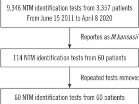
bp) and a part of rpoB gene, and subsequent Sanger sequenc- ing. Informed consent from patients was not obtained, as this was a retrospective study performed using medical records and raw data files. The patient selection process is shown in Fig. 1.
For patients with NTM identification testing being requested two or more times, we preferentially selected the tests with drug susceptibility testing results. For patients with no drug suscepti- bility testing results or two or more drug susceptibility testing re- sults, earlier NTM identification testing results were chosen. All samples included in the analysis were cultured colonies from sputum (53, 88.3%), bronchial wash or bronchoalveolar lavage fluid (6, 10.0%), and joint fluid (1, 1.7%).
Identification of M. kansasii subtypes
Sequencing data generated from routine identification testing were used. For the 60 patients, percent identity score was calculated based on curated reference sequences of each subtype of M.
kansasii. The reference sequences of the 16S rRNA gene and rpoB were curated from the List of Prokaryotic Names with Stand- ing in Nomenclature (http://www.bacterio.net) and NCBI Genbank (https://www.ncbi.nlm.nih.gov/genbank/), respectively. SnackNTM software (https://github.com/Young-gonKim/SnackNTM, last ac- cessed August 27, 2020) was used for aligning sequencing data to the curated reference sequences.
Grouping of patients
The patients were divided into two groups for comparison.
Group 1 comprised patients infected with M. kansasii subtype 1, the most pathogenic subtype [4, 10–13]. Group 2 comprised patients infected with other subtypes, M. kansasii subtypes 2, 3, and 6. Baseline characteristics of patients, clinical manifesta- tions, outcome, and drug susceptibility of the isolates were com- pared between the groups.
Reportes as M.kansasii
Repeated tests removed 9,346 NTM identification tests from 3,357 patients
From June 15 2011 to April 8 2020
114 NTM identification tests from 60 patients
60 NTM identification tests from 60 patients
Fig. 1. Among 9,364 isolates in the non-tuberculous mycobacterial (NTM) identification tests, 114 isolates were identified as M. kansa- sii (1.2%). After removing repeated isolates for the same patients, 60 isolates reported as M. kansasii were included in this analysis.
Review of medical records
Patient characteristics including age, sex, body mass index, and smoking history were retrieved from the medical records. Medi- cal histories of tuberculosis, malignancy, diabetes mellitus, liver diseases, kidney diseases, and immunocompromising diseases were reviewed. Radiologic findings and pulmonary function test results including forced expiratory volume in 1 second (FEV1), forced vital capacity (FVC), and FEV1/FVC were retrieved. Clini- cal course of NTM isolation, such as co-infection with other NTM organisms, presence of NTM pulmonary diseases, and treatment initiation were reviewed. The drug susceptibility test results were also reviewed.
Statistical analysis
Statistical analysis was performed using R software (version 4.0.2, R Foundation for Statistical Computing, Vienna, Austria).
For quantitative variable comparison, Shapiro test was used to evaluate the normality of data. Student’s t-test was used when the normality assumption was satisfied; otherwise, the non-
parametric Wilcoxon rank-sum test was used. Fisher’s exact test was used to compare categorical variables between groups. P <
0.05 were considered statistically significant.
RESULTS
Among the 60 isolates included in the analysis, 13 were reclas- sified as one of the newly reported M. kansasii-derived species (21.7%), including 10 (16.7%) isolates of M. persicum (former subtype 2), 2 (3.3%) isolates of M. pseudokansasii (former sub- type 3), and 1 (1.7%) isolate of M. attenuatum (former subtype 6). The remaining 47 isolates were classified as subtype 1.
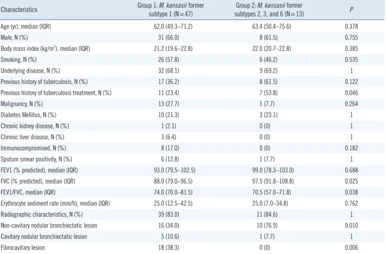
The baseline characteristics of the two patients’ groups are shown in Table 1. FVC was significantly lower in Group 1 than in Group 2 (88.0% vs. 97.5% predicted, P =0.025), leading to a significantly higher FEV1/FVC ratio in Group 1 (74.0 vs. 70.5, P =0.038). Non-cavitary nodular bronchiectatic lesions were commonly observed in Group 2 (34.0% vs. 76.9%, P =0.010) and fibrocavitary lesions were observed only in Group 1 (38.3%
Table 1. Patients’ characteristics including comorbidities, pulmonary function, and radiologic findings
Characteristics Group 1: M. kansasii former
subtype 1 (N=47)
Group 2: M. kansasii former
subtypes 2, 3, and 6 (N=13) P
Age (yr), median (IQR) 62.0 (49.3–71.2) 63.4 (50.4–75.6) 0.378
Male, N (%) 31 (66.0) 8 (61.5) 0.755
Body mass index (kg/m2), median (IQR) 21.2 (19.6–22.8) 22.0 (20.7–22.8) 0.385
Smoking, N (%) 26 (57.8) 6 (46.2) 0.535
Underlying disease, N (%) 32 (68.1) 9 (69.2) 1
Previous history of tuberculosis, N (%) 17 (36.2) 8 (61.5) 0.122
Previous history of tuberculosis treatment, N (%) 11 (23.4) 7 (53.8) 0.046
Malignancy, N (%) 13 (27.7) 1 (7.7) 0.264
Diabetes Mellitus, N (%) 10 (21.3) 3 (23.1) 1
Chronic kidney disease, N (%) 1 (2.1) 0 (0) 1
Chronic liver disease, N (%) 3 (6.4) 0 (0) 1
Immunocompromised, N (%) 8 (17.0) 0 (0) 0.182
Sputum smear positivity, N (%) 6 (12.8) 1 (7.7) 1
FEV1 (% predicted), median (IQR) 93.0 (79.5–102.5) 99.0 (78.3–103.0) 0.688
FVC (% predicted), median (IQR) 88.0 (79.0–96.5) 97.5 (91.8–108.8) 0.025
FEV1/FVC, median (IQR) 74.0 (70.0–81.5) 70.5 (57.0–71.8) 0.038
Erythrocyte sediment rate (mm/h), median (IQR) 25.0 (12.5–42.5) 25.0 (7.0–34.8) 0.762
Radiographic characteristics, N (%) 39 (83.0) 11 (84.6) 1
Non-cavitary nodular bronchiectatic lesion 16 (34.0) 10 (76.9) 0.010
Cavitary nodular bronchiectatic lesion 5 (10.6) 1 (7.7) 1
Fibrocavitary lesion 18 (38.3) 0 (0) 0.006
Abbreviations: IQR, interquartile range; FVC, forced vital capacity; FEV1, forced expiratory volume in 1 second.
vs. 0%, P =0.006).
The clinical course of the two groups is summarized in Table 2. Co-infection with M. avium complex was more frequent in Group 2 (19.1% vs. 53.8%, P =0.029). The proportion of pa- tients who satisfied the NTM pulmonary disease (NTM-PD) di- agnostic criteria did not differ between the groups (85.1% vs.
76.9%, P =0.675). However, the proportion of treated patients was significantly higher in Group 1 than in Group 2 (55.3% vs.
7.7%, P =0.003).
The in vitro drug susceptibility testing results of the available iso- lates are presented in Table 3. Among the 60 patients, 32 (25
from Group 1 and 7 from Group 2) had drug susceptibility testing results. Among the eight drugs, whose breakpoints are published in the CLSI guidelines [25], resistance to four drugs(ciprofloxacin, ethambutol, rifampin, and trimethoprim/sulfamethoxazole) was detected. Susceptibility frequencies for all the four drugs were higher in Group 1 than in Group 2. The frequency of ciprofloxacin susceptibility was significantly higher in Group 1 than in Group 2 (80.0% vs. 28.6%, P =0.019). However, there was no significant difference in the MICs of the antimicrobial drugs whose break- points are not available in the CLSI guidelines (cefoxitin, doxycy- cline, imipenem, and tobramycin) between the groups.
Table 2. Comparison of co-infection rate and clinical courses between Groups 1 and 2 Group 1: M. kansasii former
subtype 1 (N=47)
Group 2: M. kansasii former
subtypes 2, 3, and 6 (N=13) P
Co-infection with other organisms, N (%) 16 (34.0) 7 (53.8) 0.215
With M. avium complex 9 (19.1) 7 (53.8) 0.029
With M. abscessus complex 3 (6.4) 0 (0) 1
With other NTM 9 (19.1) 2 (15.4) 1
Met diagnostic criteria of NTM-PD, N (%) 40 (85.1) 10 (76.9) 0.675
Observed without treatment, N (%) 21 (44.7) 12 (92.3) 0.003
Spontaneous conversion, N (%) 6/14 (42.9) 3/7 (42.9) 1
Treatment initiation within three yrs, N (%) 26 (55.3) 1 (7.7) 0.003
Microbiologic cure* n/N (%) 18/20 (90.0) 1/1 (100) 1
Surgical treatment, N (%) 5 (10.6) 0.0 0.575
*Three or more consecutive negative results from cultures that were performed with intervals longer than one month.
Abbreviation: NTM-PD, non-tuberculous mycobacterial pulmonary disease.
Table 3. Comparison of in vitro drug susceptibilities between Groups 1 and 2
Antimicrobial Group 1 M. kansasii former subtype 1 (N=25) Group 2 M. kansasii former subtypes 2, 3, and 6 (N=7) MIC range (µg/mL) Susceptibility (N, %) MIC range (µg/mL) Susceptibility (N, %) P
Amikacin ≤1–16 25 (100.0) ≤1–16 7 (100.0) 1
Cefoxitin 64 to >256 8 to >256
Ciprofloxacin 0.5–8 20 (80.0) 0.25–8 2 (28.6) 0.019
Clarithromycin ≤0.5–1 25 (100.0) ≤0.5–1 7 (100.0) 1
Doxycycline 1 to >32 1 to >32
Imipenem 8 to >64 16 to >64
Ethambutol 0.5–16 21 (84.0) 1 to >32 5 (71.4) 0.590
Linezolid ≤2–8 25 (100.0) ≤2–4 7 (100.0) 1
Moxifloxacin ≤0.125–1 25 (100.0) ≤0.125–2 7 (100.0) 1
Rifampin ≤0.125–2 24 (96.0) 0.25–2 5 (71.4) 0.113
Tobramycin 1 to >32 4 to >32
TMP/SMX ≤0.25/4.75 to 32/608 18 (72.0) ≤0.25/4.75 to 32/608 2 (28.6) 0.074
Abbreviations: TMP/SMX, trimethoprim/sulfamethoxazole; MIC, minimum inhibitory concentration.
DISCUSSION
In this study, 21.7% of isolates previously identified as M. kan- sasii were reclassified as new species with reportedly lower pathogenicity than M. kansasii sensu stricto. As conventional line probe-based commercial kits cannot discriminate the spe- cies, a considerable proportion of isolates identified as M. kan- sasii may actually belong to different species with significantly different clinical implications [15]. Even sequencing-based methods cannot detect new species unless at least one discrim- inatory target, such as hsp65 and rpoB, is incorporated in the test.
Most reports on M. kansasii subtype 1, sensu stricto, being the most pathogenic subtype are based on its detection fre- quency in clinically relevant populations [4, 10, 12, 13]. How- ever, a recent report indicated that a specific genetic element, the espACD operon, is the main source of pathogenicity of this subtype [13]. We did not find a difference in the detection fre- quency of isolates that met the criteria for NTM-PD between Groups 1 and 2. However, other results suggested that subtype 1 isolates are more pathogenic than other subtypes. The signifi- cantly lower values of pulmonary function test parameters and FVC in Group 1 further support the higher pathogenicity of sub- type 1, considering that lung destruction can decrease the FVC.
Distinct radiographic findings were obtained for the two groups:
Group 1 showed a higher frequency of fibrocavitary lesions, and Group 2 showed a higher frequency of non-cavitary nodular bronchiectatic lesions. These results support the assumption that the disease caused by subtype 1 is more aggressive.
Only a few studies have examined the drug susceptibility of M.
kansasii subtypes [11, 12, 14]. The CLSI recommends suscep- tibility testing of clarithromycin and rifampin as first-line treat- ment, and isoniazid, ethambutol, streptomycin, amikacin, cotri- moxazole, moxifloxacin, linezolid, ciprofloxacin, and others as second-line treatment, because M. kansasii isolates are gener- ally susceptible to these drugs [12]. Indeed, all isolates included in this study were susceptible to amikacin, clarithromycin, line- zolid, and moxifloxacin. In contrast to the present study, a previ- ous study found that the drug resistance frequency was consis- tently higher for subtype 1 isolates and attributed this result to the selection pressure due to higher exposure to drugs owing to a high frequency of treatment [14].
Our study had some limitations. First, the utilization of only two target regions, the 16S rRNA gene and rpoB, would not have provided enough information for distinguishing all M. kan- sasii subtypes. No single target could accurately distinguish all
M. kansasii subtypes, requiring whole genome sequence-based approach for reliable subtyping [4]. It is possible that we have missed rarer subtypes of M. kansasii due to the limitation of the target regions used. Second, the number of isolates was limited, especially for non-subtype 1 M. kansasii isolates. We included only 2 subtype 3 M. kansasii isolates and 1 subtype 6 M. kan- sasii isolate. The adoption of laboratory methods capable of dis- tinguishing these subtypes can provide more data.
In conclusion, approximately one-fifth of the isolates identified as M. kansasii were newly designated species derived from M.
kansasii but with lower pathogenicity. Non-subtype 1 M. kansa- sii species should be identified by routine testing in clinical labo- ratories to select appropriate treatment strategies.
AUTHOR CONTRIBUTIONS
Kim YG identified patients with M. kansasii isolation, performed subtyping and drafted the manuscript. Lee HY and Kwak N re- viewed medical records of included patients. Park JH and Kim TS interpreted the data and performed the statistical analysis.
Kim MJ and Lee JS contributed to the revision of the manu- script. Park SS, Yim JJ, and Seong MW supervised the study and performed the final revision of the manuscript.
CONFLICTS OF INTEREST
No potential conflicts of interest relevant to this article were re- ported.
RESEARCH FUNDING
None declared.
ORCID
Young-gon Kim https://orcid.org/0000-0001-6840-6830 Hong Yeul Lee https://orcid.org/0000-0002-3638-8890 Nakwon Kwak https://orcid.org/0000-0002-1897-946X Jae Hyeon Park https://orcid.org/0000-0003-0261-2185 Taek Soo Kim https://orcid.org/0000-0002-2093-1721 Man Jin Kim https://orcid.org/0000-0002-9345-6976 Jee-Soo Lee https://orcid.org/0000-0002-7005-5686 Sung-Sup Park https://orcid.org/0000-0003-3754-4848 Jae-Joon Yim https://orcid.org/0000-0002-9605-0074 Moon-Woo Seong https://orcid.org/0000-0003-2954-3677
REFERENCES
1. Koh WJ, Kwon OJ, Jeon K, Kim TS, Lee KS, Park YK, et al. Clinical sig- nificance of nontuberculous mycobacteria isolated from respiratory specimens in Korea. Chest 2006;129:341-8.
2. Van Ingen J, Bendien SA, De Lange WC, Hoefsloot W, Dekhuijzen PNR, Boeree MJ, et al. Clinical relevance of non-tuberculous mycobacteria isolated in the Nijmegen-Arnhem region, The Netherlands. Thorax 2009;
64:502-6.
3. Matveychuk A, Fuks L, Priess R, Hahim I, Shitrit D. Clinical and radio- logical features of Mycobacterium kansasii and other NTM infections.
Respir Med 2012;106:1472-7.
4. Jagielski T, Borówka P, Bakuła Z, Lach J, Marciniak B, Brzostek A, et al.
Genomic insights into the Mycobacterium kansasii complex: an update.
Front Microbiol 2020;10:2918.
5. Alcaide F, Richter I, Bernasconi C, Springer B, Hagenau C, Schulze- Röbbecke R, et al. Heterogeneity and clonality among isolates of Myco- bacterium kansasii: implications for epidemiological and pathogenicity studies. J Clin Microbiol 1997;35:1959-64.
6. Picardeau M, Prod’Hom G, Raskine L, LePennec MP, Vincent V. Geno- typic characterization of five subspecies of Mycobacterium kansasii. J Clin Microbiol 1997;35:25-32.
7. Taillard C, Greub G, Weber R, Pfyffer GE, Bodmer T, Zimmerli S, et al.
Clinical implications of Mycobacterium kansasii species heterogeneity:
Swiss National Survey. J Clin Microbiol 2003;41:1240-4.
8. Kim BJ, Lee KH, Park BN, Kim SJ, Bai GH, Kim SJ, et al. Differentiation of mycobacterial species by PCR-restriction analysis of DNA (342 base pairs) of the RNA polymerase gene (rpoB). J Clin Microbiol 2001;39:
2102-9.
9. Bakuła Z, Modrzejewska M, Safianowska A, van Ingen J, Proboszcz M, Bielecki J, et al. Proposal of a new method for subtyping of Mycobacte- rium kansasii based upon PCR restriction enzyme analysis of the tuf gene. Diagn Microbiol Infect Dis 2016;84:318-21.
10. Zhang Y, Mann LB, Wilson RW, Brown-Elliott BA, Vincent V, Iinuma Y, et al. Molecular analysis of Mycobacterium kansasii isolates from the United States. J Clin Microbiol 2004;42:119-25.
11. da Silva Telles MA, Chimara E, Ferrazoli L, Riley LW. Mycobacterium kansasii: antibiotic susceptibility and PCR-restriction analysis of clinical isolates. J Med Microbiol 2005;54:975-9.
12. Bakuła Z, Modrzejewska M, Pennings L, Proboszcz M, Safianowska A, Bielecki J, et al. Drug susceptibility profiling and genetic determinants of drug resistance in Mycobacterium kansasii. Antimicrob Agents Che-
mother 2018;62:e01788-17.
13. Guan Q, Ummels R, Ben-Rached F, Alzahid Y, Amini MS, Adroub SA, et al. Comparative genomic and transcriptomic analyses of Mycobacte- rium kansasii subtypes provide new insights into their pathogenicity and taxonomy. Front Cell Infect Microbiol 2020;10:122.
14. Li Y, Pang Y, Tong X, Zheng H, Zhao Y, Wang C. Mycobacterium kansa- sii Subtype I is associated with clarithromycin resistance in China. Front Microbiol 2016;7:2097.
15. Shahraki AH, Trovato A, Mirsaeidi M, Borroni E, Heidarieh P, Hashem- zadeh M, et al. Mycobacterium persicum sp. nov., a novel species closely related to Mycobacterium kansasii and Mycobacterium gastri.
Int J Syst Evol Microbiol 2017;67:1766-70.
16. Tagini F, Aeby S, Bertelli C, Droz S, Casanova C, Prod’Hom G, et al.
Phylogenomics reveal that Mycobacterium kansasii subtypes are spe- cies-level lineages. Description of Mycobacterium pseudokansasii sp.
nov., Mycobacterium innocens sp. nov. and Mycobacterium attenuatum sp. nov. Int J Syst Evol Microbiol 2019;69:1696-704.
17. Parker CT, Tindall BJ, Garrity GM. International code of nomenclature of prokaryotes: prokaryotic code. Int J Syst Evol Microbiol 2019;69:S1-111.
18. CLSI. Laboratory detection and identification of mycobacteria. 2nd ed.
CLSI M48. Wayne, PA: Clinical and Laboratory Standards Institute. 2018.
19. Park HK, Koh WJ, Shim TS, Kwon OJ. Clinical characteristics and treat- ment outcomes of Mycobacterium kansasii lung disease in Korea. Yon- sei Med J 2010;51:552-6.
20. Moon SM, Park HY, Jeon K, Kim SY, Chung MJ, Huh HJ, et al. Clinical significance of Mycobacterium kansasii isolates from respiratory speci- mens. PLoS One 2015;10:e0139621.
21. Bloch KC, Zwerling L, Pletcher MJ, Hahn JA, Gerberding JL, Ostroff SM, et al. Incidence and clinical implications of isolation of Mycobacte- rium kansasii: results of a 5-year, population-based study. Ann Intern Med 1998;129:698-704.
22. Shitrit D, Baum GL, Priess R, Lavy A, Shitrit AB-G, Raz M, et al. Pulmo- nary Mycobacterium kansasii infection in Israel, 1999-2004: clinical features, drug susceptibility, and outcome. Chest 2006;129:771-6.
23. Bodle EE, Cunningham JA, Della-Latta P, Schluger NW, Saiman L. Epi- demiology of nontuberculous mycobacteria in patients without HIV in- fection, New York City. Emerg Infect Dis 2008;14:390-6.
24. Simons S, van Ingen J, Hsueh PR, Van Hung N, Dekhuijzen PN, Boer- ee MJ, et al. Nontuberculous mycobacteria in respiratory tract infec- tions, Eastern Asia. Emerg Infect Dis 2011;17:343-9.
25. CLSI. Susceptibility testing of mycobacteria, nocardiae, and other aero- bic actinomycetes. CLSI M24-A2. Wayne, PA: Clinical and Laboratory Standards Institute. 2011.